Compensatory orthodontic treatment of Angle
Class II malocclusion with posterior open bite
» The author reports no commercial, proprietary or financial interest in the products or companies described in this article.
» The patient displayed in this article previously approved the use of her facial and intraoral photographs.
Contact address: José Newton Torres
SCN Qd 2, bloco D, entrada A, sala 911 – Brasília / DF – Brazil CEP: 70.712-903 – Email: [email protected] INTRODUCTION
Female patient searched for treatment at the age of 18.7 months, with the chief complaint that, ater 7 years with removable appliances, she was not satisied with the inal result and was not able to “chew food with posterior teeth”, swallowing the food in big piec-es. There was no relevant data in her medical history.
DIAGNOSIS
The patient presented a Class II skeletal pattern, characterized by mandibular deiciency, with ANB = 6° (SNA = 82° and SNB = 76°) (Tab 1). The proile was
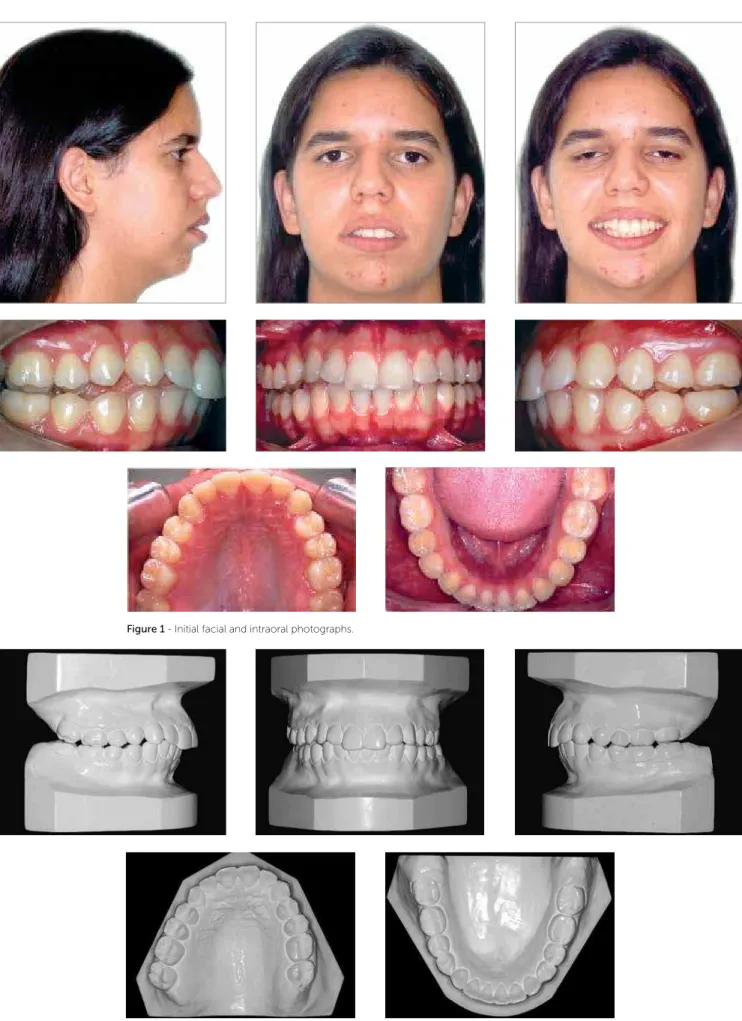
convex, with obtuse nasolabial angle, nasal apex slight-ly to the right, long face pattern, absence of passive lip sealing, increased lower facial third and short neck-chin line (Fig 1). The intraoral exam detected an Angle Class II, division 1 malocclusion, expanded and asym-metric upper and lower arches, 6-mm overjet, posterior open bite, only with second molar occlusal contact and excessive wearing on the upper and lower molar cusps, due to an attempt of occlusal adjustment (Figs 1 and 2). The upper and lower incisors were protruded and with buccal tipping, with apical rounding of the upper incisor roots (Figs 3 to 5).
José Newton Torres1
1Specialist in Orthodontics and Facial Orthopedics by the ACD/Campinas. Director
of the Brazilian Association of Orthodontics and Facial Orthopedics (ABOR) and BBO Diplomate.
How to cite this article: Torres JN. Compensatory orthodontic treatment of Angle Class II malocclusion with posterior open bite. Dental Press J Orthod. 2013 Sept-Oct;18(5):140-6.
The present case report addresses the treatment of an Angle Class II malocclusion in an adult female patient, long face pattern, with posterior open bite and dental arches extremely expanded, due to previous treatment. The patient and parents rejection to a treatment with orthognathic surgery led to orthodontic camoulage of the skeletal discrepancies. This clinical case was presented to the Brazilian Board of Orthodontics and Facial Orthopedics (BBO) as one of the requirements to become a BBO Diplomate.
Keywords:Angle Class II. Anterior open bite. Orthodontic camoulage.
O presente relato de caso aborda o tratamento de uma má oclusão de Classe II de Angle em paciente do sexo femi-nino, adulta, face longa, com mordida aberta posterior, e arcadas dentárias extremamente expandidas, decorrente de tratamento prévio. A não aceitação, por parte da paciente e de seus pais, de um tratamento envolvendo cirurgia ortog-nática levou à realização de camulagem ortodôntica das discrepâncias esqueléticas. Esse caso clínico foi apresentado à Diretoria do Board Brasileiro de Ortodontia e Ortopedia Facial (BBO) como parte dos requisitos para obtenção do título de Diplomado pelo BBO.
Torres JN BBO Case Report
Figure 3 - Initial periapical radiographs. Figure 4 - Initial panoramic radiographs.
TREATMENT PLAN
The first treatment plan option presented to the parents and the patient was the surgical-orthodontic combined treatment, which was immediately re-fused. Thus, an alternative with orthodontic camou-flage was presented, which consisted on the use of upper and lower fixed orthodontic appliances, and
Torres JN BBO Case Report
pation braided archwires and intermaxillary elastics (5/16-in) would be used. The retention planned was a wraparound removable upper plate, and lingual ca-nine-to-canine fixed retainer.
Thus, the treatment objectives were to reduce the SNA angle, by retracting the upper incisors, maintain-ing the maxilla and mandible in their vertical position, contracting upper and lower dental arches, correcting molar and canine relationship, the open bite, overbite and overjet, improving facial proile, if possible.
TREATMENT PROGRESS
The maxillary and mandibular first and second molars were banded and the other teeth were bonded. Straight-wire brackets (0.022-in x 0.028-in), Roth’s prescription, were used. A contracted palatal bar was placed in the upper first molars and the extraction of upper first premolars was requested, followed by the extraction of lower second premolars. Alignment and leveling was started after extracting these teeth with 0.014-in, 0.016-in, 0.018-in NiTi archwires; in ad-dition, Australian 0.020-in wire, 0.017 X 0.025-in NiTi and 0.019 X 0.025-in stainless steel (SS) arch-wires were used. Then, space closure was performed with the aid of 0.019 X 0.025-in SS archwires with prevalence of upper anterior teeth retraction, and an-chorage loss of lower posterior teeth. After a few ap-pointments, difficulty in moving teeth of the lower right side was observed. Thus, mini-implants place-ment in this region was performed for skeletal an-chorage, until the total closure of spaces.
Intercuspation was performed as planned, using 0.021 X 0.025-in braided archwires and 5/16-in in-termaxillary elastics (150 gf).
RESULTS
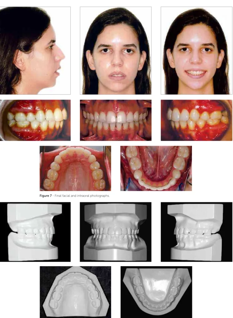
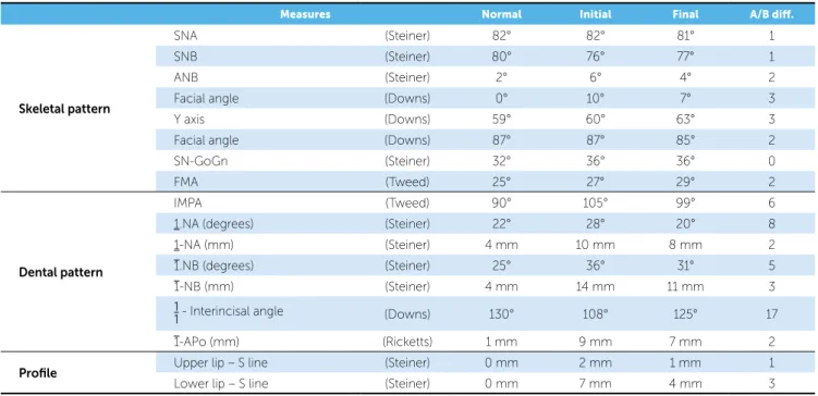
There was maintenance of the vertical position in both arches, with slight reduction of the SNA angle, which led to a reduction of the ANB angle (Table 1). The relationship for molars and canines in key of
oc-clusion was obtained on both sides, closing of the posterior open bite, correction of overjet and con-traction of upper and lower arches were also achieved (Figs 7 and 8). A decrease in upper and lower inter-molar distance of 10 mm and 11 mm, respectively, was observed, as well as a decrease of upper and lower intercanine distance of 3 mm in both arches. A slight increase of the nasolabial angle was observed, due to the retrusion of anterior upper teeth, with little al-teration of the facial profile, keeping the absence of passive lip sealing, and hyperactive mentalis muscle.
A good root parallelism was obtained at the end of treatment, as observed in the panoramic radiograph (Fig 9). Periapical radiographs showed slight increase in the apical root remodeling of incisors (Fig 10).
On cephalometric superimpositions extrusion of irst upper molar was observed, which was compensated by the mesial movement of the lower molar (Figs 13 and 14), contributing for the maintenance of the mandibu-lar plane angle. The great reduction of the intermomandibu-lar distance obtained was possible due to the mesial move-ment of these teeth, which came to occupy a narrower region on the basal bones. For retention a canine-to-canine ixed retainer was used on the lower arch, and a wraparound removable plate on the upper dental arch.
FINAL CONSIDERATIONS
Class II skeletal discrepancy, with an increased facial lower third, can be corrected by redirecting growth in young patients, or through orthognathic surgery, in adult patients.1 In both cases, it is possible to perform orthodontic camouflage when growth was not adequate or when the patient does not accept combined surgical-orthodontic treatment.2,3
Figure 7 - Final facial and intraoral photographs.
Torres JN BBO Case Report
Figure 10 - Final periapical radiographs.
Figure 9 - Final panoramic radiograph.
Figure 11 - Final lateral cephalometric radiograph. Figure 12 - Final cephalometric tracing.
B
1. Tulloch JF, Lenz BE, Phillips C. Surgical versus orthodontic correction for Class II patients: Age and severity in treatment planning and treatment outcome. Semin Orthod. 1999;5(4):231-40.
2. Bishara SE, Cummins DM, Jakobsen JR, Zaher AR. Dentofacial and soft tissue changes in Class II, division 1 cases treated with and without extractions. Am J Orthod Dentofacial Orthop. 1995;107(1):28-37. 3. Proit WR, Fields HW, Sarver DM. Contemporary orthodontics. 4th ed.
St. Louis: Mosby Elsevier; 2007.
REFERENCES
Measures Normal Initial Final A/B dif.
Skeletal pattern
SNA (Steiner) 82° 82° 81° 1
SNB (Steiner) 80° 76° 77° 1
ANB (Steiner) 2° 6° 4° 2
Facial angle (Downs) 0° 10° 7° 3
Y axis (Downs) 59° 60° 63° 3
Facial angle (Downs) 87° 87° 85° 2
SN-GoGn (Steiner) 32° 36° 36° 0
FMA (Tweed) 25° 27° 29° 2
Dental pattern
IMPA (Tweed) 90° 105° 99° 6
1.NA (degrees) (Steiner) 22° 28° 20° 8
1-NA (mm) (Steiner) 4 mm 10 mm 8 mm 2
1.NB (degrees) (Steiner) 25° 36° 31° 5
1-NB (mm) (Steiner) 4 mm 14 mm 11 mm 3
1
1- Interincisal angle (Downs) 130° 108° 125° 17
1-APo (mm) (Ricketts) 1 mm 9 mm 7 mm 2
Profile Upper lip – S line (Steiner) 0 mm 2 mm 1 mm 1
Lower lip – S line (Steiner) 0 mm 7 mm 4 mm 3