791
CLINICS 2007;62(6):791-4
LETTER TO THE EDITOR
Instituto de Ortopedia e Traumatologia, Hospital das Clínicas, Faculdade de Medicina, Universidade de São Paulo, Brazil
Email: [email protected]
TREATMENT OF OSTEOID OSTEOMA IN THE
VERTEBRAL BODY OF THE LUMBAR SPINE BY
RADIOFREQUENCY ABLATION
Alexandre Fogaça Cristante, Tarcisio Barros Filho, Reginaldo Perilo de Oliveira, Almir F Barbarini, William GJ Teixeira
INTRODUCTION
Osteoid osteoma is a rare bone tumor initially described
by Jaffe in 1935.
1It is characterized as a bone-producing
tumor that is most frequently observed in the lower
extremi-ties of children or young adults (11-22 years). Osteoid
os-teoma is differentiated from osteoblastoma according to
size. Osteoid osteoma is smaller than 1.5 centimeters in
di-ameter.
2In approximately 10% to 25% of the cases,
3-8the tumor
is observed in the vertebral column with a predilection for
posterior elements of the vertebrae.
9-11In only 10% of the
cases in which the spine is affected
12is it found in the
ver-tebral body, but involvement of the spine is more common
in the lumbar vertebrae.
12,13Here the disease is
character-ized by localcharacter-ized pain in the affected vertebra,
14,15and
pos-sibly by radiating pain similar to a disc hernia,
9,16although
without other findings in the physical and neurological
evaluation.
17The pain is generally worse during the night
and improves with the use of non-hormonal
anti-inflam-matory drugs.
18Scoliosis secondary to pain and muscular spasms
19is a
common finding in affected adolescents (63% to 70%). If
treatment is delayed, scoliosis may become a complication
since the curve may become structured through
asymmet-ric inhibition of the growth of the vertebral
epiphy-sis.
13,17,20,21The tumor is generally located at the apex of
the deformity.
18,19When the fourth or fifth lumbar vertebra
is involved, it is generally associated with pelvic obliquity.
18The tumor niche, even when small, can generally be
observed by means of scintigraphy with technetium.
22It is
possible to identify the lesion on tomographic sections of
thickness less than 1.5 centimeters and magnetic resonance
images (MRI). Osteoid osteoma is better seen on MRI
be-cause of its high signal in the bone around the lesion in
sections with T2 weighting, thus demonstrating local
edema.
22CASE REPORT
A 44-year-old female patient sought orthopedic
attend-ance because of constant lumbar pain that had lasted for
two years with progressive worsening. She reported that the
pain was more intense at night, but did not worsen with
movement or any pain crises during the day. Her pain did
not radiate to the lower limbs or other regions. There was
no history of trauma, fever, weight loss, or sphincter
al-terations. She had previously undergone treatment with
non-hormonal anti-inflammatory drugs, with improvement
only while she was using the medications. On physical
evaluation, she had pain on palpation of the fourth lumbar
vertebra, without deformity. Neurological and vascular
ex-aminations did not present alterations.
Radiography of the lumbar spine did not reveal
abnor-malities. Scintigraphy using Tc
99mdemonstrated increased
uptake on the left side of the vertebral body of the fourth
vertebra (figure 1). Computed tomography of the lumbar
spine revealed an area of hypoattenuation surrounded by an
area of hyperattenuation (bone sclerosis), suggestive of an
osteogenic tumor (figures 2 and 3). Complementary
792
CLINICS 2007;62(6):791-4 Treatment of osteoid osteoma in the vertebral body of the lumbar spine by radiofrequency ablation
Cristante af et al.
nation using MRI demonstrated a signal alteration of 1 cm
diameter in the vertebral body of the fourth lumbar
verte-bra, close to the base of the left pedicle, surrounded by an
area of signal compatible with bone edema (figures 4 and
5). The anamnesis data, physical evaluation, and
complemen-tary examinations suggested the presence of osteoid osteoma
in the vertical body of the fourth lumbar vertebra.
A tomography-guided biopsy was performed, and
ma-terial was collected for cultures, pathological studies in
par-affin, and fast freezing (
in print
). Pathological study of
fro-zen sections ruled out the presence of neoplastic cells. At
the same time, minimally invasive destruction of the tumor
was performed through a pedicullar approach, via an
Arthrocare® radiofrequency probe set at 80
o. The correct
positioning of the probe was confirmed with computed
to-mography (figure 6). The histopathological examination of
paraffin sections confirmed the diagnosis of osteoid
os-teoma (figure 7). There was no bacterial growth in the
cul-tures collected.
Figure 2 - Computed tomography demonstrating an area of hypoattenuation surrounded by an area of hyperattenuation (bone sclerosis).
Figure 3 - Tomographic reconstruction demonstrating the tumor niche and adjacent bone sclerosis.
Figure 4 - Magnetic resonance showing an axial section through the fourth lumbar vertebra in a T1 sequence, demonstrating alteration in the vertebral body at the base of the left pedicle.
Figure 5 - Magnetic resonance showing a sagittal section through the lumbar column in a T2 sequence, demonstrating the tumor niche and the sclerosis halo.
793
CLINICS 2007;62(6):791-4 Treatment of osteoid osteoma in the vertebral body of the lumbar spine by radiofrequency ablation Cristante af et al.
After the procedure, the patient was allowed to walk
with the use of a Putti jacket. The jacket was used for six
weeks, and the patient’s pain progressively improved. One
year after the procedure, computed tomography did not
demonstrate any tumor, and the patient did not report any
lumbar pain.
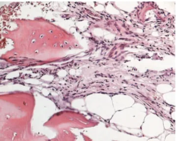
Figure 7 - Histological appearance of the material collected from the percutaneous biopsy, processed in paraffin for staining with hematoxylin and eosin. Observe the osteoid osteoma niche and the eosinophilic areas of newly formed bone without a trabecular pattern.
DISCUSSION
Osteoid osteoma in the vertebral column is more
fre-quent in young adults (11-22 years)
1and is also more
com-mon in the posterior elements of the vertebra.
10The case
in question did not present the typical epidemiology.
The natural history of osteoid osteoma demonstrates the
possibility of spontaneous cure of the lesion after two to
eight years. However, intense pain and the risk of
second-ary scoliosis
20justify surgical treatment in selected cases.
The surgical treatment consists of
en-bloc
resection to
re-move the niche and the sclerosis halo.
10,23,24Osteoid osteoma can usually be diagnosed through
com-puted tomography. It demonstrates a radiolucent nidus
sur-rounded by a dense reactive rim of cortical bone which is
usually less than 1 centimeter in diameter.
25However, the
definitive diagnosis must be made by histopathological
ex-amination.
22Biopsy of the lesion and resection of the tumor
niche aided by intraoperative computed tomography has
been described by several authors.
26-28Radiofrequency ablation was initially described by
Rosenthal et al. Recently, it has been successfully used in
minimally invasive treatment of osteoid osteoma,
15,29-33with
fewer complications compared to surgical treatment.
15However, the efficacy of this procedure still needs to be
analyzed with a larger case series.
REFERENCES
1. Jaffe HL. Osteoid-osteoma. A benign osteoblastic tumor of osteoid and atypical bone. Arch Surg. 1935;31:709-28.
2. McLeod RA, Dahlin DC, Beabout JW. The spectrum of osteoblastoma. Am J Roentgenol. 1976;126:321-5.
3. Barei DP, Moreau G, Scarborough MT, Neel MD. Percutaneous radiofrequency ablation of osteoid osteoma. Clin Orthop Relat Res. 2000;373:115-24.
4. Boriani S, Weinstein JN. Differential diagnosis and surgical treatment of primary benign and malignant neoplasms. In: Frymoyer JW, editor. The adult spine. New York: Raven Press; 1997. p. 950-87.
5. Jackson RP, Reckling PW, Mants FA. Osteoid Osteoma and osteoblastoma: similar histology lesions with different natural histories. Clin Orthop Relat Res. 1977;128:303-13.
6. Lindner NJ, Ozaki T, Roedl R, Gosheger G, Winkelmann W, Wortler K. Percutaneous radiofrequency ablation in osteoid osteoma. J Bone Joint Surg Br. 2001;83:391-6.
7. Raskas DS, Graziano GP, Herzenberg JE, Heidelberger KP, Hensinger RN. Osteoid osteoma and osteoblastoma of the spine. J Spinal Disord. 1992;5:204-11.
8. Azouz EM, Korolowski K, Marton D, Sprague P, Zerhouni A, Asselah F. Osteoid osteoma and osteoblastoma of the spine in children: report of 22 cases with brief literature review. Pediatr Radiol. 1986;16:25-31.
9. Fountain EM, Burge CH. Osteoid osteoma of the cervical spine. A review and case report. J Neurosurg. 1961;18:380-3.
10. Marcove RC, Heelan RT, Huvos AG, Healey J, Lindeque BG. Osteoid osteoma: diagnosis, localization and treatment. Clin Orthop Relat Res. 1991;267:197-201.
11. Crouzet G, Mnif J, Vasdev A, Pascal-Ortiz D, Chirossel JP, Pasquier B. Osteoid osteoma of the spine: radiological aspects and value of arteriography. Four cases. J Neuroradiol. 1989;16:45–59.
12. Heiman ML, Cooley CJ, Bradford DS. Osteoid osteoma of a vertebral body: report of a case with extension across the intervertebral disk. Clin Orthop Relat Res. 1976;118:159–63.
13. Keim HA, Reine FG. Osteoid osteoma as a cause of scoliosis. J Bone Joint Surg Am. 1975;57:159–63.
794
CLINICS 2007;62(6):791-4 Treatment of osteoid osteoma in the vertebral body of the lumbar spine by radiofrequency ablation
Cristante af et al.
15. Rosenthal DI, Hornicek FJ, Wolfe MW, Jennings LC, Gebhardt MC, Mankin HJ. Percutaneous radiofrequency coagulation of osteoid osteoma compared with operative treatment. J Bone Joint Surg Am. 1998;80:815-21.
16. Dahlin DC. Bone tumors: general aspects and data on 6,221 cases. Philadelphia: Lippincott-Raven; 1996. p. 463
17. Mehta MH. Pain provoked scoliosis. Observations on the evolution of the deformity. Clin Orthop. 1978;135:58-65.
18. Kirwan EO, Hutton PA, Pozo JL, Ransford AO. Osteoid osteoma and benign osteoblastoma of the spine. Clinical presentation and treatment. J Bone Joint Surg Br. 1984;66:21-6.
19. Lundeen MA, Herren JA. Osteoid-osteoma of the spine: sclerosis in two levels. A case report. J Bone Joint Surg Am. 1980;62:476-8.
20. MacLellan DT, Wilson FC. Osteoid osteoma of the spine. A review of the literature of the literature and report of six new cases. J Bone Joint Surg Am. 1967;49:111-21.
21. Saifuddin A, White J, Sherazi Z, Shaikh MI, Natali C, Ransford AO. Osteoid osteoma and osteoblastoma of the spine. Factors associated with the presence of scoliosis. Spine. 1998;23:47-53.
22. Kchouk M, Mrabet A, Touibi S, Douik M, Siala M, Slimen N. Osteoid osteoma of the spine. Radiological study of 21 cases. J Radiol. 1993;74:135-42.
23. Lee D, Malawer M. Staging and treatment of primary and persistent (recurrent) osteoid osteoma: evaluation of intraoperative nuclear scanning, tetracycline fluorescence and tomography. Clin Orthop Relat Res. 1992;281:229-38.
24. Ozaki T, Liljenqvist U, Hillmann A, Halm H, Lindner N, Gosheger G, et al. Osteoid osteoma and osteoblastoma of the spine: experience with 22 patients. Clin Orthop Relat Res. 2002;397:394-402.
25. McGarry SV, Gibbs CP. Radiofrequency ablation in bone neoplasia. Curr Opin Orthop. 2005;16:484-8.
26. Poey C, Clement JL, Baunin C, Assoun J, Puget-Mechinaud C, Giron J, et al. Percutaneous extraction of osteoid osteoma of the lumbar spine under CT guidance. J Comput Assist Tomogr. 1991;15:1056-8
27. Baunin C, Puget C, Assoun J, Railhac JJ, Cahuzac JP, Clement JL, et al. Percutaneous resection of osteoid osteoma under CT guidance in eight children. Pediatr Radiol. 1994;24:185-8.
28. Labbe JL, Clement JL, Dubarc B, Poey C, Raihac JJ. Percutaneous extraction of vertebral osteoid osteoma under computed tomography guidance. Eur Spine J. 1995;4:368-71.
29. Rosenthal DI, Springfield DS, Gebhardt MC, Rosenberg AE, Mankin HJ. Osteoid osteoma: percutaneous radiofrequency ablation. Radiology. 1995;197:451-4.
30. Rosenthal DI, Alexander A, Rosenberg AE, Springfield D. Ablation of osteoid osteomas with a percutaneously placed electrode: a new procedure. Radiology. 1992;183:29-33.
31. Tillotson C, Rosenberg A, Rosenthal D. Controlled thermal injury of bone: report of a percutaneous technique using radiofrequency electrode and generator. Invest Radiol. 1989;24:888-92.
32. De Berg J, Pattynama P, Obermann W, Bode PJ, Vielvoye GJ, Taminiau AH. Percutaneous computed-tomography guided thermocoagulation for osteoid osteomas. Lancet. 1995;346:350-1.