CASE REPORT
MSX2
copy number increase and craniosynostosis:
copy number variation detected by array comparative
genomic hybridization
Karla de Oliveira Pelegrino,ISofia Sugayama,II Karina Lezirovitz,IIIAna Lu´cia Catelani,IFernando Kok,I Maria de Lourdes ChauffailleI
IInstituto de Pesquisa e Desenvolvimento, Grupo Fleury, Sa˜o Paulo/SP, Brazil.IIHospital das Clı´nicas da Faculdade de Medicina da Universidade de Sa˜o
Paulo, Departamento de Pediatria, Sa˜o Paulo/SP, Brazil.IIIHospital das Clı´nicas da Faculdade de Medicina da Universidade de Sa˜o Paulo, Departamento de
Otorrinolaringologia (LIM32), Sa˜o Paulo/SP, Brazil.
Email: [email protected] Tel.: 55 11 5014-7621.
INTRODUCTION
Craniosynostosis is a disorder characterized by the premature fusion of the calvarial sutures, causing an abnormal skull shape. Because this disorder occurs with a relatively high frequency, estimated at 1 in 2500 individuals, craniosynostosis represents a relevant medical problem (1). More than 100 syndromes have been shown to be associated with this disorder. It is believed that at least 20% of cases are due to single gene mutations or chromosome abnormalities (2). Although most cases can be considered both clinically and genetically heterogeneous, there is evidence that six genes are involved in many cases: MSX2, FGFR1, FGFR2, FGFR3, FBN1,andTWISTY(1).
The Msh homeobox 2 (Msx2) gene encodes a home-odomain transcription factor protein and is expressed in migrating cranial neural crest cells during development (3). Msx2 has central roles in craniofacial development (4) and limb and tissue formation (6). Furthermore, Msx2 over-expression was demonstrated to be associated with cranio-synostosis in mice (5,7), indicating that normal craniofacial formation is dependent on the Msx2 dosage. Corroborating these findings, increases in the copy number in theMSX2
region have been reported in craniosynostosis patients (8-11). In the present study, we describe the findings from a whole-genome array comparative genomic hybridization (aCGH) analysis of a four-year-old patient exhibiting cranio-synostosis, microcephaly, psychomotor development delay, short stature, and cognitive impairment, among other ab-normalities. We found a gain in a region of chromosome 5q35.2 that contains MSX2 and has been shown to be associated with craniosynostosis. Quantitative PCR con-firmed the increase in theMSX2copy number.
CASE DESCRIPTION
This study was approved by the Hospital das Clı´nicas Institutional Ethics Committee, and written informed consent
was obtained from the family. The propositus (a girl) was the first live born child of a healthy and non-consanguineous couple (the mother was 38 years old and the father was 40 years old at the time of conception). The mother is of Japanese descent, and the father is of African descent. The mother had experienced two previous miscarriages due to incontinence of the endocervical isthmus. The pregnancy was uneventful except for mild vaginal bleeding during the first trimester. The patient was born preterm (31 weeks) after vaginal delivery, with Apgar scores of 8 and 9, a weight of 1550 g, a length of 38.5 cm, and an OFC of 28.0 cm. At birth, the patient exhibited respiratory distress and underwent orotracheal intubation (3 days). The patient was hypotonic, had poor suction, and exhibited neonatal jaundice and metabolic disturbances (hyponatremia and hypocalcemia). She was discharged at 2 months of age at a weight of 1910 g. The patient also exhibited failure to thrive, recurrent otitis and global developmental delay. She was able to sit without support at 15 months and able to walk with no support and say simple words at 24 months.
Physical examination at 19 months showed a weight of 6500 g, a length of 68.5 cm, and an OFC of 42.5 cm. The patient’s face was peculiar, with a narrowing of the frontal diameter, upslanting palpebral fissures, an enlarged nasal root, low and posteriorly rotated ears, bilateral convergent strabismus, a high arched palate and mild retrognathia. Neuroimaging studies (CT) revealed coronary craniosynos-tosis. No heart defects were observed. Abdominal ultra-sonography showed a mild abnormality of the renal parenchyma, but the patient’s kidney function is normal.
Phytohemagglutinin (PHA)-stimulated lymphocytes were subjected to G-banding karyotyping (500 bands) of the propositus and progenitors. The results were described according to the recommendations of the ISCN (2008). G-banding karyotype analysis of the patient showed a normal 46,XX pattern with no structural or numeric variations. The same was observed for both progenitors.
For the aCGH experiments, DNA from the patient and progenitors was extracted from blood samples using a QIAamp DNA Blood Midi kit (Qiagen, Germany). Labeling and hybridization reactions were performed as recommended by the manufacturer (Perkin Elmer, Norwalk). aCGH was performed with Constitutional Chip 4.0 (Perkin Elmer, Norwalk), which includes 5000 BAC clones spotted in
Copyrightß2012CLINICS– This is an Open Access article distributed under the terms of the Creative Commons Attribution Non-Commercial License (http:// creativecommons.org/licenses/by-nc/3.0/) which permits unrestricted non-commercial use, distribution, and reproduction in any medium, provided the original work is properly cited.
Table 1 -Clones that exhibited copy number variation detected through aCGH screening of the patient’s genome.
Clones Status of alteration Cytogenetic location Genomic coordinates (GRCh36/ hg18) Genes present in the region
RP11-203P12 Loss 4q22.1 4:88,282,368- 88,336,595 KLHL8
RP11-603O17 Gain 5q35.2 5:173,984,926- 174,145,340 MSX2
RP11-606P24 Gain 5q35.2 5:174,718,444- 174,907,513 DRD1, SFXN1 RP11-10J21 Loss 8q24.3 8:142,215,092- 142,412,727 DENND3, SLC45A4 RP11-153P4 Loss 9q34.2 9:135,531,566- 135,710,693 SARDH, VAV2
RP11-311J21 Loss 9q34.3 9:136,982,786- 137,166,802 OLFM1
duplicate with a resolution of at least 650 kb and human DNA segments of 100,300 kb distributed through the whole genome.
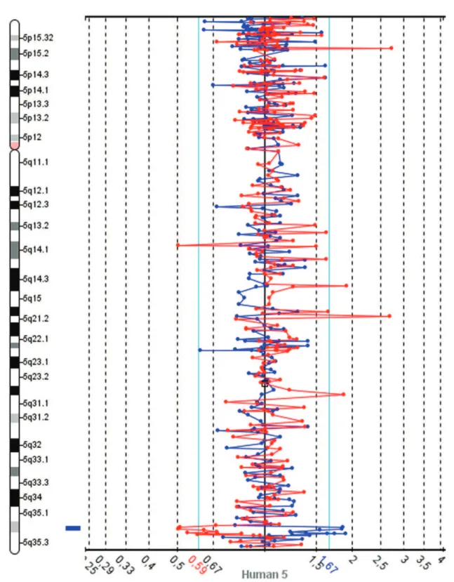
The slides were scanned with InnoScan 710 (Innopsys, Carbonne, France), and MAPIX 4.5 software was used to generate GPR files. Data analysis was conducted with SpectralWareHv2.3.3 aCGH Analysis System software (12) from Perkin Elmer. The computational parameters used were pin linear normalization, a threshold between 0.7 and 1.3, a Loess alpha of 0.1 for normalization and a confidence level of 95%. Median values were used for the interpretation of the results. The Database of Genomics Variants was consulted to determine the size of chromosome regions and to identify CNVs (13). These analyses revealed gains in two neighboring BAC clones on 5q32.2 and losses in 4q22.1, 8q24.3 and 9q34.2-34.3 (Table 1). A spectral image of chromosome 5 from the patient demonstrating the gain in 5q35.2 is presented in Figure 1. The patients revised karyotype was 46,XX.arr 4q22.1(88,282,368-88,336,595)x1, 5q35.2(173,984,926-174,145,340)(174,718,444-174,907,513)x5, 8q24.3(142,215,092-142,412,727)x1, 9q34.2q34.3(136,982,786-137,166,802)x1[hg18].
DNA samples from the patient and progenitors were used for MSX2 gene copy number assessment. A TaqMan copy number assay (Applied Biosystems, Foster City, CA) consist-ing of an MGB probe labeled with FAM and unlabeled PCR primers for MSX2 (assay Hs01821094_cn) was employed. Unlabeled primers and a VIC dye-labeled TAMRA probe for RNase P gene, which is known to be present in two copies in human diploid genome, were used for normalization.
Reactions were performed as recommended by the manufac-turer in triplicate. Duplex real-time polymerase chain reactions were conducted in a 7500 Real-Time PCR System, and the data were analyzed with CopyCallerTMSoftware v1.0, both from
Applied Biosystems. Cycle thresholds (CT) were calculated
using the relative quantification method (Applied Biosy-stems). Copy numbers were calculated by determining the difference in the CT between the target and control probes
(DCT). Cycle thresholds greater than 32 were excluded from
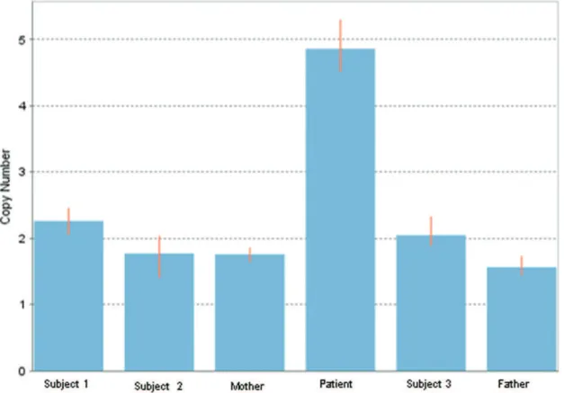
analyses. A quantitative PCR assay to detect MSX2 CNVs revealed two copies for both progenitors and five copies for the patient (Figure 2). Phenotypically normal subjects unre-lated to the studied family also had two copies. The mean delta Ct, calculated as the FAM Ct divided by the VIC Ct, and the standard deviation of the delta Ct were 0.2192¡0.13 for Subject 1, 0.5772¡0.29 for Subject 2, 0.5807¡0.1 for the patient’s mother, -0.8858¡0.12 for the patient, 0.3630¡0.16 for Subject 3 and 0.7518¡0.12 for the patient’s father.
DISCUSSION
A search for similar abnormalities in DECIPHER (Database of Chromosomal Imbalance and Phenotype in Humans Using Ensembl Resources) (14) returned three cases with chromosome 4 alterations related to that observed in the present study (cases 753, 994, and 249573). The phenotypes in these cases included mental retardation/developmental delay, short stature, atrial septum defects, hypotonia and eyelid defects, among other abnormalities. However, it is important to stress that no correlation between a loss in the
Figure 2 -Assessment of MSX2 copy number variation. The estimated copy number variations for the patient, the progenitors, and
4q22.1 region and craniosynostosis has been described. In addition, no patient with case data reported in DECIPHER who had alterations in 8q24.3 or 9q34.2-34.3 similar to those in our patient exhibited craniosynostosis.
In a detailed study that investigated common CNVs in Asian population, it was found that the KLHL8 (4q22.1), VAV2 (9q34.2) and OLFM1 (9q34.3) genes exhibited copy number variations in phenotypically normal subjects (15), indicating that copy number losses in these genes most likely do not contribute to the craniosynostosis phenotype. Corro-borating these findings, a search of the Online Mendelian Inheritance in Man (OMIM) database (16) returned no correlations between the genes KLHL8, VAV2, DRD1, SFXN1, DENND3, SLC45A4 and OLFM1 and clinical pheno-types. The SARDH gene, located in the 9q34.2 region, was shown to be associated with sarcosinemia, which is generally considered a benign condition unrelated to neurologic symptoms or significant clinical problems (17).
Among the CNVs found in the present work, the gain in the 5q35.2 chromosome region where theMSX2gene is located has already been reported to be associated with craniosynos-tosis (8); however, the MSX2 gain was confirmed using molecular techniques in only four cases (8-11). MSX2 is a member of the homeobox MSX family and is related to
Drosophila muscle segment homeobox (msh). MSX2 has important roles in tissue and organ development (3) and is also expressed in several regions of the developing skull. In mice, Msx2 is strongly expressed in osteogenic cells from calvarial sutures, andMsx2activation is thought to initiate the release of factors from the dura mater that affect osteoblastic suture cells (18,19).
Consistent with this role of Msx2, transgenic mice that over expressed this gene exhibited premature suture closure and craniofacial abnormalities (5), whereas other evidence suggests thatMSX2haploinsufficiency leads to a delayed or incomplete closure of the opening between the frontal and parietal bones. MSX2haploinsufficiency has been demon-strated to be responsible for the majority of foramina parietalia permagna (FPP) cases (20-22).
There are 39 reported cases of 5q distal trisomy, but craniosynostosis was observed in only ten of these cases. Because most craniosynostosis patients exhibit a distal 5q duplication, it is hypothesized that in patients with larger duplications, MSX2 expression may be inhibited due to extra copies of other genes (8). Additional cases may help to elucidate the validity of this hypothesis.
In conclusion, our analysis indicates that an increase in
MSX2 copy number is correlated with this disorder, corroborating previous findings that subjects with gains in 5q35.2 in theMSX2gene region exhibit craniosynostosis. In addition, the aCGH technique was shown to be useful for the detection of CNVs throughout the whole genome. Further studies should now be undertaken to determine which of these CNVs are normal individual variations and which are of clinical relevance, e.g., associated with specific syndromes.
ACKNOWLEDGMENTS
We would like to thank the patient and her parents for their participation in our research. This study used data generated by the DECIPHER Consortium. A full list of the centers that contributed to the generation of the data is available from http://decipher.sanger.ac.uk and via e-mail from: [email protected]. We would specifically like to thank Dr. J. M. Friedman and Dr. McGillivray from the University of British Columbia for
additional data on Decipher patient 994, Dr. Anne Philippe from Universite´ Rene´ Descartes for data on Decipher patient 753 and Dr. Koen Devriendt for data on patient 249573. This study was supported by Fleury Group.
AUTHOR CONTRIBUTIONS
Pelegrino KO performed the aCGH experiments, the qPCR assays and the data analysis and interpretation and participated in the preparation and revision of the manuscript. Sugayama S was responsible for the patient examination and clinical description and participated in the preparation of the manuscript. Lezirovitz K performed the DNA extraction and participated in the aCGH experiments. Catelani AL performed the karyotype analysis. Kok F participated in the writing of the discussion section and in the revision of the manuscript. Chauffaille ML coordinated the study, designed the project, was responsible for fund obtaining, contributed to the data interpretation, manuscript writing and review.
REFERENCES
1. Wilkie AO. Craniosynostosis: genes and mechanisms. Hum Mol Genet. 1997;6(10):1647-56, http://dx.doi.org/10.1093/hmg/6.10.1647. 2. Johnson D and Wilkie AO. Craniosynostosis. Europ J Hum Genet.
2011;19(4):369-76, http://dx.doi.org/10.1038/ejhg.2010.235.
3. Finnerty JR, Mazza ME, Jezewski PA. Domain duplication, divergence, and loss events in Msx paralogs reveal phylogenomically diseases markers. BMC Evolutionary Biology. 2009;9-18.
4. Davidson D. The function and evolution of Msx genes: pointers and paradoxes. Trends Genet. 1995;11(10):405-11, http://dx.doi.org/ 10.1016/S0168-9525(00)89124-6.
5. Liu YH, Kundu R, Wu L, Luo W, Ignelzi MA Jr, Snead ML, et al. Premature suture closure and ectopic cranial bone in mice expressing Msx2 transgenes in the developing skull. Proc Natl Acad Sci USA. 1995;92(13):6137-41, http://dx.doi.org/10.1073/pnas.92.13.6137. 6. Satokata I, Ma L, Ohshima H, Bei M, Woo I, Nishizawa K, et al. Msx2
deficiency in mice causes pleiotropic defects in bone growth and ectodermal organ formation. Nat Genet. 2000;24(4):391-5.
7. Winograd J, Reilly MP, Roe R, Lutz J, Laughner E, Xu X, et al. Perinatal lethality and multiple craniofacial malformations in MSX2 transgenic mice. Hum Mol Genet. 1997;6(3):369-79, http://dx.doi.org/10.1093/hmg/6.3.369. 8. Kariminejad A, Kariminejad R, Tzschach A, Ullmann R, Ahmed A, Asghari-Roodsari A, et al. Craniosynostosis in a patient with 2q37.3 deletion 5q34 duplication: Association of extra copy of MSX2 with craniosynostosis. Am J Med Genet. 2009;149A(7):1544-9, http:// dx.doi.org/10.1002/ajmg.a.32949.
9. Shiihara T, Kato M, Kimura T, Hayasaka K, Yamamori S, Ogata T. Craniosynostosis with extra copy of MSX2 in a patient with partial 5q-trisomy. (Letter) Am J Med Genet. 2004;128A(2):214-6, http:// dx.doi.org/10.1002/ajmg.a.20552.
10. Bernardini L, Castori M, Capalbo A, Mokini V, Mingarelli R, Simi P, et al. Syndromic craniosynostosis due to complex chromosome 5 rearrange-ment and MSX2 gene triplication. Am J Med Genet Part A. 2007; 143A(24):2937-43.
11. Wang JC, Steinraths M, Dang L, Lomax B, Eydoux P, Stockley T, et al. Craniosynostosis associated with distal 5q-trisomy: Further evidence that extra copy of MSX2 gene leads to craniosynostosis. 2007;143A(24):2931-6. 12. SpectralWareH v2.3.3 aCGH Analysis System software, available at
http://service.spectralgenomics.com.
13. Database of Genomics Variants, available at http://projects.tcag.ca/ variation/.
14. Park H, Kim JI, Ju YS, Gokcumen O, Mills RE, Kim S, et al. Discovery of common Asian copy number variants using integrated high-resolution array CGH and massively parallel DNA sequencing. Nat Genet. 2010;42(5):400-5, http://dx.doi.org/10.1038/ng.555.
15. Database of Chromosomal Imbalance and Phenotype in Humans Using Ensembl Resources, available at http://decipher.sanger.ac.uk. 16. Online Mendelian Inheritance in Man (OMIM), available at http://
omim.org/.
17. Scott CR. Sarcosinemia. In: Scriver CR, Beaudet AL, Sly WS, Valle D (eds.): The Metabolic and Molecular Bases of Inherited Disease. Vol. II New York: McGraw-Hill (pub.) (8th ed.). 2001;2057-63.
18. Jabs EW, Muller U, Li X, Ma L, Luo W, Haworth IS, et al. A mutation in the homeodomain of the human MSX2 gene in a family affected with autosomal dominant craniosynostosis. Cell. 1993;75(3):443-50, http:// dx.doi.org/10.1016/0092-8674(93)90379-5.
20. Wuyts W, Reardon W, Preis S, Homfrey T, Rasore-Quartino A, Christians H, et al. Identification of mutations in theMSX2homeobox gene in families affected with foramina parietalia permagna. Hum. Mol. Genet. 2000;9(8):1251-5, http://dx.doi.org/10.1093/hmg/9.8.1251. 21. Wilkie AO, Tang Z, Elanko N, Walsh S, Twigg SR, Hurst JA, et al.
Functional haploinsufficiency of the human homeobox gene MSX2
causes defects in skull ossification. Nat Genet. 2000;24(4):387-90, http:// dx.doi.org/10.1038/74224.