Sir2 and Yap1 Are Essential for Hsf1 Activation by Heat
Shock and Oxidative Stress, Respectively
Inbal Nussbaum., Esther Weindling., Ritta Jubran., Aviv Cohen, Shoshana Bar-Nun*
Department of Biochemistry and Molecular Biology, George S. Wise Faculty of Life Sciences, Tel Aviv University, Tel Aviv, Israel
Abstract
Stationary-phase cultures have been used as an important model of aging, a complex process involving multiple pathways and signaling networks. However, the molecular processes underlying stress response of non-dividing cells are poorly understood, although deteriorated stress response is one of the hallmarks of aging. The budding yeastSaccharomyces cerevisiaeis a valuable model organism to study the genetics of aging, because yeast ages within days and are amenable to genetic manipulations. As a unicellular organism, yeast has evolved robust systems to respond to environmental challenges. This response is orchestrated largely by the conserved transcription factor Hsf1, which inS. cerevisiaeregulates expression of multiple genes in response to diverse stresses. Here we demonstrate that Hsf1 response to heat shock and oxidative stress deteriorates during yeast transition from exponential growth to stationary-phase, whereas Hsf1 activation by glucose starvation is maintained. Overexpressing Hsf1 does not significantly improve heat shock response, indicating that Hsf1 dwindling is not the major cause for Hsf1 attenuated response in stationary-phase yeast. Rather, factors that participate in Hsf1 activation appear to be compromised. We uncover two factors, Yap1 and Sir2, which discretely function in Hsf1 activation by oxidative stress and heat shock. InDyap1mutant, Hsf1 does not respond to oxidative stress, while inDsir2 mutant, Hsf1 does not respond to heat shock. Moreover, excess Sir2 mimics the heat shock response. This role of the NAD+
-dependent Sir2 is supported by our finding that supplementing NAD+precursors improves Hsf1 heat shock response in
stationary-phase yeast, especially when combined with expression of excess Sir2. Finally, the combination of excess Hsf1, excess Sir2 and NAD+precursors rejuvenates the heat shock response.
Citation:Nussbaum I, Weindling E, Jubran R, Cohen A, Bar-Nun S (2014) Deteriorated Stress Response in Stationary-Phase Yeast: Sir2 and Yap1 Are Essential for Hsf1 Activation by Heat Shock and Oxidative Stress, Respectively. PLoS ONE 9(10): e111505. doi:10.1371/journal.pone.0111505
Editor:Jorge Luis Folch-Mallol, Universidad Auto´noma del estado de Morelos, Mexico
ReceivedAugust 7, 2014;AcceptedOctober 2, 2014;PublishedOctober 30, 2014
Copyright:ß2014 Nussbaum et al. This is an open-access article distributed under the terms of the Creative Commons Attribution License, which permits unrestricted use, distribution, and reproduction in any medium, provided the original author and source are credited.
Data Availability:The authors confirm that all data underlying the findings are fully available without restriction. All relevant data are within the paper and its Supporting Information files.
Funding:Israel Science Foundation (www.isf.org.il) grant no. 384/11. The funders had no role in study design, data collection and analysis, decision to publish, or preparation of the manuscript.
Competing Interests:The authors have declared that no competing interests exist. * Email: [email protected]
.These authors contributed equally to this work.
Introduction
The prevailing and most prominent theories on the process of aging were formulated many years ago, yet the molecular basis and proximal cause(s) of aging remain largely unknown [1]. This gap in knowledge reflects the complexity of longevity, which even in unicellular model organisms such as yeast, involves many and likely interconnected intracellular pathways [2]. The multiple factors and pathways that contribute to lifespan extension are conserved in evolution [3].
The budding yeast Saccharomyces cerevisiae emerges as a convenient model organism to study aging at the cellular level because yeast ages within days. Two aging model systems are commonly accepted inS. cerevisiae. The replicative lifespan (RLS) is the number of daughter cells a single mother cell can produce. This parameter represents the length of time a single cell remains mitotically active. The chronological lifespan (CLS) is the length of time in which stationary-phase cells remain viable, monitoring the long-term survival of non-dividing, non-mitotic cells [4]. CLS is typically measured by growing yeast culture into the post-diauxic
phase that begins,24 hrs after inoculation, which is followed by
stationary-phase that begins between day 2 and 7 [4]. As yeast undergoes transition from exponential growth to stationary-phase, the thickened cell wall, the decreased metabolism, transcription and translation, and the increased stress resistance, which are characteristic of quiescent cells, are also shared by cells under other conditions. Therefore, the proposed hallmark for quiescent cells is their ability to retain viability and reproduce. Although stationary-phase cultures contain quiescent and nonquiescent cells, which are distinguished by more than 260 transcripts, transcrip-tional changes occurring during exponential-growth-to-stationary-phase transition are likely common to both quiescent and nonquiescent cells [5]. Hence, chronological aging can be monitored in non-dividing stationary-phase yeast [6].
widely accepted that one of the hallmarks of aging is the deteriorating capacity to cope with stresses [8,9]. The response of S. cerevisiae to multiple stresses is largely orchestrated by a single transcription factor, heat shock factor 1 (Hsf1). Hsf1 is conserved from yeast to humans and functions by binding to heat shock element (HSE). The HSE, which was initially discovered in Drosophila as an upstream element of the Hsp70 promoter required for heat-induction [10], has now been recognized in the promoter region of Hsf1 target genes in various organisms [11– 13]. InS. cerevisiae, HSE is composed of at least three inverted repeats of the nGAAn/nTTCn module. There is some divergence in the HSEs with respect to number of modules and their spacing, which appears to confer response specificity to the numerous Hsf1-regulated genes [14,15]. Although Hsf1 binds to HSE as a homotrimer [16], it has been demonstrated in S. cerevisiaethat HSEs with different architectures are distinctly regulated [17]. The single Hsf1 inS. cerevisiae, encoded by the essentialHSF1gene, is constitutively bound to HSEs, thus maintains basal transcription levels required for viability [16,18–23]. However, heat-induced binding of Hsf1 to specific HSEs has been demonstrated, and genome-wide search for Hsf1 targets has identified global heat-stimulated binding of Hsf1 to multiple target genes. These studies reveal that in response to stress, Hsf1 binding is strengthened and stabilized via Hsf1 hyperphosphorylation and cooperative Hsf1-HSE interactions, resulting in increased transcriptional activity of Hsf1 and enhanced expression of Hsf1 target genes [24–28].
Since Hsf1 controls the expression of multiple genes, its response must be regulated yet modular. Modularity in Hsf1 activation can be achieved by the HSE divergence discussed above. Namely, various targets of Hsf1 can respond differently to the same stress, reflecting the Hsf1-HSE mode of interaction, as dictated by the distinct architecture of the HSEs. An additional layer of modularity is the activation of Hsf1 by multiple stresses. Hsf1 responds to heat shock, oxidative stress, glucose starvation, ethanol exposure and osmotic stress [29–31]. Hence, to adequately respond to the various stresses, Hsf1 must integrate diverse stimuli. Combining this stress-specific mode of Hsf1 activation with HSEs distinct architectures may lead to fine tuning of the Hsf1 response. Indeed, distinct patterns of Hsf1 hyperphosphorylation have been observed in response to either heat shock or oxidative stress, and kinetic studies suggest differential phosphorylation under different stress conditions [29]. Furthermore, this altered Hsf1 response includes phosphorylation sites that are involved in Hsf1 activation, while other sites contribute to attenuation of Hsf1 activity [20,32– 34]. Although the components and signaling pathways that participate in modulating Hsf1 activity remain largely unidenti-fied, it appears that exclusive regulators are involved in activating Hsf1 in response to its various stimuli. For example, the AMP kinase Snf1 is required for Hsf1 activation in response to low glucose but plays no role in Hsf1 activation by heat shock [35].
Hsf1 is considered a pro-longevity gene. The link between stress resistance and longevity is underscored by the fact that genetic and physiological manipulations that extend lifespan in S. cerevisiae also confer resistance to various stresses [6]. A genetic screen in Caenorhabditis elegans has shown that genes essential for regulating cytoprotective pathways are also required for lifespan extension, establishing the notion that cytoprotection is central to lifespan extension [36]. Hence, if increased stress resistance leads to longevity, Hsf1, which is activated by various stresses, is a plausible candidate for linking stress with longevity. Indeed, inC. elegans, overexpression of HSF1 or its target genes extends lifespan, and Hsf1 is essential for lifespan extension caused by inactivation of the insulin/IGF-1 signaling [37,38].
Hsf1 is also linked to genes involved in lifespan extension in response to dietary restriction, as Hsf1 inS. cerevisiaeis activated upon exposure to low glucose [35]. Mechanisms conserved from yeast to humans are implicated in the contribution of dietary restriction to longevity [39–41]. Among the genes that are required for this effect are the sirtuins, a family of class III NAD+
-dependent deacetylases [42]. Whether yeast SIR2, the founding member of the sirtuins family, is a pro-longevity gene depends on the yeast aging model system. WhileSIR2is beneficial for RLS,Dsir2cells show prolonged CLS and higher resistance to different stresses [4,6,43,44], including increased aggregation and reduced toxicity of polyglutamine-containing proteins [45]. Also, a functional role of sirtuins in Hsf1 activation was reported in mammals and recently in worms [46,47].
To address the potential pro-longevity functions of Hsf1, here we examined the activity of Hsf1 during transition of yeast from exponential growth to stationary-phase. The first hint that Hsf1 activity might be compromised came from our study on the aging-dependent aggregation of polyglutamine-containing proteins in yeast. We showed that aggregation of a protein with 47 glutamine residues (47Q) occurred progressively with chronological aging, an effect that was ameliorated by overexpressing Hsf1 [45]. To gain a broader view on the link between aging and Hsf1 functions, here we measure directly Hsf1 activity in response to different stresses during yeast transition from exponential growth to stationary-phase, a scenario representing early stages of chronological aging. Our results show that Hsf1 response to either heat shock or oxidative stress deteriorates in stationary-phase yeast, whereas the response to sugar starvation is maintained. We further show that distinct factors are involved in the activation of Hsf1 by the different stresses; while Yap1 is required for the response to oxidative stress, Sir2 is essential for the heat shock response, and overexpression of Sir2 mimics heat shock. Heat shock response in stationary-phase yeast is restored by a combination of excess HSF1, excess SIR2 and supplementation of NAD+ precursor.
Taken together with the effect of aging on protein aggregation [45], the results establishS. cerevisiaeas a suitable model organism not merely for studying lifespan ending in cell death, but also for research addressing the aging process.
Materials and Methods
Strains and plasmids
The S. cerevisiae wild-type strains used in this study were BY4741 (MATahis3D1 leu2D0 met15D0 ura3D0) and W303–1b (MATaura3-52 trp1D2 leu2–3,112 his3–11 ade2–1 can1–100). Mutants deleted in individual non-essential genes generated from BY4741 [48] included Dyap1 and Dsir2. The additional Dsir2 mutant (RS1717; W303–1b sir2D::his5+
) and a centromeric plasmid for expressing excess SIR2 (pRS313-SIR2; pCLW21) [49] were generously provided by Rolf Sternglanz (Stony Brook University, USA). The library of endogenously expressed GFP-tagged proteins [50] was used to follow two Hsf1 targets harboring different HSEs, Hsp26-GFP (HSE:-888tttttcatttttttatgttttTTCta-GAAccTTCtttacgtgattctcgc-839) and Btn2-GFP (HSE:-365taaagt-tactgacacttttttTTCtaGAAagTTCcgGAAaattgcgacac-316). The cdc48-10temperature-sensitive strain (KFY194; MATa lys2-801 leu2-3,112 ura3-52 cdc48-10ts) and its wild-type strain (KFY100;
were generously provided by Dennis Thiele (Duke University, USA) and Dennis Winge (University of Utah, USA).
Growth
Yeast used for theb-galactosidase assay were grown in synthetic complete (SC) media containing 2% (w/v) glucose, and yeast used for GFP detection were grown in SC media containing either 2% (w/v) galactose or 2% (w/v) glucose. Drop-out media were used for selecting transformants. Cells were grown at 30uC in 20 ml medium in 100 ml loosely-capped bottles with constant shaking (200 rpm). An overnight culture was inoculated at a specific A600
(1 A600= 1.5610 7
cells/ml), aiming at exponential growth phase (0.2–0.8 A600) or stationary-phase (2.0–6.0 A600). Where indicated,
H2O2, nicotinamide (NAM; Sigma) or nicotinamide riboside (NR;
generously provided by Charles Brenner, University of Iowa, USA) were added to the media at concentrations and timing specified in the figure legends. For glucose starvation experiments, cells were transferred to fresh media supplemented with either standard 2% (w/v) or low 0.05% glucose. For heat shock, cells were exposed for 20 min to 42uC in either water bath or heating block.
b-galactosidase assay
The assay is based on a protocol described by Guarente et al [53]. Frozen cell pellets were thawed, washed in 1 ml of ice-cold breaking buffer (100 mM Tris-HCl pH 8.0/20% (v/v) glycerol/ 38.5 mM freshly added b-mercaptoethanol), centrifuged (13,000 rpm; 1 min) and resuspended in screw-capped tubes in 400ml of ice-cold breaking buffer supplemented with 1.25 mM phenylmethylsulfonyl fluoride (PMSF). Acid-washed glass beads were added, followed by incubation on ice for 30 min. Cells were broken in a mini-bead beater by 2 rounds of 1 min each at maximal speed. Breaking buffer (100ml) was added and samples were centrifuged (13,000 rpm; 15 min; 4uC). Supernatants were transferred to fresh tubes, andb-galactosidase activity was assayed, as follows: 100ml of supernatant were added to 900ml Z-buffer (60 mM Na2HPO4/40 mM NaH2PO4/10 mM KCl/1 mM
MgSO4 (pH 7.0)/38.5 mM freshly added b-mercaptoethanol)
and pre-incubated for 10 min at 28uC. After adding 200ml of ortho-nitrophenyl-b-galactoside (ONPG; 4 mg/ml in Z-buffer), yellow color was allowed to develop by further incubation at 28uC. Reaction was stopped by adding 1 M Na2CO3 (500ml), and absorbance at 420 nm of theortho-nitrophenol (ONP) produced was measured in Genesys 10UV spectrophotometer. Protein Figure 1. Hsf1 response to heat shock is lost in stationary-phase yeast.BY4741 cells harboring the HSE2-LacZplasmid (A) or expressing Hsp26-GFP (B) or Btn2-GFP (C) were grown at 30uC to the indicated growth stages ((A, B) indicated as A600; (C) EG, exponentially-growing; SP stationary-phase). Cells were either incubated for 20 min at 30uC (blue bars) or subjected to a 20 min heat shock at 42uC (red bars). (A) Hsf1 activity was measured asb-galactosidase specific activity. Mann-Whitney rank sum test indicates that the difference between 30uC and 42uC is statistically significant (p,0.001) up to 1.3 A600and not later (A600$1.8). (B, C) Hsf1 activity was measured as levels of Hsp26-GFP or Btn2-GFP relative to actin as a loading control, determined by SDS-PAGE and immunoblotting (upper panels). Blots were visualized and quantified by the Odyssey Infrared Imaging System (LI-COR Biosciences). The data are the mean plus standard error of 7–15 independent experiments. The fold induction by heat shock (A–C, insets) is the ratio ofb-galactosidase specific activity or levels of Hsp26-GFD or Btn2-GFP relative to actin, at 42uC and at 30uC. (D) Wild-type BY4741 cells were grown at 30uC and on the indicated days were spotted on rich agar plates as 10-fold serial dilutions starting with 0.5A600. (E) Wild-type CDC48strain and two independent colonies of thecdc48-10temperature-sensitive strain were grown at 30uC either exponentially (EG) or kept in culture for 2 days (SP). Ten-fold serial dilutions starting with 0.5A600were spotted on rich agar plates and incubated for 2 days at either 30uC or 39uC, as indicated.
concentration was determined with Bradford reagent, using bovine serum albumin as a standard. Specific b-galactosidase activity is calculated as nmol ONP/min/mg protein.
SDS-PAGE and immunoblotting
The levels of GFP-tagged proteins were estimated by immuno-blotting. Equal number of cells (2 A600) were lyzed by 30 min
incubation on ice in lysis buffer (0.2 M NaOH/0.5% (v/v) b -mercaptoethanol), pH was adjusted to 8.0 with 5 N HCl and samples were boiled for 5 min as previously described [45]. Samples were resolved by SDS-PAGE, transferred to nitrocellu-lose membranes and probed with rabbit anti-GFP antibody (ab290, Abcam), using mouse anti-actin antibody (ab3280, Abcam) as a loading control. Primary antibodies were followed by DyLight 680-labled goat anti-rabbit IgG (072-06-15-06, KPL) or IRDye 800CW-conjugated goat anti-mouse IgG (LI-COR Biosciences). Secondary antibodies were visualized and quantified by the Odyssey Infrared Imaging System (LI-COR Biosciences).
Statistics
Experiments with marked differences between treatments were repeated at least 3 times and in most cases 6–9 times. Quantified data are presented as the mean with bars representing the standard errors. Experiments with marginal differences between treatments were repeated more than 10 times and analyzed using SigmaStat software. Statistical tests (paired t-test, Mann-Whitney rank sum test, Kruskal-Wallis one way analysis of variance on ranks) were applied, as detailed in figure legends.
Results
Hsf1 response to heat shock deteriorates in stationary-phase yeast
To gain broad insights into the effect of stationary-phase transition on Hsf1 function, we elicited three different stimuli, heat shock, oxidative stress and glucose starvation, and followed Hsf1 activity using three different reporters: one exogenously intro-duced, with a synthetic HSE (HSE2-LacZ), and two endogenously expressed targets with native HSEs (Hsp26-GFP and Btn2-GFP). These reporters were selected because HSE2-LacZ contains 4 inverted repeats of the nGAAn/nTTCn module and is commonly used, 4 and 3 repeats are located in the BTN2 and HSP26 promoters, respectively, and both HSEs are of the perfect type [15]. The expression of the HSE2-LacZ was followed by measuringb-galactosidase activity (Figure 1A), whereas the levels of the endogenously expressed GFP-tagged Hsp26 and Btn2 [50] were followed by immunoblotting with anti-GFP antibodies (Figure 1B,C). To activate Hsf1 by heat shock, cells were exposed to 42uC for 20 min. Exponentially-growing cells (A600,0.8),
responded robustly to heat shock, as indicated by the impressive increase inb-galactosidase activity (Figure 1A) or in the levels of Hsp26-GFP (Figure 1B) or Btn2-GFP (Figure 1C). In multiple experiments in exponentially-growing cells, the three Hsf1 reporters responded to heat shock with fold induction ranging from 6 to 50 (Figure 1A–C, insets). However, the response to heat shock progressively declined along the transition (post-diauxic
,24 hrs culture at 0.8–1.5 A600) and it was completely abolished
in stationary-phase yeast (day 2–3 at A600.2.0) (Figure 1 A–C).
These stationary-phase cells were mostly quiescent, since they Figure 2. Overexpression of Hsf1 improves its activity in
stationary-phase yeast. BY4741 cells expressing HSE2-LacZ (A), Hsp26-GFP (B), or Btn2-GFP (C) were transformed with an empty vector (EV) or with centromeric plasmids encoding either wild-typeHSF1or its R206Sconstitutively-active mutant (only B). Cells grown at 30uC either exponentially (EG) or kept in culture for 2 days (SP). were either incubated for 20 min at 30uC (blue bars) or subjected to a 20 min heat shock at 42uC (red bars). Hsf1 activity in (A) was measured as b -galactosidase specific activity. Hsf1 activity in (B, C) was measured as levels of Hsp26-GFP or Btn2-GFP, respectively, relative to actin (a loading control), as determined by quantified immunoblotting (upper panels). The data are the mean plus standard error of at least 4 independent experiments. Kruskal-Wallis one way analysis of variance on ranks (pairwise multiple comparison with Dunn’s method) applied
on data in (B) indicates a statistically significant difference between the basal activity in EG cells (p = 0.016) or in stationary-phase cells (p = 0.002) expressing an empty vector (EV) or theR206Splasmid, but not between the wild-typeHSF1and theR206Splasmids.
remained fully viable for up to 3 days under non-dividing conditions (Figure 1D), and their diminished response to heat shock was obvious with the three reporters of Hsf1 activity. Actually, in stationary-phase yeast there were high basal levels of Hsp26-GFP even without heat shock, and exposure to elevated temperature was somewhat inhibitory (Figure 1B). Since neither b-galactosidase nor Btn2-GFP showed high basal levels in stationary-phase yeast, we interpreted this result to reflect mechanisms specific for Hsp26 induction that were independent of Hsf1 but were activated in stationary-phase yeast. Indeed, while Hsf1, as well as the responsive Msn2 and the oxidative stress-responsive Yap1 regulate both Btn2 and Hsp26, transcription of the latter is activated by additional factors, including Gis1, which responds to nutrient depletion, and stress-responsive Msn4 and Cad1, as well as Zap1, Pho2 and Abf1 (SGD project. http://www. yeastgenome.org/download-data).
As stationary-phase cultures are thermotolerant [54], it was important to rule out the remote possibility that such cells are somehow thermally insulated, hence do not sense elevated temperatures. To that end, we tested the effect of high temperature on the growth of thecdc48-10temperature-sensitive mutant [51]. Clearly, similar to exponentially-growing cells, two-days oldcdc48-10mutants failed to grow at 39uC, whereas both exponentially-growing and stationary-phase wild-type cells grew at 39uC, and all cells grew well at 30uC (Figure 1E). We conclude that the inability of stationary-phase yeast to respond to heat shock (Figure 1A–C) is not due to their inability to sense elevated
temperature. Rather, it reflects the failure of stationary-phase yeast to activate Hsf1.
Excess Hsf1 improves Hsf1 activity but hardly restores heat shock response in stationary-phase yeast
The inability of stationary-phase yeast to respond to heat shock could be the consequence of diminished Hsf1 levels. To test this possibility, we transformed yeast with plasmids to express excess of either wild-type Hsf1 or its constitutively active R206S mutant [55]. Increased basal activity of Hsf1 was observed in exponen-tially-growing as well as in stationary-phase yeast when excess wild-type Hsf1 was expressed (Figure 2A; 2B, compare lanes 1,5 and lanes 7,11; 2C, compare lanes 5,7). However, excess Hsf1 had a slight effect on the response of stationary-phase yeast to heat Figure 3. Hsf1 activation by glucose starvation is maintained in
stationary-phase yeast.Exponential BY4741 cells expressing Btn2-GFP were grown at 30uC in SC medium containing 2% (w/v) glucose. Cells were transferred to fresh media supplemented with the standard 2% or low 0.05% glucose and were either maintained at exponential growth (EG) or allowed to reach stationary-phase (SP) in these media. Prior to heat shock, cells were transferred to fresh media supplemented with the respective 2% or 0.05% glucose and further incubated at 30uC for 3 hrs. Cells were either incubated for 20 min at 30uC (2) or subjected to a 20 min HS at 42uC (+). Hsf1 activity was measured as levels of Btn2-GFP relative to actin (a loading control), as determined by quantified immunoblotting (upper panel). The data are the mean plus standard error of at least 4 independent experiments.
doi:10.1371/journal.pone.0111505.g003
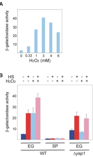
Figure 4. Hsf1 response to oxidative stress is lost in stationary-phase yeast and depends on Yap1.(A) Wild-type BY4741 cells harboring the HSE2-LacZplasmid grown exponentially at 30uC were incubated for 30 min with the indicated concentrations of H2O2. (B) Wild-type andDyap1BY4741 cells harboring the HSE2-LacZplasmid grown at 30uC either exponentially (EG) or to stationary-phase (SP) were incubated for 30 min with (+) or without (2) H2O2(3 mM) prior to heat shock. Cells were either incubated further for 20 min at 30uC (2) or subjected to a 20 min to heat shock (HS) at 42uC (+). Hsf1 activity was
measured asb-galactosidase specific activity. The data are the mean plus standard error of at least 3 independent experiments. Kruskal-Wallis one way analysis of variance on ranks (pairwise multiple comparison with Tukey test) applied on data of EG cells in (B) indicates a statistically significant difference (p,0.001) between the activity in untreated cells and in cells exposed to HS or H2O2.
shock (Figure 2). This suggests that Hsf1 is somewhat dwindled in stationary-phase yeast but even when overexpressed, other component(s) in the heat shock activation pathway is either missing or becomes limiting. This possibility was corroborated by expressing the constitutively active Hsf1 R206S mutant, which could partially bypass this activation pathway [55]. As shown also here, the R206S mutant could not recapitulate fully the heat shock response, as exponentially-growing yeast still responded to heat shock (Figure 2B, lanes 3, 4). In stationary-phase yeast, thea priori higher basal levels of Hsp26-GFP increased further upon expressing the R206S mutant (Figure 2B, lanes 7,9). More importantly, in stationary-phase cells the excess wild-type Hsf1 enabled a weak but discernable heat shock response, reflected by a 1.2-fold increase in b-galactosidase activity (Figure 2A) and in Hsp26-GFP levels (Figure 2B, lanes 11,12) and a 2-fold increase in Btn2-GFP levels (Figure 2C, lanes 7,8). Expression of the R206S mutant abolished the response of stationary-phase yeast to elevated temperature (Figure 2B, lanes 9,10). To conclude, our findings suggest that although the levels of Hsf1 are compromised, the stationary-phase largely impacts on Hsf1 activation. The effects of the constitutively active R206S mutant in
exponentially-growing cells was similar to that of the wild-type Hsf1, both still responding to heat shock, whereas in stationary-phase cells the R206S mutant could compensate more efficiently than the wild-type protein for the hampered Hsf1 activation (Figure 2B). This may indicate that R206S cannot bring about its full effect in exponentially-growing cells, where Hsf1 activation is intact and functional. When this activation deteriorates, as in stationary-phase cells, the R206S can partially bypass the need for such activation. This deteriorated Hsf1 activation is indicated by the modest 1.2–2-fold heat shock induction in stationary-phase yeast expressing excess Hsf1, as compared to the substantial 10.7-, 4.1-and 5.1-fold induction upon heat shock in exponentially-growing cells expressing an empty vector, wild-type Hsf1 or R206S, respectively.
Hsf1 response to glucose starvation is maintained in stationary-phase yeast
Since Hsf1 itself did not appear to be a major limiting factor in stationary-phase yeast (Figure 2), we next examined whether the lack of response to heat shock in these cells was manifested also in other modes of Hsf1 activation. In addition to heat shock, Hsf1 is Figure 5. Precursors of NAD+ affect Hsf1 activity.
BY4741 cells expressing Hsp26-GFP (A,B) or Btn2-GFP (C,D) grown at 30uC either exponentially (EG) or to stationary-phase (SP) were incubated for 30 min with either NR (10mM; A,C (+)) or NAM (10 mM; B,D (+)) prior to the heat
shock. Cells were either incubated further for 20 min at 30uC (2) or subjected to a 20 min to heat shock (HS) at 42uC (+). Hsf1 activity was measured
as levels of Hsp26-GFP (A,B) or Btn2-GFP (C,D) relative to actin (a loading control), as determined by quantified immunoblotting. Insets in C, D, levels of proteins in SP yeast drawn to a smaller scale. The data shown are mean plus standard error of at least 10 independent experiments. Kruskal-Wallis one way analysis of variance on ranks (pairwise multiple comparison with Tukey test) applied on data of EG cells in (A) indicates a statistically significant difference (P = 0.001) between untreated cells and cells exposed to either HS or to HS plus NR, and between 30uC and 42uC in cells exposed to NR. Paired t-test applied to data of SP cells in (A) indicates a statistically significant difference (p = 0.07) between untreated cells and cells exposed to NR (*), and between 30uC or 42uC only in cells exposed to NR (**). There is no statistically significant difference between 30uC and 42uC in cells not exposed to NR (***).
also activated in response to low glucose [8]. For glucose starvation, exponential cells growing at 2% (w/v) glucose were transferred to fresh media supplemented with the standard 2% or low 0.05% glucose and were either maintained at exponential growth or allowed to reach stationary-phase in these media. Prior to heat shock, cells were transferred to fresh media supplemented with the respective 2% or 0.05% glucose. Clearly, stationary-phase yeast, which did not respond to heat shock, maintained their response to glucose starvation by increasing the Btn2-GFP levels when exposed to 0.05% glucose (Figure 3). This response was not accompanied by cell division. Hsf1 responded slightly to glucose starvation in exponential cells but most effectively in
stationary-phase cells (Figure 3, lanes 1,3 and 5,7, respectively). Similar results were obtained with Hsp26-GFP at either low glucose or low galactose (Figure S1A), in agreement with the increased Hsp26 mRNA levels in response of exponential yeast to low glucose [35]. Unlike HSP26 and BTN2, the synthetic HSE2-LacZ reported poorly on glucose starvation (Figure S1B). Our finding that stationary-phase yeast cannot respond to elevated temperature but maintain their response to low glucose agree with the previously reported independent Hsf1 response to glucose starvation and heat shock [35]. Moreover, it indicates that the inability of Hsf1 in stationary-phase yeast to respond to heat shock at any glucose concentration (Figure 3) is neither due to defects in global protein synthesis nor to considerable depletion of Hsf1 itself. Instead, in stationary-phase cells the heat shock activation of Hsf1 is impaired elsewhere. Although some nutrient depletion was observed, as reflected by improved heat shock response of cells transferred to fresh media, this effect was apparent mostly in exponentially-growing yeast and much less so in stationary-phase cells (Figures 3 and S1). This indicated that the inability of stationary-phase yeast to respond to heat shock was not the consequence of nutrients depletion, since these cells were transferred to fresh media prior to heat shock and still could neither divide nor respond to the elevated temperature (Figure 3, lanes 5–8). Nonetheless, glucose starvation did impose limiting resources for de novo Btn2-GFP synthesis, since the intact heat shock response in the exponential yeast was still weaker at low glucose as compared to standard glucose concentration (Figure 3, compare lanes 4 and 2, respec-tively). Therefore, the ability of stationary-phase yeast to respond to glucose starvation by increasing the Btn2-GFP levels under such limiting conditions was indeed impressive. Yet, such cells did not respond to heat shock (Figure 3, lanes 7,8). We conclude that the inability of stationary-phase yeast to produce b-galactosidase, Hsp26-GFP or Btn2-GFP upon heat shock (Figure 1) reflects an intrinsic modification of their heat shock response, resulting in their failure to activate Hsf1. Since these stationary-phase yeast can still activate Hsf1 by glucose starvation, it rules out the possibility that Hsf1 itself and/or its ability to activate transcription of its target genes is lost in stationary-phase yeast. Instead, it points to factors that participate in Hsf1 activation as components that are compromised.
Hsf1 response to oxidative stress is lost in stationary-phase yeast and depends on Yap1
Since stationary-phase yeast lost the Hsf1 response to heat shock (Figure 1) but maintained its response to glucose starvation (Figure 3), we tested in these cells the activation of Hsf1 by yet a third stressor, the oxidative stress. Exponentially-growing yeast harboring HSE2-LacZ were exposed for 30 min to increasing concentrations of H2O2and the measuredb-galactosidase activity
showed that 3 mM H2O2yielded maximal response (Figure 4A), a
concentration that was used in subsequent experiments. Notably, the expression of neither Hsp26-GFP nor Btn2-GFP was significantly upregulated by H2O2itself, although the heat shock
response was augmented in the presence of this oxidant (Figure S2). Theb-galactosidase activity revealed that in exponentially-growing yeast H2O2activated Hsf1 by itself and the combination
of H2O2 and heat shock generated a stronger activation
(Figure 4B). This suggests that oxidative stress and heat shock utilize distinct activation pathways. Conversely, stationary-phase yeast responded neither to heat shock nor to H2O2 or to their
combination (Figure 4B), reflecting deteriorated Hsf1 activation by both stresses.
Next, we examined a potential factor that might play a role in Hsf1 response to oxidative stress. We focused on Yap1, a major Figure 6. Sir2 is required for Hsf1 response to heat shock but
not to oxidative stress.Wild-type andDsir2BY4741 cells (A), or wild-type andDsir2W303-1b cells (B), harboring HSE2-LacZplasmid, were grown at 30uC either exponentially (EG) or to stationary-phase (SP). Cells were either incubated for 20 min at 30uC (blue bars) or subjected to 20 min heat shock at 42uC (red bars). (C) Exponentially growing wild-type and Dsir2 BY4741 cells harboring HSE2-LacZ plasmid were incubated for 30 min with (+) or without (2) H2O2(3 mM) prior to the heat shock. Cells were either incubated further for 20 min at 30uC (2) or subjected to a 20 min heat shock (HS) at 42uC (+). Hsf1 activity was measured asb-galactosidase specific activity. The data are mean plus standard error of at least 3 independent experiments.
transcription factor in the yeast’s protective response against oxidative challenges [56]. Yap1 is found in the cytoplasm, and upon exposure to oxidants, this member of the AP-1 family of transcription factors translocates to the nucleus to activate anti-oxidant genes transcription [57–62]. Stress regulatory networks studies suggest that Hsf1 and Yap1 operate in parallel pathways, independently activatingPDR3expression, which leads toRPN4 andSNQ2production [63]. Intrigued by the delayed age-induced cell death uponYAP1overexpression [64] and by the notion that Hsf1 and Yap1, two master regulators of stress responses, are also pro-longevity genes [28], we revisited the possible link between Yap1 and Hsf1.
Using the HSE2-lacZ reporter, we found that exponentially-growing wild-type cells responded to either heat shock or oxidative stress by augmenting Hsf1 activity, and an even stronger effect was observed in cells challenged with both stresses (Figure 4B). However, while Hsf1 inDyap1cells was activated by heat shock as efficiently as in wild-type cells, in the absence of YAP1 the mutant cells no longer responded to oxidative stress, either by itself or in combination with heat shock (Figure 4B). Since Yap1 is not known to directly bind and activate HSEs, these results demonstrate that Yap1 is required to allow activation of Hsf1 by oxidative stress but not by heat shock. Thus, Yap1 must function not only in parallel to, but also upstream of, Hsf1.
Hsf1 response to heat shock depends on Sir2, is mimicked by excess Sir2, and is improved in stationary-phase yeast by NAD+precursors
The activation of Hsf1 by glucose starvation links Hsf1 to metabolism and possibly to the established lifespan extension by dietary restriction [39–41]. In particular, it has been shown that levels of NADPH and NAD+ decline upon yeast aging, but
NADPH levels are maintained when yeast cells are starved [65– 67]. Decline in NAD+
levels with aging were also reported in mice [68]. Moreover, nicotinamide riboside (NR), which increases NAD+
levels [69,70], extends lifespan [65,66], directly linking NAD+
levels to aging.
To investigate the possible contribution of NAD+
to Hsf1 activation, we supplied yeast with NR or nicotinamide (NAM) in order to increase NAD+levels [69,70]. Addition of NR or NAM to
exponentially-growing cells had no significant effect on the basal activity or the heat shock response of Hsf1, as manifested by the levels of Hsp26-GFP or Btn2-GFP (Figure 5). In stationary-phase yeast, NR or NAM also exerted similar responses. On one hand, they attenuated the basal activity of Hsf1, as reflected by the levels of Hsp26-GFP (Figure 5A,B) or Btn2-GFP (Figure 5C,D, insets). More importantly, both NAD+
precursors inverted the effect of heat shock. Instead of its inhibitory effect in untreated stationary-phase yeast, supplementing either NR or NAM allowed a slight activation of Hsf1 by heat shock (Figure 5). These marginal effects are statistically significant, showing a difference in the basal Hsf1 Figure 7. Activation of Hsf1 by heat shock is mimicked by excess Sir2 and improved by the NAD+precursor.
(A) Wild-type BY4741 cells harboring HSE2-LacZplasmid were transformed with an empty vector (2) or a centromeric pSIR2plasmid (+). Cells grown at 30uC either exponentially (EG) or to stationary-phase (SP) were either incubated for 20 min at 30uC (2) or subjected to a 20 min HS at 42uC (+). (B) Wild-type BY4741 cells harboring HSE2-LacZplasmid were transformed with an empty vector (2) or a pSIR2plasmid (+). Cells grown at 30uC to the indicated growth phase were incubated for 30 min with (+) or without (2) NR (10mM) prior to the heat shock. Cells were either incubated further for 20 min at 30uC (2) or subjected to a 20 min heat shock (HS) at 42uC (+). (C) Activity in SP yeast from (B) drawn to a smaller scale. Hsf1 activity was measured asb
activity in stationary-phase yeast between untreated cells and cells supplemented with either NAD+ precursor, as well as between
30uC and 42uC only in cells exposed to NAD+precursor, but not
in cells not supplemented with NAD+(Figure 5).
A role for NAD+
in aging makes sense in light of the involvement of sirtuins in lifespan determination. These class III protein deacetylases that consume NAD+
are implicated in lifespan extension in many model organisms and in particular in mediating the beneficial effects of dietary restriction [39–41]. Sir2, the founding member of the sirtuins family, exerts opposite effects on S. cerevisiae aging, depending on the yeast aging model system. While RLS is extended by excessSIR2and shortened uponSIR2 deletion, CLS is prolonged in Dsir2 mutant under dietary restriction [4,6,43,44]. Despite the enigmatic contribution of Sir2 to yeast aging, we examined whether activation of Hsf1 was modified in mutants lacking the SIR2 gene. Following b -galactosidase activity, we found that although exponentially-growing Dsir2 cells (two different strains, BY4741 (Figure 6A) and W303-1b (Figure 6B)) exhibited somewhat higher basal Hsf1 activity than their wild-type counterparts, these mutants totally failed to respond to heat shock. In stationary-phase yeast,SIR2 deletion had no effect on the residual basal or heat shock-induced activities of Hsf1 (Figure 6A).
Our data implicate Yap1 in the Hsf1 response to oxidative stress but not to heat shock (Figure 4), suggesting two distinct Hsf1 activation pathways. Therefore, it was interesting to determine whether Sir2 was restricted to the heat shock activation mode of Hsf1. Clearly, Dsir2 cells failed to respond to heat shock but maintained their full response to oxidative stress, similarly augmenting theirb-galactosidase activity when exposed to H2O2
alone or in combination with heat shock (Figure 6C). This was in contrast to wild-type cells, which responded independently to either stress, and with augmented activity when both stressors were combined (Figure 6C; see also Figure 4). These findings exclude Sir2 from the activation of Hsf1 by oxidative stress and demonstrate that it functions in the heat shock activation pathway. To substantiate the role of Sir2 in Hsf1 activation by heat shock, we expressed in wild-type yeast excess SIR2 from a plasmid. Clearly, in exponentially-growing yeast excess Sir2 mimicked the effect of heat shock and there was no further increase in Hsf1 activity by heat shock (Figures 7A and S3A). However, while the NAD+ precursor NR had no effect on Hsf1 activity in
SIR2by itself increased Hsf1 activity by two-fold (Figures 7 and S3), supplementing stationary-phase yeast expressing excessSIR2 with NR increased Hsf1 activity nearly four-fold, yet there was no additional effect of heat shock (Figures 7B,C and S3 B,C).
Excess Hsf1, Sir2 and NAD+precursor rejuvenate heat
shock response in stationary-phase yeast
Our findings in Figures 5–7 suggested that both Sir2 and NAD+
were limiting in stationary-phase yeast. Yet, additional factors seemed to be limiting in the Hsf1 activation cascade, as the heat shock response in these cells fell short of that in exponentially-growing yeast (Figures 7 and S3). Moreover, while the effect of excess Sir2 was further augmented by NR, neither in exponen-tially-growing nor in stationary-phase yeast was the excess Sir2 (with or without NR) further increased by heat shock (Figures 7 and S3). A plausible candidate for such a limiting factor was Hsf1 itself, since excess Hsf1 improved Hsf1 activity in stationary-phase yeast (Figure 2). Indeed, overexpressing Hsf1 together with Sir2 and providing the cells with NR restored the heat shock response of stationary-phase yeast to nearly 70% of that of exponentially-growing cells (Figure 8). Thus, limiting levels of three factors appear to impair the ability of stationary-phase yeast to respond to heat shock: Hsf1, Sir2 and NAD+
.
Discussion
The current studies establish Hsf1 as a longevity-related gene also in yeast, as its activation by heat shock or oxidative stress deteriorates in stationary-phase cells. We also provide evidence for two mediators of Hsf1 activation, Sir2 and Yap1, which operate in two discrete activation pathways: Sir2 in the heat shock response
and Yap1 in the oxidative stress response (Figure 9). Our direct measurements of Hsf1 activity are based on three reporters with distinct HSEs, which respond differently to the three stressors tested. All three reporters respond to heat shock by increasing the levels of the proteins encoded by them. However, only HSE2-lacZ, driven by the synthetic HSE, is activated by oxidative stress yet it is indifferent to glucose starvation. Conversely, the genes driven by the perfect type endogenous HSEs, HSP26 and BTN2, are activated by glucose starvation, but are indifferent to oxidative stress. This differential reaction to stress challenges emphasizes the specificity and modularity of the Hsf1 response, which is reflected by distinct subsets of responsive genes but more importantly, by unique modes of Hsf1 activation (Figure 9).
Clearly, Hsf1 response to either heat shock or oxidative stress declines progressively and is completely lost in stationary-phase yeast (Figure 1). Our findings inS. cerevisiae, showing that yeast Hsf1 is a longevity-related gene, echo studies inC. elegans, where Hsf1 has been shown to be essential for lifespan extension and to extend lifespan when overexpressed [37,38]. Declined response to heat shock and oxidative stress has also been reported in old flies, aged rat tissues and senescent human cells [9,46,71,72]. These effects on Hsf1 in stationary-phase yeast are neither due to considerable decline in Hsf1 levels nor to impaired ability to upregulate its target genes. This is indicated by the marginal increase in Hsf1 activity in stationary-phase yeast overexpressing HSF1 (Figure 2) and by the activation of Hsf1 by glucose starvation that is maintained in stationary-phase yeast (Figure 3). To conclude, here we show that also in yeast, Hsf1 links responses to stress with lifespan, but it remains to be determined if the failure of Hsf1 to undergo activation is a cause or consequence of aging and whether lifespan extension requires Hsf1 and/or maintains activation-competent Hsf1.
The modularity of the Hsf1 activity is underscored by its three targets that respond differently to the three stresses elicited (Figure 9). Our model indicates that Hsf1 undergoes distinct modes of activation (denoted by different shapes and superscripts) by discrete and independent pathways. The different response of Hsf1 in stationary-phase yeast to the three stresses we employ indicates that specific factors operate in each stress pathway to mediate Hsf1 activation. Thus, the factor(s) involved in the response to glucose starvation appears to survive the transition from exponential growth to stationary-phase and is therefore distinct from factors that participate in the response to heat shock or oxidative stress, which are compromised during this transition. Indeed, Snf1 has been shown to be essential for Hsf1 activation by glucose starvation (as monitored by elevation of Hsp26 mRNA), but this kinase is not required for Hsf1 response to heat shock [35]. Significantly, although both responses decline in stationary-phase yeast, our data distinguish between the heat shock pathway and the oxidative stress pathway (Figure 9). The transcription factor Yap1 is implicated in the response of Hsf1 to oxidative stress but excluded from the heat shock response (Figure 4). Conversely, the NAD+
-dependent Sir2 is implicated in Hsf1 response to heat shock but excluded from the oxidative stress (Figure 6).
Of particular interest is our finding that Hsf1 response to heat shock stringently depends on Sir2 (Figure 6) and is mimicked by excess Sir2 (Figures 7 and S3). The role of Sir2 in Hsf1 activation is also supported by the small but consistent increased heat shock response in stationary-phase yeast supplemented with NR or NAM (Figure 5). Indeed, NAM, unlike NR, was reported to be a noncompetitive inhibitor of Sir2 [73] and was shown by us to affect protein aggregation in a manner resemblingSIR2deletion [45]. However, the similar effects on Hsf1 heat shock response exerted by NAM or NR (Figure 5) suggest that both compounds Figure 9. A schematic presentations of the various Hsf1
activation pathways.The three stresses, heat shock, oxidative stress and sugar starvation, activate the inactive Hsf1 through different mediators, Sir2, Yap1 and Snf1, respectively. Consequently, three distinct types of active Hsf1 are generated, HSF1HS, Hsf1OSand Hsf1SS, respectively. These, in turn, transactivate the transcription of the indicated subsets of target genes.
replenish intracellular NAD+
, the levels of which are reported to decline in aging yeast and mice [65–68]. Furthermore, the effect of excess Sir2 is augmented in the presence of NR, and combination of excess Sir2 and NR partially restores Hsf1 activation also in stationary-phase yeast (Figures 7 and S3). Since sirtuins consume NAD+ in their deacetylation reaction, it is possible that their
activity is regulated by cellular [NAD+
]/[NADH] ratios, hence responds to changes in cellular metabolism, another hallmark of aging [74]. Such modulation of sirtuins’ activity by the metabolic status of the cell adds yet another layer of regulation to Hsf1 functions in orchestrating stress responses. The clear involvement of Sir2 in the activation of yeast Hsf1 by heat shock (Figures 6–8 and S3) corresponds with similar findings regarding Sirt1 in mammals. It has been shown that Sirt1, the closest mammalian homolog of Sir2, deacetylates the mammalian Hsf1 as one of its activation modes [46]. By contrast, inC. elegansthe heat shock response is independent of the Sir2/Sirt1 homologue Sir2.1, although the synergistic effect of dietary restriction and heat shock requires this sirtuin [47]. In our hands, the enhanced Hsf1 activation by the NAD+
precursor NR, which is further augmented when excess Sir2 is expressed, strongly suggests that yeast Sir2 acts in the heat shock response as Hsf1 deacetylase, as does Sirt1 in mammals.
To conclude, although much about cellular aging in yeast and in general remains obscure, the current work unveils some of the players and pathways that affect and are affected by aging. Our data indicate that at least three components in the Hsf1 heat shock activation pathway are limiting in stationary-phase yeast, Hsf1 itself, Sir2 and its cofactor NAD+
. When supplemented in combination, excessHSF1, excessSIR2and the NAD+
precursor NR can rejuvenate to a large extent the heat shock response in stationary-phase yeast (Figure 8). If restoring the heat shock response also slows down aging, it would indicate that its decline is a cause rather than a consequence of aging. Finally, the aging-dependent changes in Hsf1 response described here, combined with the effects of aging on the aggregation of a polyglutamine-containing protein we previously reported [45], establish S. cerevisiae as a suitable model organism not merely for lifespan studies ending in cell death, but also for research addressing various molecular aspects of the aging process.
Supporting Information
Figure S1 Hsf1 activation by sugar starvation is main-tained in stationary-phase yeast but poorly reported by
HSE2-LacZ. Exponential BY4741 cells expressing Hsp26-GFP
were grown at 30uC in SC medium containing 2% (w/v) galactose (A), and exponential BY4741 cells harboring HSE2-LacZplasmid were grown at 30uC in SC medium containing 2% (w/v) glucose (B). Cells were transferred to fresh media supplemented with the standard 2% or low 0.05% sugar and were either maintained at exponential growth (EG) or allowed to reach stationary-phase (SP) in these media. Prior to heat shock, cells were transferred to fresh media supplemented with the respective 2% or 0.05% sugar and further incubated at 30uC for 3 hrs. Cells were either incubated
for 20 min at 30uC (2) or subjected to a 20 min HS at 42uC (+). Hsf1 activity was measured as (A) levels of Hsp26-GFP relative to actin (a loading control), as determined by quantified immuno-blotting or (B)b-galactosidase specific activity. The data are the mean of 2–3 independent experiments. Similar Hsp26-GFP levels were obtained in cells grown in either galactose or glucose. (TIF)
Figure S2 Hsf1 response to oxidative stress is poorly
reported by Hsp26-GFP or Btn2-GFP. BY4741 cells
expressing Hsp26-GFP (A) or Btn2-GFP (B) grown at 30uC either exponentially (EG) or to stationary-phase (SP) were incubated for 30 min with (+) or without (2) H2O2(3 mM) prior to heat shock.
Cells were either incubated further for 20 min at 30uC (2) or subjected to a 20 min to heat shock (HS) at 42uC (+). Hsf1 activity was measured as levels of Hsp26-GFP (A) or Btn2-GFP (B) relative to actin (a loading control), as determined by quantified immunoblotting. The data are the mean plus standard error of at least 5 independent experiments.
(TIF)
Figure S3 Activation of Hsf1 by heat shock is mimicked
by excess Sir2 and improved by the NAD+
precursor.(A)
Wild-type W303-1b cells harboring HSE2-LacZ plasmid were transformed with an empty vector (2) or a centromeric pSIR2 plasmid (+). Cells grown at 30uC either exponentially (EG) or to stationary-phase (SP) were either incubated for 20 min at 30uC (2 ) or subjected to a 20 min HS at 42uC (+). (B) Wild-type W302-1b cells harboring HSE2-LacZ plasmid were transformed with an empty vector (2) or a pSIR2plasmid (+). Cells grown at 30uC to the indicated growth phase were incubated for 30 min with (+) or without (2) NR (10mM) prior to the heat shock. Cells were either incubated further for 20 min at 30uC (2) or subjected to a 20 min heat shock (HS) at 42uC (+). (C) Activity in SP yeast from (B) drawn to a smaller scale. Hsf1 activity was measured as b -galactosidase specific activity. The data are mean plus standard error of at least 3 independent experiments.
(TIF)
Acknowledgments
We would like to thank Rolf Sternglanz (Stony Brook University), Ian Dawes (University of New South Wales), Dennis Thiele (Duke University) and Dennis Winge (University of Utah), for generously providing strains and plasmids. We thank Charles Brenner (University of Iowa) for sharing with us his vast knowledge on NAD+metabolism and his generous gift of
nicotinamide riboside (NR). We are grateful to Ian Dawes, Anita Ayer and Bethany Pillay for helping us in the early stages of this study. We thank Yossi Roitelman for insightful discussions and critical reading of the manuscript and Aviv Shaish for helping us with statistical tests.
Author Contributions
Conceived and designed the experiments: IN EW RJ AC SB. Performed the experiments: IN EW RJ AC SB. Analyzed the data: IN EW RJ AC SB. Wrote the paper: SB.
References
1. Gems D, Partridge L (2013) Genetics of longevity in model organisms: debates and paradigm shifts. Annu Rev Physiol 75:621–644.
2. Lorenz DR, Cantor CR, Collins JJ (2009) A network biology approach to aging in yeast. Proc Natl Acad Sci U S A 106:1145–1150.
3. Longo VD (2004): Search for methuselah genes heats up. Sci Aging Knowledge Environ 2004:e6.
4. Longo VD, Shadel GS, Kaeberlein M, Kennedy B (2012) Replicative and chronological aging in Saccharomyces cerevisiae. Cell Metab 16:18–31.
5. Allen C, Buttner S, Aragon AD, Thomas JA, Meirelles O, et al. (2006) Isolation of quiescent and nonquiescent cells from yeast stationary-phase cultures. J Cell Biol 174:89–100.
6. Fabrizio P, Longo VD (2003) The chronological life span of Saccharomyces cerevisiae. Aging Cell 2:73–81.
8. Kourtis N, Tavernarakis N (2011) Cellular stress response pathways and ageing: intricate molecular relationships. EMBO J 30:2520–2531.
9. Labbadia J, Morimoto RI (2014) Proteostasis and longevity: when does aging really begin? F1000Prime Rep 6:7.
10. Pelham HR (1982) A regulatory upstream promoter element in the Drosophila hsp 70 heat-shock gene. Cell 30:517–528.
11. Amin J, Ananthan J, Voellmy R (1988) Key features of heat shock regulatory elements. Mol Cell Biol 8:3761–3769.
12. Xiao H, Lis JT (1988) Germline transformation used to define key features of heat-shock response elements. Science 239:1139–1142.
13. Tuite MF, Bossier P, Fitch IT (1988) A highly conserved sequence in yeast heat shock gene promoters. Nucleic Acids Res 16:11845.
14. Torres FA, Bonner JJ (1995) Genetic identification of the site of DNA contact in the yeast heat shock transcription factor. Mol Cell Biol 15:5063–5070. 15. Yamamoto A, Mizukami Y, Sakurai H (2005) Identification of a novel class of
target genes and a novel type of binding sequence of heat shock transcription factor in Saccharomyces cerevisiae. J Biol Chem 280:11911–11919. 16. Sorger PK, Nelson HC (1989) Trimerization of a yeast transcriptional activator
via a coiled-coil motif. Cell 59:807–813.
17. Hashikawa N, Yamamoto N, Sakurai H (2007) Different mechanisms are involved in the transcriptional activation by yeast heat shock transcription factor through two different types of heat shock elements. J Biol Chem 282:10333– 10340.
18. Sorger PK, Lewis MJ, Pelham HR (1987) Heat shock factor is regulated differently in yeast and HeLa cells. Nature 329:81–84.
19. Jakobsen BK, Pelham HR (1988) Constitutive binding of yeast heat shock factor to DNA in vivo. Mol Cell Biol 8:5040–5042.
20. Sorger PK, Pelham HR (1988) Yeast heat shock factor is an essential DNA-binding protein that exhibits temperature-dependent phosphorylation. Cell 54:855–864.
21. Gross DS, English KE, Collins KW, Lee SW (1990) Genomic footprinting of the yeast HSP82 promoter reveals marked distortion of the DNA helix and constitutive occupancy of heat shock and TATA elements. J Mol Biol 216:611– 631.
22. McDaniel D, Caplan AJ, Lee MS, Adams CC, Fishel BR, et al (1989) Basal-level expression of the yeast HSP82 gene requires a heat shock regulatory element. Mol Cell Biol 9:4789–4798.
23. Verghese J, Abrams J, Wang Y, Morano KA (2012) Biology of the heat shock response and protein chaperones: budding yeast (Saccharomyces cerevisiae) as a model system. Microbiol Mol Biol Rev 76:115–158.
24. Giardina C, Lis JT (1995) Dynamic protein-DNA architecture of a yeast heat shock promoter. Mol Cell Biol 15:2737–2744.
25. Erkine AM, Magrogan SF, Sekinger EA, Gross DS (1999) Cooperative binding of heat shock factor to the yeast HSP82 promoter in vivo and in vitro. Mol Cell Biol 19:1627–1639.
26. Sekinger EA, Gross DS (2001) Silenced chromatin is permissive to activator binding and PIC recruitment. Cell 105:403–414.
27. Hahn JS, Hu Z, Thiele DJ, Iyer VR (2004) Genome-wide analysis of the biology of stress responses through heat shock transcription factor. Mol Cell Biol 24:5249–5256.
28. Gibney PA, Lu C, Caudy AA, Hess DC, Botstein D (2013) Yeast metabolic and signaling genes are required for heat-shock survival and have little overlap with the heat-induced genes. Proc Natl Acad Sci U S A 110:E4393–E4402. 29. Liu XD, Thiele DJ (1996) Oxidative stress induced heat shock factor
phosphorylation and HSF-dependent activation of yeast metallothionein gene transcription. Genes Dev 10:592–603.
30. Tamai KT, Liu X, Silar P, Sosinowski T, Thiele DJ (1994) Heat shock transcription factor activates yeast metallothionein gene expression in response to heat and glucose starvation via distinct signalling pathways. Mol Cell Biol 14:8155–8165.
31. Amoros M, Estruch F (2001) Hsf1p and Msn2/4p cooperate in the expression of Saccharomyces cerevisiae genes HSP26 and HSP104 in a gene- and stress type-dependent manner. Mol Microbiol 39:1523–1532.
32. Hoj A, Jakobsen BK (1994) A short element required for turning off heat shock transcription factor: evidence that phosphorylation enhances deactivation. EMBO J 13:2617–2624.
33. Hashikawa N, Sakurai H (2004) Phosphorylation of the yeast heat shock transcription factor is implicated in gene-specific activation dependent on the architecture of the heat shock element. Mol Cell Biol 24:3648–3659. 34. Ferguson SB, Anderson ES, Harshaw RB, Thate T, Craig NL, et al. (2005)
Protein kinase A regulates constitutive expression of small heat-shock genes in an Msn2/4p-independent and Hsf1p-dependent manner in Saccharomyces cere-visiae. Genetics 169:1203–1214.
35. Hahn JS, Thiele DJ (2004) Activation of the Saccharomyces cerevisiae heat shock transcription factor under glucose starvation conditions by Snf1 protein kinase. J Biol Chem 279:5169–5176.
36. Shore DE, Carr CE, Ruvkun G (2012) Induction of cytoprotective pathways is central to the extension of lifespan conferred by multiple longevity pathways. PLoS Genet 8:e1002792.
37. Hsu AL, Murphy CT, Kenyon C (2003) Regulation of aging and age-related disease by DAF-16 and heat-shock factor. Science 300:1142–1145.
38. Morley JF, Morimoto RI (2004) Regulation of longevity in Caenorhabditis elegans by heat shock factor and molecular chaperones. Mol Biol Cell 15:657– 664.
39. Bishop NA, Guarente L (2007) Genetic links between diet and lifespan: shared mechanisms from yeast to humans. Nat Rev Genet 8:835–844.
40. Kaeberlein M, Burtner CR, Kennedy BK (2007) Recent developments in yeast aging. PLoS Genet 3:e84.
41. Bitterman KJ, Medvedik O, Sinclair DA (2003) Longevity regulation in Saccharomyces cerevisiae: linking metabolism, genome stability, and hetero-chromatin. Microbiol Mol Biol Rev 67:376–99, table.
42. Lin SJ, Defossez PA, Guarente L (2000) Requirement of NAD and SIR2 for life-span extension by calorie restriction in Saccharomyces cerevisiae. Science 289:2126–2128.
43. Fabrizio P, Gattazzo C, Battistella L, Wei M, Cheng C, et al. (2005) Sir2 blocks extreme life-span extension. Cell 123:655–667.
44. Kennedy BK, Smith ED, Kaeberlein M (2005) The enigmatic role of Sir2 in aging. Cell 123:548–550.
45. Cohen A, Ross L, Nachman I, Bar-Nun S (2012) Aggregation of polyQ proteins is increased upon yeast aging and affected by Sir2 and Hsf1: novel quantitative biochemical and microscopic assays. PLoS One 7:e44785.
46. Westerheide SD, Anckar J, Stevens SM Jr., Sistonen L, Morimoto RI (2009) Stress-inducible regulation of heat shock factor 1 by the deacetylase SIRT1. Science 323:1063–1066.
47. Raynes R, Leckey BD, Jr., Nguyen K, Westerheide SD (2012) Heat shock and caloric restriction have a synergistic effect on the heat shock response in a sir2.1-dependent manner in Caenorhabditis elegans. J Biol Chem 287:29045–29053. 48. Giaever G, Chu AM, Ni L, Connelly C, Riles L, et al. (2002) Functional
profiling of the Saccharomyces cerevisiae genome. Nature 418:387–391. 49. Wang CL, Landry J, Sternglanz R (2008) A yeast sir2 mutant temperature
sensitive for silencing. Genetics 180:1955–1962.
50. Huh WK, Falvo JV, Gerke LC, Carroll AS, Howson RW, et al. (2003) Global analysis of protein localization in budding yeast. Nature 425:686–691. 51. Rabinovich E, Kerem A, Frohlich KU, Diamant N, Bar-Nun S (2002)
AAA-ATPase p97/Cdc48p, a cytosolic chaperone required for endoplasmic reticulum-associated protein degradation. Mol Cell Biol 22:626–634. 52. Sorger PK, Pelham HR (1987) Purification and characterization of a heat-shock
element binding protein from yeast. EMBO J 6:3035–3041.
53. Guarente L (1983) Yeast promoters and lacZ fusions designed to study expression of cloned genes in yeast. Methods Enzymol 101:181–191. 54. Gray JV, Petsko GA, Johnston GC, Ringe D, Singer RA, et al. (2004) ‘‘Sleeping
beauty’’: quiescence in Saccharomyces cerevisiae. Microbiol Mol Biol Rev 68:187–206.
55. Sewell AK, Yokoya F, Yu W, Miyagawa T, Murayama T, et al. (1995) Mutated yeast heat shock transcription factor exhibits elevated basal transcriptional activation and confers metal resistance. J Biol Chem 270:25079–25086. 56. Moye-Rowley WS, Harshman KD, Parker CS (1989) Yeast YAP1 encodes a
novel form of the jun family of transcriptional activator proteins. Genes Dev 3:283–292.
57. Kuge S, Jones N, Nomoto A (1997) Regulation of yAP-1 nuclear localization in response to oxidative stress. EMBO J 16:1710–1720.
58. Okazaki S, Tachibana T, Naganuma A, Mano N, Kuge S (2007) Multistep disulfide bond formation in Yap1 is required for sensing and transduction of H2O2 stress signal. Mol Cell 27:675–688.
59. Toone WM, Jones N (1999) AP-1 transcription factors in yeast. Curr Opin Genet Dev 9:55–61.
60. Drakulic T, Temple MD, Guido R, Jarolim S, Breitenbach M, et al. (2005) Involvement of oxidative stress response genes in redox homeostasis, the level of reactive oxygen species, and ageing in Saccharomyces cerevisiae. FEMS Yeast Res 5:1215–1228.
61. Temple MD, Perrone GG, Dawes IW (2005) Complex cellular responses to reactive oxygen species. Trends Cell Biol 15:319–326.
62. Morano KA, Grant CM, Moye-Rowley WS (2012) The response to heat shock and oxidative stress in Saccharomyces cerevisiae. Genetics 190:1157–1195. 63. Hahn JS, Neef DW, Thiele DJ (2006) A stress regulatory network for
co-ordinated activation of proteasome expression mediated by yeast heat shock transcription factor. Mol Microbiol 60:240–251.
64. Herker E, Jungwirth H, Lehmann KA, Maldener C, Frohlich KU, et al. (2004) Chronological aging leads to apoptosis in yeast. J Cell Biol 164:501–507. 65. Belenky P, Racette FG, Bogan KL, McClure JM, Smith JS, et al. (2007)
Nicotinamide riboside promotes Sir2 silencing and extends lifespan via Nrk and Urh1/Pnp1/Meu1 pathways to NAD+. Cell 129:473–484.
66. Belenky P, Bogan KL, Brenner C (2007) NAD+metabolism in health and disease. Trends Biochem Sci 32:12–19.
67. Brandes N, Tienson H, Lindemann A, Vitvitsky V, Reichmann D, et al. (2013) Time line of redox events in aging postmitotic cells. Elife 2:e00306. 68. Gomes AP, Price NL, Ling AJ, Moslehi JJ, Montgomery MK, et al. (2013)
Declining NAD(+) induces a pseudohypoxic state disrupting nuclear-mitochon-drial communication during aging. Cell 155:1624–1638.
69. Bieganowski P, Brenner C (2004) Discoveries of nicotinamide riboside as a nutrient and conserved NRK genes establish a Preiss-Handler independent route to NAD+in fungi and humans. Cell 117:495–502.
70. Tempel W, Rabeh WM, Bogan KL, Belenky P, Wojcik M, et al. (2007) Nicotinamide riboside kinase structures reveal new pathways to NAD+. PLoS Biol 5:e263.
72. Blake MJ, Fargnoli J, Gershon D, Holbrook NJ (1991) Concomitant decline in heat-induced hyperthermia and HSP70 mRNA expression in aged rats. Am J Physiol 260:R663–R667.
73. Imai S, Armstrong CM, Kaeberlein M, Guarente L (2000) Transcriptional silencing and longevity protein Sir2 is an NAD-dependent histone deacetylase. Nature 403:795–800.