Oxygen Effect Contributes to the Radiosensitizing Effect
of Paclitaxel
Fabienne Danhier1., Pierre Danhier2., Nicolas Magotteaux1
, Ge´raldine De Preter2, Bernard Ucakar1, Oussama Karroum2, Be´ne´dicte Jordan2, Bernard Gallez2, Ve´ronique Pre´at1*
1Pharmaceutics and Drug Delivery, Louvain Drug Research Institute, Universite´ Catholique de Louvain, Brussels, Belgium,2Laboratory of Biomedical Magnetic Resonance, Louvain Drug Research Institute, Universite´ Catholique de Louvain, Brussels, Belgium
Abstract
Background:Paclitaxel (PTX) is a potent anti-cancer chemotherapeutic agent and is widely used in the treatments of solid tumors, particularly of the breast and ovaries. An effective and safe micellar formulation of PTX was used to administer higher doses of PTX than TaxolH(the current commercialized drug). We hypothesize that PTX-loaded micelles (M-PTX) may enhance tumor radiosensitivity by increasing the tumor oxygenation (pO2). Our goals were (i) to evaluate the contribution
of the ‘‘oxygen effect’’ to the radiosensitizing effect of PTX; (ii) to demonstrate the therapeutic relevance of the combination of M-PTX and irradiation and (iii) to investigate the underlying mechanisms of the observed oxygen effect.
Methodology and Principal Findings: We used (PEG-p-(CL-co-TMC)) polymeric micelles to solubilize PTX. pO2 was
measured on TLT tumor-bearing mice treated with M-PTX (80 mg/kg) using electron paramagnetic resonance (EPR) oximetry. The regrowth delay following 10 Gy irradiation 24 h after M-PTX treatment was measured. The tumor perfusion was assessed by the patent blue staining. The oxygen consumption rate and the apoptosis were evaluated by EPR oximetry and the TUNEL assay, respectively. EPR oximetry experiments showed that M-PTX dramatically increases the pO224 h post
treatment. Regrowth delay assays demonstrated a synergy between M-PTX and irradiation. M-PTX increased the tumor blood flow while cells treated with M-PTX consumed less oxygen and presented more apoptosis.
Conclusions:M-PTX improved the tumor oxygenation which leads to synergy between this treatment and irradiation. This increased pO2can be explained both by an increased blood flow and an inhibition of O2consumption.
Citation:Danhier F, Danhier P, Magotteaux N, De Preter G, Ucakar B, et al. (2012) Electron Paramagnetic Resonance Highlights That the Oxygen Effect Contributes to the Radiosensitizing Effect of Paclitaxel. PLoS ONE 7(7): e40772. doi:10.1371/journal.pone.0040772
Editor:Krisztian Stadler, Pennington Biomedical Research Center, United States of America
ReceivedMay 2, 2012;AcceptedJune 13, 2012;PublishedJuly 12, 2012
Copyright:ß2012 Danhier et al. This is an open-access article distributed under the terms of the Creative Commons Attribution License, which permits
unrestricted use, distribution, and reproduction in any medium, provided the original author and source are credited.
Funding:This work is supported by grants from the FNRS, the Fonds Joseph Maisin, the Belgian Foundation against Cancer, the ‘‘Actions de Recherches Concerte´es-Communaute´ Franc¸aise de Belgique’’, and the «Poˆle d’attraction Interuniversitaire». The funders had no role in study design, data collection and analysis, decision to publish, or preparation of the manuscript.
Competing Interests:The authors have declared that no competing interests exist. * E-mail: [email protected]
.These authors contributed equally to this work.
Introduction
Paclitaxel (PTX) is a potent anti-cancer chemotherapeutic agent and is widely used in the treatments of solid tumors, particularly of the breast and ovaries [1]. PTX promotes the polymerization of tubulin causing the death of the cell by disrupting the normal tubule dynamics required for the mitotic cell division [2].
Radiotherapy is most effective in controlling local-regional disease whereas chemotherapy can attack distant metastases. Furthermore, many chemotherapeutic agents are able to increase the sensitivity of tumors to radiation [3]. Besides its anti-tumor activity, PTX has been reported as a radiosensitizing agent bothin vitroandin vivo[3,4]. This was firstly based on the principle that the PTX blocks cells in the G2/M phase which is the most
radiosensitive of all cell cycle phases. Moreover, Milas et al. suggested that the PTX-induced enhancement of tumor radio-response was also mediated by reoxygenation of hypoxic tumor
cells [3,5]. Oxygen deficiency in tumors reduces the efficacy of cancer treatments such as chemotherapy or radiotherapy. Hypoxia has been described as an important factor for develop-ment of tumor aggressiveness and there is an overlap between this phenomenon and the poor outcome of hypoxic tumors to radiotherapy [6]. Using radiotherapy, the presence of molecular oxygen increases DNA damage through the formation of oxygen-derived free radicals, which occurs primarily after the interaction of radiation with intracellular water. Because of this so-called ‘‘oxygen effect’’, the radiation dose required to achieve the same biologic effect is about three times higher in the absence of oxygen than in the presence of normal levels of oxygen [7]. The level of tumor oxygenation depends on the balance between oxygen supply, as result of inadequate tumor perfusion, and consumption (mainly via mitochondrial respiration) [8].
responsible for a lot of serious side effects such as hypersensitivity reaction and neuropathy [1]. Recently, novel polymeric micelles have been developed as safe and effective delivery system for PTX [9]. Removing CremophorH EL of the formulation, these polymeric micelles have allowed the enhancement of the Maximum Tolerated Dose (MTD) of TaxolH from 13.5 mg/kg to 80 mg/kg, after intraperitoneal injection. Polymeric micelles are known to extravasate from blood vessels to tumors through fenestrations presented in the tumor endothelium and to be retained into the tumor tissue, by the well-known Enhanced Permeation and Retention effect [10,11]. Because of the increased resident time in the tumor resulting from the Enhanced Permeation and Retention effect and because of the solubilization of PTX in polymeric micelles (avoiding the CremophorH EL-associated toxicity of TaxolH), we hypothesized that the use of polymeric micelles could be considered as a innovative tool to study the radiosensitivity of PTX.
The aims of this work were (i) to evaluate if the ‘‘oxygen effect’’ using electron paramagnetic resonance (EPR) oximetry could be one of the mechanisms involved in the radiosensitizing effect of PTX-loaded micelles; (ii) to demonstrate the therapeutic pre-clinical relevance of the combination of PTX-loaded micelles and irradiation and (iii) to investigate the underlying mechanisms of the observed oxygen effect. All experiments were performed on TLT tumor-bearing mice. This tumor model was chosen because it is a fast growing model which presents intrinsic hypoxia-mediated resistance which is described as an important cause of unsuccess-fulness of radiotherapy [6,7].
Methods
1. Preparation and Characterization of Micelles Loaded with PTX
PTX-loaded micelles were prepared as previously described [12,9]. The amount of solubilised PTX in micelles was determined by HPLC. The particle size and zeta potential were determined, respectively, by photon correlation spectroscopy (PCS) and laser Doppler velocimetry [9].
2. Tumor Model
Syngeneic TLT tumors [13,14] were injected intramuscularly in the gastrocnemius muscle in the rear leg of 8 week old male NMRI mice. Before injection, animals were anesthetized with a Keta-mine/Xylazine dose mixing (62.5 and 6.25 mg/kg, respectively). All experiments were performed in compliance with guidelines set by national regulations and were approved by the Ethical Committee for animal care of the Universite´ Catholique de Louvain (Permit number: UCL/MD/2008/025). All tumor implantation were performed under anesthesia and all efforts were made to minimize suffering.
3. pO2Measurements by Electronic Paramagnetic Resonance (EPR) Oximetry
Tumor oxygen measurements were performed on TLT tumors bearing mice, using Electron Paramagnetic Resonance (EPR) oximetry. Treatments began when tumors reached 8.060.5 mm in diameter. PTX-loaded micelles were injected intravenously (13.5 mg/kg) or intraperitonealy (80 mg/kg); 2, 4, 6, 20 and 24 h before pO2measurements. These times were chosen based on the
literature [3,5]. Control mice were not treated. EPR spectra were acquired by an EPR spectrometer (Magnettech) with a low-frequency microwave bridge operating at 1.2 GHz and extended loop resonator. For oxygen detection in vivo, charcoal (Charcoal wood powder, CX0670-1, EM Science) was used as suitable sensor
interacting with oxygen. Mice were injected 24 h before EPR analysis in the center of the tumor using a suspension of charcoal (100 mg/ml, 1–25mm particle size). The tumor was placed in the center of the extended loop resonator which had a sensitive volume extending 10 mm3into the tumor mass, using a protocol previously described [14,15].
4. Irradiation and Tumor Regrowth Delay Assay
The effect of PTX-loaded micelles combined with X-rays applied 24 h after chemotherapy on TLT growth was assessed by daily measurements of the diameter of the tumors with an electronic calliper. The tumor-bearing leg was locally irradiated with a single dose of 10 Gy of 250-kV X-rays (RT 250, Philips medical Systems). When TLT tumors reached 7.060.5 mm in diameter, the mice were randomly assigned to a treatment group: group 1, PBS injection; group 2, PTX-loaded micelles (PTX dose of 80 mg/kg); Group 3, irradiation with 10 Gy of X-rays; Group 4, PTX-loaded micelles (PTX dose of 80 mg/kg) + irradiation with 10 Gy of X-rays 24 h post chemotherapy; Group 5, PTX-loaded micelles + irradiation with 10 Gy of X-rays 24 h post chemotherapy (mice leg ligatured).
5. Underlying Mechanisms of the Improvement of Tumor Oxygenation
5.1 Evaluation of tumor perfusion by Patent blue staining. The Patent blue (Sigma-Aldrich) was used to estimate the tumor perfusion (adapted from [16,17]) of mice pre-treated with PTX-loaded micelles (80 mg/kg) 24 h before. We injected 50ml of patent blue stain (1.25%) into the tail vein of mice. After 1.0 min, mice were sacrificed and the tumor was carefully excised and cut into two halves. Pictures of each tumor cross section were taken with a digital camera. The comparison between the stained and the unstained area was performed using an in-house program running on Interactive Data Language (Research Systems Inc. Boulder). For each tumor, the stained area was defined on two pictures and the percentage of stained area of the whole cross section was calculated.
5.2Ex vivooxygen consumption rate evaluation by EPR
oximetry. The oxygen consumption was assessed using an EPR method [18]. The spectra were recorded on a Bruker EMX EPR spectrometer operating at 9 GHz. TLT tumor-bearing mice were treated with PTX-loaded micelles (80 mg/kg) (untreated mice were used as control) and were sacrificed 24 h post-treatment. Tumors were excised, trysinized for 30 min and the cell viability was determined. Cells (1.56107/ml) were suspended in 20%
dextran in complete medium. A neutral nitroxide, 15N 4-oxo-2,2,6,6-tetramethylpiperidine-d16-15N-1-oxyl at 0.2 mM (CDN
Isotopes), was added to 100ml of tumor cells that were then drawn into glass capillary tubes. The probe was calibrated at varying oxygen levels between 100% nitrogen and air so that the linewidth measurements could be related to oxygen at any value. The sealed tubes were placed into quartz EPR tubes and the samples maintained at 37uC. Oxygen consumption rates were calculated by measuring the pO2in the closed tubes as a function
of time and subsequently finding the slope of the resulting linear plot [19].
were then fixed with a paraformaldehyde 4% solution and permeabilized with Triton X-100 0.1%. The fluorescent TUNEL assay was then conducted by following instructions of In Situ Cell Death Detection kit (Roche). Nuclear apoptosis is accompanied by the segmentation of nucleus into dense nuclear parts further distributed into apoptotic bodies. These DNA breaks can be visualized by red-labeled TdT staining. Nuclei were stained using Hoechst 33342 (Sigma-Aldrich), mounted with Vectashield (Vector Laboratories) and examined under a fluorescent micro-scope with 350 nm (blue) and 515–560 nm (red) excitation filters.
6. Statistics
All results are expressed as mean6standard error of the mean (SEM) except for Fig. 1 where results are expressed as mean 6
standard deviation (SD). Two-way ANOVA and Bonferroni post test, t-test, linear regression and Kaplan-Meier survival rate were performed using the software GraphPad Prism 5 for Windows to demonstrate statistical differences (p,0.05).
Results
1. Preparation and Characterization of Micelles Loaded with PTX
A solution of 10% of PEG-p(CL-co-TMC) allowed injections of PTX at a dose of 13.5 mg/kg, intravenously and 80 mg/kg,
intraperitonealy. PTX-loaded micelles had a size and a zeta potential of 24.160.5 nm and22.860.6 mV, respectively [9].
2. Enhancement of Tumor pO2Induced by PTX-loaded Micelles
EPR oximetry relies on the oxygen-dependent broadening of the EPR line width of a paramagnetic oxygen sensor implanted in the tumor. This technique allows repeated measurements from the same site for days and weeks [7]. Intravenous injection of PTX-loaded micelles (13.5 mg/kg) did not change the tumor pO2when
compared to non treated mice (p.0.05). Nevertheless, there was a slight enhancement (not significant) for mice treated 24 h before the pO2measurement (Fig. 1A). Based on the literature [20] and
this result, we hypothesized that an increase of pO2 could be
observed at 24 h with a higher dose of PTX. Intraperitoneal injection of PTX-loaded micelles (80 mg/kg) dramatically mod-ified the tumor pO2 in TLT tumors as shown in Fig. 1B. The
tumor pO2 before the treatment was 3.161.5 mm Hg. A
significant increase (p,0.001) in pO2 was observed from 20 h
(10.1563.07 mm Hg) and remained elevated until at least 24 h (10.9764.8 mm Hg).
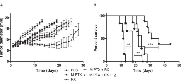
3. Synergy between PTX-loaded Micelles and Irradiation Based on the observed increased tumor oxygenation assessed by EPR oximetry, we checked whether PTX-loaded micelles may
Figure 1. Effect of the administration of PTX-loaded micelles on tumor pO2.Mean tumor pO2(6SD) monitored by EPR oximetry. A. Mice
treated with PTX-loaded micelles (13.5 mg/kg) (#); Non treated mice (&) (n= 4 for 0, 2, 4 and 6 h;n= 3 for 24 h) B. Mice treated with PTX-loaded micelles (80 mg/kg) (#); Non treated mice (&) (n= 7 except for control mice at 24 h:n= 4). C. Effect of the ligation of leg of mice on tumor pO2
enhance tumor radioresponse. Hence, the in vivo anti-tumor efficacy of the combination of PTX-loaded micelles and irradia-tion was performed (Fig. 2). PTX-loaded micelles were adminis-tered 24 h before irradiation, based on EPR oximetry results. All treatments were significantly different as compared to the PBS control group (p,0.001). Previously, it was demonstrated that unloaded micelles did not present any sign of cytotoxicity bothin vitroand in vivo[11]. The time to reach 18 mm was 14 days for mice treated with PTX-loaded micelles and 21 days for irradiated mice (p,0.001). The combination of these two treatments allowed a significant enhancement of the time to reach 18 mm until 28 days when compared with single treatment (PTX-loaded micelles or irradiation,p,0.001 and p,0.01, respectively). Mice were also treated with the combination of treatments with a transient ligation, using a rubber band, (just before and during the irradiation) of their leg to abolish the oxygen effect caused by PTX-loaded micelles. Interestingly, with the ligation, the time to reach 18 mm was decreased to 19 days which is similar to mice treated only with irradiation (p.0.05). The effect of ligation was also confirmed by pO2 measurements (Fig. 1C). On the other
hand, no significant difference was observed when TLT-bearing mice were irradiated and when mice were irradiated with the ligation of their leg (p.0.05). Indeed, TLT tumors are hypoxic (pO2,5 mm Hg [6]. Consequently, the ligation of the legs did not significantly decrease the oxygenation of hypoxic tumors. Togeth-er these results indicate that the ligation of PTX-treated tumors induced anoxia and counteracted the radiosensitizing effect of PTX and that the oxygen effect may contribute to the radiosensitizing properties of PTX (Fig. 2A).
The survival rate was significantly higher for mice treated with the combination of PTX-loaded micelles and irradiation as compared to other groups (p,0.001). After 45 days, for the half of mice, it was not anymore possible to detect any tumor (Fig. 2B).
4. Underlying Mechanisms of the Improvement of Tumor Oxygenation
To understand which mechanisms are responsible of the observed improvement of tumor oxygenation 24 h after PTX-loaded micelles treatment, additional experiments were per-formed: (i) the patent blue staining was used to evaluate the tumor perfusion, (ii) the oxygen consumption rate of tumor was assessed ex vivo by EPR oximetry and (iii) the apoptosis induced by PTX-loaded micelles was examined by fluorescent microscopy, using the TUNEL assay.
4.1 Tumor perfusion. The tumor perfusion was assessed by observing colored area after injection of Patent blue. This method, previously validated in comparison with DCE-MRI, provides an approximate of relative blood flow [21]. Tumors pre-treated 24 h before with PTX-loaded micelles showed a higher stained area, expressed in percentage of perfusion, (n= 8; 55.266.5%) than non-treated tumors (n= 7; 25.467.6%) (p,0.05), indicating that PTX-loaded micelles increased the tumor blood flow (Fig. 3).
4.2 Tumor oxygen consumption. We measured by EPR the oxygen consumption rate of tumor cells extracted from TLT tumors of mice treated 24 h before with PTX-loaded micelles (80 mg/kg), compared to the rate of non-treated mice as control group. The mean slopes were found to be significantly different (–4.3760.05 and –7.3460.14 for control and treated groups, respectively) (p,0.001) (Fig. 4). This means that tumor cells pretreated with PTX-loaded micelles consumed oxygen 1.7 times slower than non-treated tumor cells.
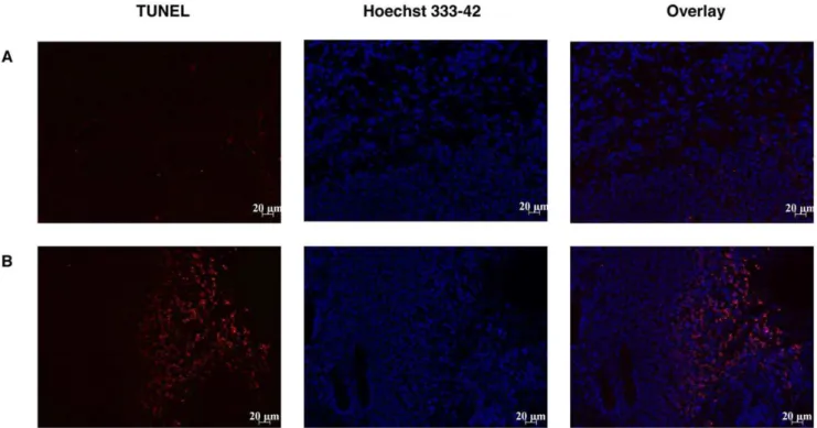
4.3 Apoptosis in TLT tumor treated by PTX-loaded micelles. Using the TUNEL method to assess apoptosis, visualization of total apoptotic cells (TUNEL positive cells labeled in red) indicated that higher apoptosis was obtained when mice were treated with PTX-loaded micelles compared to untreated mice (Fig. 5).
Discussion
PTX was previously described as a radiosensitizer agent. Because PTX-treated cells are arrested in the G2/M phase and
because this phase of the cell cycle is known to be the more radiosensitive, a combination of PTX with ionizing radiation has been proposed [3]. Moreover, tumor reoxygenation has been suggested to be another important mechanism by which PTX potentiates the radioresponse of tumors [5]. Elsewhere, it has been previously suggested that these two mechanisms are strongly linked: the tumor reoxygenation resulting from the G2/M block
induced by PTX [5].
PEG-p-(CL-co-TMC) polymeric micelles loaded with micelles were recently described as safe and effective PTX delivery system, which drastically enhance the maximum tolerated dose (MTD) of TaxolH [9]. Two PTX-loaded micellar systems are currently in clinical trials: Genexol-PM (phase IV for breast cancer) and NK105 (phase II for stomach cancer). For example, Genexol allows a 3-fold enhancement of the MTD while our system allows a 6-fold enhancement of the MTD. Moreover, compared to the most of polymeric micelles described in the literature, the major advantage of our PEG-p-(CL-co-TMC) micelles is that they are self-assembling upon contact with water and that not organic solvent neither dialysis or freeze-dried or evaporation steps is required leading this system feasible for scaling-up.
To evaluate if the radiosensitizing effect of PTX-loaded micelles results from an oxygen effect, the evaluation of reoxygenation over the time should be established. Therefore, we assessed EPR
oximetry after injection of PTX-loaded micelles (80 mg/kg). After 20 h, a drastic increase in pO2was observed and was maintained
at least until 24 h (the critical cut off of pO2is situated at 5 mm
Hg). Since cytotoxic effects, after a single PTX dose, decrease after 3 days [9], we can thus suppose that the increased pO2does not
last more than 3 days. At the MTD of TaxolH(13.5 mg/kg), no increase in pO2 was observed. Polymeric micelles may thus be
considered as an innovative tool to administer higher doses of PTX. It is important to note that at this dose of PTX, its was impossible to compare our polymeric micelles formulation with TaxolH because of the death of mice within 2 h treated with TaxolHat the equivalent PTX dose. Moreover, it is important to note that it was previously demonstrated that unloaded micelles did not present any sign of cytotoxicity bothin vitroandin vivo[11]. These Electron Paramagnetic Resonance results allow the determination of the time window of reoxygenation, which was used to design the irradiation experiments. To our knowledge, for the first time, we used the timing sequence of reoxygenation by measurement of pO2 to design experiments of PTX treatment
combined with irradiation. Using the regrowth delay assay, we found a synergy between PTX and irradiation during the reoxygenation window. The hypothesis that a synergy between PTX and irradiation was mediated with an oxygen effect was clearly confirmed with the regrowth delay assay on TLT tumor-bearing mice treated with PTX-loaded micelles 24 h before irradiation. Tumor clamping by temporary ligation of the irradiated leg induced complete hypoxia at the time of irradiation and prevents enhancement of tumor pO2 induced by PTX
treatment.The fact that mice treated by the combination of PTX and X-rays, with ligation of their leg, responded similarly to mice treated only with irradiation demonstrates that the radiosensitizing effect of PTX is mainly due to an oxygen effect. We did not expect any difference between irradiation alone group and X-rays +
ligature group because the chosen tumor model TLT is already described as highly hypoxic. Using such radioresistant model is thus well adapted to demonstrate oxygen-mediated radiosensitiz-ing properties of PTX [22].
The enhancement of tumor oxygenation can be explained by either an enhancement in blood supply which is associated with an increased blood flow or/and by a decrease in oxygen consumption (Fig. 6). We demonstrated that PTX-loaded micelles contribute to reoxygenation effect by both of these mechanisms. To our
Figure 3. Effect of PTX-loaded micelles on tumor perfusion
24 h post treatment by Patent blue staining. (A) Each bar
represents the mean value of tumor percentage of colored area6SEM for control group and PTX-loaded micelles group. *p,0.05. (B) Control. (C) PTX-loaded micelles.
doi:10.1371/journal.pone.0040772.g003
knowledge it is the first study which evaluates the mechanisms underlying the ‘‘oxygen effect’’ for PTX. The radiosensitivity of PTX-incorporating micelles was also studied by Negishi et al., focusing their attention to the ability of PTX to arrest cells in G2/
M phase [23]. The cytotoxic effect of PTX-loaded micelles was previously demonstrated bothin vitroandin vivo[9]. The decrease in oxygen consumption is the result of two distinct mechanisms. First, as we observed an important apoptosis after treatment (Fig. 5), the total number of tumor cells consuming oxygen is decreased. Moreover, we also observed that the PTX treatment induced a decrease in oxygen respiration by the living tumor cells (Fig. 4). In addition to the effect on oxygen consumption, we observed that the PTX treatment led to an increase in tumor perfusion (Fig. 3). This effect could be linked to the decrease of tumor cells leading to a decrease in interstitial fluid pressure and a consequent increase in tumor perfusion.
The radiosensitizing effect of PTX can thus be explained by at least two mechanisms: the G2/M blockade and the tumor
reoxygenation. The first one was largely described in the literature [3,23]; while we highlighted and explained the tumor reoxygena-tion in the present paper. Moreover, these two mechanisms are likely linked: the G2/Mblock leads to apoptosis of cells which is
partly responsible from the tumor reoxygenation.
In conclusion, we demonstrated that PEG-p(CL-co-TMC) polymeric micelles loaded with PTX radiosensitized TLT tumors by improving the tumor oxygenation. Combining polymeric drug chemotherapy with radiation may have important clinical implications in terms of scheduling and optimization of the therapeutic ratio. We also showed that the increased pO2can be
explained both by an increased blood flow and an inhibition of O2
consumption.
Acknowledgments
The authors wish to thank Janssen Pharmaceutica, Belgium for providing PEG-p(CL-co-TMC).
Author Contributions
Conceived and designed the experiments: FD PD BJ BG VP. Performed the experiments: FD PD NM GDP BU. Analyzed the data: FD PD BG VP. Contributed reagents/materials/analysis tools: GDP OK BJ. Wrote the paper: FD PD.
Figure 5. Effect of PTX-loaded micelles 24 h pre-treatment on TLT tumor apoptosis assessed by TUNEL assay.(A) Control mice (n= 3). (B) Mice treated 24 h before analysis with PTX-loaded micelles (80 mg/kg). DAPI staining was used to label nuclei (blue) and TUNEL staining was used to label apoptotic cells (red).
doi:10.1371/journal.pone.0040772.g005
Figure 6. Mechanisms of tumor oxygenation induced by PTX (80 mg/kg). Mechanistically, tumor oxygenation results from an balance between oxygen delivery (perfusion) and local oxygen consumption (which depends on the cellular viability and mitochon-drial respiration) (Adapted from [7]).
References
1. Singla AK, Garg A, Aggarwal D (2002) Paclitaxel and its formulations, Int. J. Pharm.235: 179–92.
2. Adams JD, Flora KP, Goldspiel BR, Wilson JW, Arbuck SG, et al. (1993) Taxol: a history of pharmaceutical development and current pharmaceutical concerns, J. Natl. Cancer Inst. Monogr. 141–47.
3. Milas L, Hunter N, Mason KA, Milross C, Peters LJ (1995) Tumor reoxygenation as a mechanism of taxol-induced enhancement of tumor radioresponse, Acta Oncol. 34: 409–412.
4. Tishler RB, Geard CR, Hall EJ, Schiff PB (1992) Taxol sensitizes human astrocytoma cells to radiation, Cancer Res. 52: 3495–97.
5. Milas L, Hunter NR, Mason KA, Milross CG, Saito Y, et al. (1995) Role of reoxygenation in induction of enhancement of tumor radioresponse by paclitaxel, Cancer Res. 55: 3564–68.
6. Overgaard J (2007) Hypoxic radiosensitization: adored and ignored. J. Clin. Oncol. 25: 4066–74.
7. Gallez B, Baudelet C, Jordan BF (2004) Assessment of tumor oxygenation by electron paramagnetic resonance: principles and applications, NMR Biomed. 17: 240–62.
8. Vaupel P, Kelleher DK, Thews O (1998) Modulation of tumor oxygenation, Int. J. Radiat. Oncol. Biol. Phys. 42: 843–48.
9. Danhier F, Magotteaux N, Ucakar B, Lecouturier N, Brewster M, et al. (2009) Novel self-assembling PEG-p-(CL-co-TMC) polymeric micelles as safe and effective delivery system for Paclitaxel, Eur. J. Pharm. Biopharm. 73: 230–38. 10. Maeda H (2001) The enhanced permeability and retention (EPR) effect in tumor
vasculature: the key role of tumor-selective macromolecular drug targeting, Adv. Enzyme Regul.41: 189–207.
11. Danhier F, Feron O, Pre´at V (2010) To exploit the tumor microenvironment: passive and active tumor targeting of nanocarriers for anti-cancer drug delivery. J. Control. Rel. 148: 135–146.
12. Ould-Ouali L, Arien A, Rosenblatt J, Natan A, Twaddle T, et al. (2004) Biodegradable self-assembling PEG-copolymer as vehicle for poorly water-soluble drugs, Pharm. Res. 21: 1581–90.
13. Taper HS, Woolley GW, Teller MN, Lardis MP (1966) A new transplantable mouse liver tumor of spontaneous origin, Cancer Res. 26: 143–48.
14. 14. Gallez B, Jordan BF, Baudelet C, Misson PD (1999) Pharmacological modifications of the partial pressure of oxygen in murine tumors: evaluation using in vivo EPR oximetry, Magn Reson. Med., 42: 627–30.
15. Jordan BF, Misson P, Demeure R, Baudelet C, Beghein N, et al. (2000) Changes in tumor oxygenation/perfusion induced by the no donor, isosorbide dinitrate, in comparison with carbogen: monitoring by EPR and MRI, Int. J. Radiat. Oncol. Biol. Phys. 48: 565–70.
16. Ansiaux R, Baudelet C, Jordan BF, Beghein N, Sonveaux P, et al. (2005) Thalidomide radiosensitizes tumors through early changes in the tumor microenvironment. Clin. Cancer Res. 11: 743–50.
17. Crokart N, Jordan BF, Baudelet C, Beghein N, Sonveaux P, et al. (2005) Early reoxygenation in tumors after irradiation: determining factors and consequences for radiotherapy regimens using daily multiple fractions, Int. J. Radiat. Oncol. Biol. Phys. 63: 901–10.
18. James PE, Jackson SK, Grinberg OY, Swartz HM (1995) The effects of endotoxin on oxygen consumption of various cell types in vitro: an EPR oximetry study, Free Radic. Biol. Med. 18: 641–47.
19. Jordan BF, Sonveaux P, Feron O, Gregoire V, Beghein N, et al. (2003) Nitric oxide-mediated increase in tumor blood flow and oxygenation of tumors implanted in muscles stimulated by electric pulses, Int. J. Radiat. Oncol. Biol. Phys. 55: 1066–73.
20. 20. Milross CG, Mason KA, Hunter NR, Terry NH, Patel N, et al. (1997) Enhanced radioresponse of paclitaxel-sensitive and -resistant tumours in vivo, Eur. J. Cancer 33: 1299–308.
21. Ansiaux R, Baudelet C, Cron G, Segers J, Dessy C, et al. (2006) Botulinium toxin potentiates cancer radiotherapy and chemotherapy. Clin. Cancer Res. 12: 1276–83.
22. Diepart C, Karroum O, Magat J, Feron O, Verrax J, et al. (2012) Arsenic trioxide treatment decreases the oxygen consumption rate of tumor cells and radiosensitizes solid tumors. Cancer Res. 72: 482–90.