Fluorogenic Substrates for
Monitoring
of Caspase-3 Activity in Live Cells
Ana M. Pérez-López1, M. Lourdes Soria-Gila2, Emma R. Marsden1,
Annamaria Lilienkampf1, Mark Bradley1*
1School of Chemistry, EaStCHEM, University of Edinburgh, Joseph Black building, West Mains Road, Edinburgh EH9 3FJ, United Kingdom,2Department of Medicinal and Organic Chemistry, University of Granada, School of Pharmacy, Campus Cartuja s/n–18071, Granada, Spain
Abstract
Thein situdetection of caspase-3 activity has applications in the imaging and monitoring of
multiple pathologies, notably cancer. A series of cell penetrating FRET-based fluorogenic substrates were designed and synthesised for the detection of caspase-3 in live cells. A variety of modifications of the classical caspase-3 and caspase-7 substrate sequence Asp-Glu-Val-Asp were carried out in order to increase 3 affinity and eliminate caspase-7 cross-reactivity. To allow cellular uptake and good solubility, the substrates were conju-gated to a cationic peptoid. The most selective fluorogenic substrate27, FAM-Ahx-Asp-Leu-Pro-Asp-Lys(MR)-Ahx, conjugated to the cell penetrating peptoid at the C-terminus, was able to detect and quantify caspase-3 activity in apoptotic cells without cross-reactivity by caspase-7.
Introduction
Fluorogenic substrates and activity-based probes enable the study of protease function and have been used to elucidate the role of caspases in the progression of diseases such as cancer [1–6], neurodegenerative disorders [7–10], and sepsis [11,12]. Caspases are an important fam-ily of cysteine-dependent aspartate proteases that exist within cells as inactive zymogens with their cleavage giving active enzymes initiating cellular apoptosis [13–15]. Inappropriate control of this apoptotic machinery is implicated in many diseases [14,15], notably cancer [16,17]. As a part of the apoptotic cascade, executioner caspase-3 activates several important cellular sub-strates [14–20], such as PARP and ICAD, and its decreased activity is a prognostic indicator of chemoresistance in breast and ovarian cancer [21,22]. The ability to monitor caspase-3 activity
in situcould provide a means, not only to elucidate its complex role in biological processes, but
also to monitor the efficacy of anticancer drugs and to identify patients for whom discontinua-tion of ineffective toxic treatment is warranted, for example, due to acquired drug resistance [23].
Current methods are able to detect caspase-3 activityin vitroalthough they often display
promiscuity and cannot be used to monitor caspase-3 within cells [24–26]. The majority of
a11111
OPEN ACCESS
Citation:Pérez-López AM, Soria-Gila ML, Marsden ER, Lilienkampf A, Bradley M (2016) Fluorogenic Substrates forIn SituMonitoring of Caspase-3 Activity in Live Cells. PLoS ONE 11(5): e0153209. doi:10.1371/journal.pone.0153209
Editor:Shawn B Bratton, The University of Texas MD Anderson Cancer Center, UNITED STATES
Received:November 17, 2015
Accepted:March 26, 2016
Published:May 11, 2016
Copyright:© 2016 Pérez-López et al. This is an open access article distributed under the terms of the
Creative Commons Attribution License, which permits unrestricted use, distribution, and reproduction in any medium, provided the original author and source are credited.
Data Availability Statement:All relevant data are within the paper and its Supporting Information files.
Funding:This work was supported by the Ramon Areces and Caja Madrid Foundations to AMPL and Spanish Ministry of Economy and Competitiveness to MLSG (graduate student fellowships FPI BES-2010-030257 and EEBB-I-13-07131). The funders had no role in study design, data collection and analysis, decision to publish, or preparation of the manuscript.
fluorogenic caspase-3 substrates are based on a four-residue recognition sequence Asp-Glu-Val-Asp (DEVD) [23,27,28], established via combinatorial library methods [29,30]; however, this sequence is also efficiently cleaved by caspase-7, which shares very similar substrate speci-ficities with caspase-3. In mouse macrophages, 46 out of the 55 identified protein cleavage sites (within 48 proteins) were cleaved by both enzymes with only 3 sites specifically cleaved by cas-pase-3 [31]. Incorporation of unnatural amino acids into the recognition sequence has yielded caspase-3 substrates with increased selectivity [32,33]. Recently, Wolan achieved live cell imag-ing of caspase-3 activity in apoptotic cells, with selectivity over caspase-7, with a near-infrared fluorogenic pentapeptide substrate (incorporating the unnatural amino acidβ-homo-Leu) cou-pled to a cell penetrating peptide derived from the viral SV40 Large T-antigen nuclear localis-ing signal [34].
Here, FRET-based fluorogenic substrates, incorporating a tetrapeptide recognition sequence Asp-X3-X2-Asp, were designed and synthesised for the selective,in situmonitoring of caspase-3 activity. To allow detection in live cells, the substrates were conjugated to a cationic peptoid-based cellular delivery vehicle.
Results and Discussion
Substrate design and synthesis
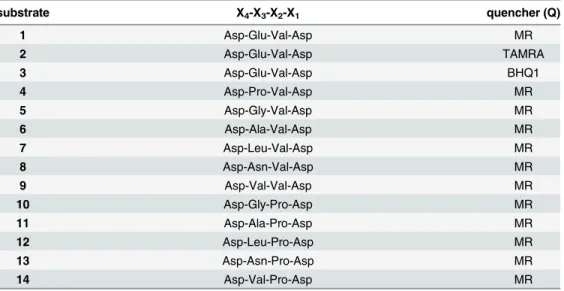
In order to improve selectivity towards caspase-3 over caspase-7, permutations of the classical tetrapeptide substrate Asp-Glu-Val-Asp (X4-X3-X2-X1) were explored. All known caspase-3 substrates contain Asp at position X1and 80% contain an Asp at the X4position of the sequence. The positions X3and X2are more varied, with no clear amino acid preference being reported for the X3position (~20% of the substrates contain Glu and ~15% Phe or Val at this position). Approximately 40% of known caspase-3 substrates contain Val at the X2position; however, Pro at the X2position is known to increase specificity for caspase-3 over caspase-7 [35]. With the aim of improving caspase-3 selectivity, X3and X2modifications of the tetrapep-tide sequence were carried out, retaining Asp at X1and X4positions. The X3position was changed to Pro, Gly, Ala, Leu, Asn and Val, and the X2position had Val (substrates1–9) or Pro (substrates10–14) (Table 1) [36,37]. For each substrate, the corresponding d-amino acid sequence was synthesised as a control (compounds15–24, respectively, see ESI). 5(6)-Carboxy-fluorescein was coupled to the N-terminus of the substrates via a 6-aminohexanoic acid (Ahx) spacer, and a quencher moiety was introduced next to the caspase cleavage site via Lys side chain modification (separated by an Ahx spacer from the Asp-X3-X2-Asp) (Fig 1). As the choice of the quencher can affect the rate of cleavage and level of background fluorescence, three different quenchers, methyl red (MR), Black Hole Quencher1
-1 (BHQ1), and 5(6)-car-boxytetraethylrhodamine (TAMRA) were evaluated. A cationic,“lysine-like”nonaresidue pep-toid was incorporated onto the C-terminus to ensure cellular uptake of the substrates [38]. Unlike many common cell penetrating peptides [39–41] this peptoid is resistant to proteolysis, non-toxicin vivo, and has demonstrated a highly efficient cell entry profile [42–44]. In
addi-tion, peptoid-based delivery systems are not prone producing immunogenic responses associ-ated with virus-derived sequences [45,46].
The peptides1–14and controls15–24were synthesised on a Rink amide-functionalised
aminomethyl polystyrene resin (1% DVB, 100–200 mesh, loading 1.2 mmol/g) using an Fmoc/tBu-based strategy with microwave heating (S1 Fig) [47]. First, the nonapeptoid was synthesised usingN-Fmoc-(6-Boc-aminohexyl)glycine [48] and DIC and Oxyma. Fmoc-Lys
of the carboxy-functionalised quencher. After deprotection and cleavage from the resin with TFA–TIS–DCM, peptides1–24were purified by preparative HPLC and analysed by MALDI--TOF MS.
The effect of the quencher on caspase-3 cleavage
To optimise the fluorogenic substrates, MR (λAbs480 nm), TAMRA (λAbs555 nm), and BHQ1 (λAbs534 nm) were evaluated as quenchers for 5(6)-carboxyfluorescein (λEx/Em488/528 nm) in compounds1,2and3, using the classical substrate Asp-Glu-Val-Asp. Only1(MR as quencher) showed notable time-dependent increase in fluorescence upon incubation with cas-pase-3 and 7 (2and3, incorporating TAMRA and BHQ1, did not demonstrate significant increase in fluorescence) (Fig 2).
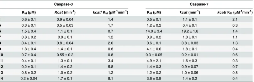
Kinetic studies with Caspase-3 and 7
The ability of1and5–24(bearing MR as the quencher of choice) to act as s substrate for cas-pase-3, as well as caspase-7, was investigated by determining the catalytic efficiency (kcat/KM) for each substrate with both enzymes (Table 2). None of the d-amino acids containing sequences15–24or4, which has a Pro residue at position X3, showed any change in fluores-cence intensity over time. With caspase-3, all the substrates with Val in X2position (substrates
5–9) showed similar catalytic efficiency (kcat/KM0.7–2.0μM-1min-1) as Asp-Glu-Val-Asp
(sub-strate1, 1.4μM-1min-1). Substrate5(Asp-Gly-Val-Asp) showed 5-fold selectivity over
caspase-7 (Table 2). Pro at the X2position increased specificity for caspase-3 over caspase-7 [49], par-ticularly with substrates11(Asp-Ala-Pro-Asp),12(Asp-Leu-Pro-Asp) and14 (Asp-Val-Pro-Asp), which exhibited 8–20-fold selectivity over caspase-7, along with increased caspase-3 affinity (KM0.2–0.4μM) and catalytic effiency (kcat/KM3.4–8.1μM-1min-1).
To confirm how specific11,12and14were for caspase-3, these substrates were incubated with high enzyme concentrations (10–20μM substrate, 0.4μM enzyme) and the
caspase-medi-ated cleavage analysed by MALDI-TOF MS. As expected, all the substrates were cleaved by
Table 1. The recognition sequence is a tetrapeptide (X4-X3-X2-X1) with two variable positions.X1and
X4was Asp in all peptides. Three different quenchers, methyl red (MR), Black Hole Quencher1-1 (BHQ1),
and 5(6)-carboxytetraethylrhodamine (TAMRA) were evaluated. As controls, substrates were also synthe-sised with the corresponding d-amino acid sequence (compounds15–24, respectively, see supporting infor-mation). For full structures, seeFig 1.
substrate X4-X3-X2-X1 quencher (Q)
1 Asp-Glu-Val-Asp MR
2 Asp-Glu-Val-Asp TAMRA
3 Asp-Glu-Val-Asp BHQ1
4 Asp-Pro-Val-Asp MR
5 Asp-Gly-Val-Asp MR
6 Asp-Ala-Val-Asp MR
7 Asp-Leu-Val-Asp MR
8 Asp-Asn-Val-Asp MR
9 Asp-Val-Val-Asp MR
10 Asp-Gly-Pro-Asp MR
11 Asp-Ala-Pro-Asp MR
12 Asp-Leu-Pro-Asp MR
13 Asp-Asn-Pro-Asp MR
14 Asp-Val-Pro-Asp MR
caspase-3 at the X1position (between Asp and the Ahx spacer), with the parent compound no longer detected after 2h; however, these substrates were also (partially) cleaved by caspase-7. Remarkably, MS analysis revealed that that caspase-7 exhibited a different cleavage pattern cleaving the substrates between the Ahx spacer and the Lys(MR) (S2–S4Figs).
Substrate optimisation
To eliminate the caspase-7 cross reactivity, three new substrates, all bearing Asp-Leu-Pro-Asp, were synthesised using d-Lys orN-Methyl-Lys as the quencher attachment point (25and26,
respectively) and switching the position of the Ahx spacer (27) (Fig 3). Substrate25was cleaved by both caspase-3 and 7, whereas theN-methylated substrate26was not cleavage
by either enzyme (S6andS7Figs). Substrate27showed good affinity for caspase-3 (KM 1.1 ± 0.3μM) with kcatof 2.1 ± 0.8 min-1and a catalytic efficiency of 0.5 ± 0.08μM-1min-1.
Remarkably, caspase-7 (0.4μM) did not show any cleavage of this substrate (S7andS8Figs).
Detecting caspase-3 activity in live cells
Caspase-3 activity in HEK293T cells was evaluated using the caspase-3 selective substrate27
with apoptosis induced by staurosporine. Flow cytometry analysis of cells treated with27
Fig 1. The design of the FRET-based fluorogenic tetrapeptide substrates for caspase-3 detection in live cells.The substrates bear 5(6)-carboxyfluorescein (λEx/Em488/528 nm) at the amino-terminus and a
quencher coupled via a Lys side-chain. The recognition sequence is a tetrapeptide (Asp-X3-X2-Asp) with two
variable positions (seeTable 1). The C-terminus bears a“lysine-like”nonaresidue peptoid to enable cellular uptake.
doi:10.1371/journal.pone.0153209.g001
Fig 2. Relative increase in fluorescence intensity of the FRET-based peptides, bearing different quenchers (MR, TAMRA or BHQ1) on the Lys side chain, after incubation with caspase-3 and 7.The FRET-based peptides1,2and3(6μM) were incubated with caspase-3 and 7 (20 nM) and fluorescence recorded at 10, 20 and 40 min (n = 3, normalised to zero).
(10μM) showed a 2.5-fold increase in the fluorescence intensity (λEx/Em488/530 nm) of the cells after apoptotic stimulation by staurosporine (1μM), with no increase observed in
fluo-resce without it (Fig 4). This increase in fluorescence with substrate27suggested that the con-centration of caspase-3 in apoptotic cells was approximately 15.7 ± 0.5 nM per cell (28271 molecules per cell) in the execution phase of apoptosis (based on theVmaxand thekcatof27,
see supporting information) [50]. In live-cell confocal imaging of caspase-3 activation with27
(10μM), fluorescence“turn-on”was only detected in the cytoplasm of apoptotic HEK293T
cells, with no increase in fluorescence observed in non-apoptotic cells (Fig 5A, 5BandS9 Fig). No fluorescence“turn-on”was observed in apoptotic MCF-7 cells (Fig 5C), which lack func-tional caspase-3 but express caspase-7 [34,51,52], confirming the isoform selectivity. Substrate
27was nontoxic in an MTT assay at 10μM concentration (S10 Fig).
Table 2. Kinetic analysis of the fluorogenic substrates.The kinetic parameters (n = 3) were determined for the fluorogenic substrates1and5–14 (mea-sured using substrate range of 0.1–8μM) with caspase-3 and caspase-7. Substrate4or the d-amino acids containing15–24were not cleaved by either enzyme.
Caspase-3 Caspase-7
KM(μM) Kcat(min-1) kcat/ KM(μM-1min-1) KM(μM) kcat(min-1) kcat/ KM(μM-1min-1)
1 0.6±0.1 0.9±0.04 1.4 0.5±0.1 1.1±0.1 2.1
5 0.3±0.1 0.5±0.03 1.7 1.2±0.2 0.4±0.1 0.3
6 1.5±0.4 1.1±0.1 0.7 14.0±3.4 19.2±1.6 1.4
7 0.8±0.2 0.9±0.1 1.2 0.9±0.2 1.0±0.1 1.1
8 0.4±0.1 0.8±0.04 2.0 0.6±0.1 0.8±0.03 1.3
9 1.8±0.4 1.4±0.1 0.8 4.1±0.6 1.8±0.1 0.4
10 0.7±0.4 0.55±0.2 0.8 0.3±0.05 0.2±0.01 0.6
11 0.4±0.1 1.3±0.1 3.4 4.9±2.1 1.6±0.3 0.3
12 0.2±0.1 1.4±0.2 5.8 1.4±0.3 0.9±0.07 0.7
13 0.8±0.2 1.0±0.2 1.2 1.2±0.2 1.0±0.06 0.8
14 0.2±0.04 1.7±0.1 8.1 3.6±0.9 1.4±0.2 0.4
doi:10.1371/journal.pone.0153209.t002
Fig 3. Structural modifications to the fluorogenic substrates with the aim of eliminating caspase-7 cross-reactivity.Substrate25
has a D-Lys residue,26anN-Methyl-Lys, and in substrate27the Ahx spacer has been moved between the Lys and the peptoid moiety.
Caspase-3 selectivity was achieved with27.
Conclusions
Fluorogenic, FRET-based substrates for monitoring the enzymatic activity of caspase-3in situ
were synthesised with permutations of the substrate sequence Asp-X2-X3-Asp with the aim of improving selectivity for caspase-3 over caspase-7. The fluorogenic substrates had 5(6)-carbox-yfluorescein in the amino-terminus and an optimised quencher, methyl red, introduced via Lys
Fig 4. Flow cytometry analysis of healthy and apoptotic HEK293T cells treated with substrate 27.The cells were incubated 5 h with fluorogenic substrate27(10μM), detached, and analysed by flow cytometry (λEx/Em488/530 nm, x-axis = fluorescence intensity). (A) Healthy, non-apoptotic cells. (B) Apoptotic cells
(induced by 1μM staurosporine).
doi:10.1371/journal.pone.0153209.g004
Fig 5. Confocal microscopy images of HEK293T and MCF-7 cells treated with substrate 27.Confocal microscopy images (objective HCX PL APO ×63/1.40–0.6 Oil CS) of HEK273T cells with substrate27
(10μM) without staurosporine (STS) (A) and with staurosporine (1μM) (B)induced apoptosis (green fluorescence is from fluorescein“turned on”by caspase-3 cleavage of the substrate, blue is DAPI nuclear stain). (C) Staurosporine treated MCF-7 cells with substrate27.
side chain modification. These fluorogenic substrates were conjugated at the C-terminus to a cationic cell delivery vehicle to allow efficient cellular uptake. Substrates with Pro in the X2 position (instead of Val), showed selectivity for caspase-3 over caspase-7, along with increased caspase-3 affinity, particularly when X3was Ala, Leu or Val. Mass spectrometry studies revealed unexpected cleavage pattern of the fluorogenic substrates with caspase-7, while opti-misation of the substrate via spacer relocation yielded a caspase-3 selective substrate27
(FAM-Ahx-Asp-Leu-Pro-Asp-Lys(MR)-Ahx-peptoid). In apoptotic cells, the optimised sub-strate27allowed imaging of caspase-3 activityin situ. Flow cytometry analysis gave
approxi-mate quantification of the concentration of caspase-3 in a cell to be 16 nM (28271 molecules per cell). Future work is aimed at the use of this caspase-3 selective sequence inin vivo
near-infrared fluorescence imaging techniques, especially for cancer, which require stable, highly specific, and sensitive fluorogenic substrates.
Materials and Methods
Synthesis of fluorogenic substrates 1
–
27
A highly optimised microwave-based solid–phase strategy was used to synthesise the cell pene-trating peptide–peptoids [38]. All solvents and reagents were obtained from commercial sup-pliers and used without purification. A Rink-amide functionalised aminomethyl polystyrene (1% DVB, 100–200 mesh, loading 1.2 mmol/g) resin was used for the synthesis of the peptides with an Fmoc-based strategy (S1 Fig).Coupling of the Fmoc-amino acids and fluorophores:
The resin (1 eq) was pre-swollen in DCM, washed with DMF, and added a pre-activated mix-ture (10 min) of the carboxylic acid (3 eq), DIC (3 eq) and Oxyma (3 eq) in DMF (0.1 M). This reaction mixture was stirred in the microwave (Biotage Initiator) for 20 minutes at 60°C after which the resin was washed with DMF, DCM and MeOH.Fmoc deprotection:The resin was shaken with 20% piperidine in DMF (2 ×10 min), and subsequently washed with DMF, DCM and MeOH.Dde deprotection:The resin was shaken with 2% hydrazine in DMF (v/v) (2 ×10 min), and subsequently washed with DMF, DCM and MeOH.Cleavage from the resin and deprotection:A solution of TFA/TIS/DCM (90:5:5) was added to the resin (20μL of the
cleav-age cocktail per mg of resin) and left to shake for 5 hours. The resin was filtrated and washed with DCM, and the collected filtrate was evaporated under reduced pressure and the com-pound precipitated using cold diethyl ether. Peptides1–24were purified by preparative HPLC and analysed by MALDI-TOF MS. For the characterisation of the peptides, seeS1 Table.
Kinetic assays with caspase-3 and 7
Caspase-3 or caspase-7 (R&D systems, USA) was added to 100μL of caspase assay buffer with
substrates1–14at concentrations from 0.1μM to 8μM in a 96-well plate (n = 3) to give final
enzyme concentration of 20 nM (1–9) or 15 nM (10–14). Fluorescence (λEx/Em485/528 nm) was recorded on a Biotek Synergy HT Multi-Mode Microplate Reader every 2 min. Control samples had the same composition but no enzyme. The rate (μM/min) was calibrated using a
5(6)-carboxyfluorescein conversion factor (0.0055μM/RFU) and data plotted against time
(min). For initial cleavage rate (0–5 min), plots were fitted using linear regression analysis and the Michaelis-Menten data generated using GraphPad Prism 5.
Caspase-3 detection in live cells
streptomycin and 25 mg/mL amphotericin B, and 10% FBS) were seeded onto a 48-well plate at a density of 104cells per well. After 12 hours, the media was removed and substrates1–14
added at 10μM in fresh media. Selected wells were also treated with staurosporine (1μM).
After 5 hours, the cells were washed twice with PBS, detached with trypsin/EDTA, harvested with 2% FBS in PBS supplemented with Trypan Blue (0.04%) for analysis on a BD FACSAria1 flow cytometer. Fluorescence was evaluated as mean fluorescence intensity (MFI) and esti-mated<100 u.a. for untreated control cells (consistent values independently of staurosporine addition). Apoptotic cells treated with substrate1were used as a positive control to obtain the maximum of fluorescence signal. 50,000 events per sample were plotted in two-dimensional dot plots based on forward and side scattering. The cellular size and complexity (SSC-H vs. FSC-H) were used to gate two populations (alive/apoptotic cells) (debri excluded). The data were analysed using the software Flowjo17.5.
For confocal microscopy, the cells were fixed with 4% paraformaldehyde in PBS and the nuclei stained with Hoechst-33342 (1% w/v in PBS). Cellular fluorescence of cells was analysed using an Inverted Leica DM IRB with filter I3 (450–490 nm) and a Leica SP5 Confocal (FITC and DAPI channel).
Supporting Information
S1 Fig. Solid phase synthesis of the fluorogenic substrates 1–24.
(PDF)
S2 Fig. MALDI-TOF MS spectra of substrate 11.
(PDF)
S3 Fig. MALDI-TOF MS spectra of substrate 12.
(PDF)
S4 Fig. MALDI-TOF MS spectra of substrate 14.
(PDF)
S5 Fig. MALDI-TOF MS spectra of substrate 25.
(PDF)
S6 Fig. MALDI-TOF MS spectra of 26.
(PDF)
S7 Fig. Analysis of substrate 27 with caspase-3 and caspase-7.
(PDF)
S8 Fig. MALDI-TOF MS spectra of 27.
(PDF)
S9 Fig. Substrate 27 selectively labels apoptotic cells.
(PDF)
S10 Fig. Cell viability.
(PDF)
S1 File. Quantification of caspase-3 in apoptotic cells by flow cytometry.
(PDF)
S1 Table. MALDI-TOF MS and HPLC analysis of substrates 1–27.
Author Contributions
Conceived and designed the experiments: AMPL MLSG AL MB. Performed the experiments: AMPL MLSG ERM. Analyzed the data: AMPL MLSG AL MB. Wrote the paper: AL AMPL MB.
References
1. Tsai FY, Greenbaum DC, Hager JH, Bogyo M, Hanahan D. Cathepsin cysteine proteases are effectors of invasive growth and angiogenesis during multistage tumorigenesis. Cancer Cell 2004; 5: 443–453. PMID:15144952
2. Speers AE, Cravatt BF. Profiling enzyme activities in vivo using click chemistry methods. Chem Biol. 2004; 11: 535–546. PMID:15123248
3. Paulick MG, Bogyo M. Application of activity-based probes to the study of enzymes involved in cancer progression. Curr Opin Genet Dev. 2008; 18: 97–106. doi:10.1016/j.gde.2007.12.001PMID: 18294838
4. Edgington LE, Berger AB, Blum G, Albrow VE, Paulick MG, Lineberry N, et al. Noninvasive optical imaging of apoptosis by caspase-targeted activity-based probes. Nat Med. 2009; 15: 967–973. doi:10. 1038/nm.1938PMID:19597506
5. Nomura DK, Dix MM, Cravatt BF. Activity-based protein profiling for biochemical pathway discovery in cancer. Nat Rev Cancer. 2010; 10: 630–638. doi:10.1038/nrc2901PMID:20703252
6. Edgington LE, Verdoes M, Ortega A, Withana NP, Lee J, Syed S, et al. Functional imaging of legumain in cancer using a new quenched activity-based probe. J Am Chem Soc. 2013; 135: 174–182. doi:10. 1021/ja307083bPMID:23215039
7. Graham RK, Deng Y, Slow E, Haigh B, Bissada N, Lu G, et al. Cleavage at the caspase-6 site is required for neuronal dysfunction and degeneration due to mutant huntingtin. Cell. 2006; 125: 1179– 1191. PMID:16777606
8. Leyva MJ, Degiacomo F, Kaltenbach LS, Holcomb J, Zhang N, Gafni J, et al. Identification and evalua-tion of small molecule pan-caspase inhibitors in Huntington's disease models. Chem Biol. 2010; 17: 1189–1200. doi:10.1016/j.chembiol.2010.08.014PMID:21095569
9. Edgington LE, van Raam BJ, Verdoes M, Wierschem C, Salvesen GS, Bogyo M. An optimized activity-based probe for the study of caspase-6 activation. Chem Biol. 2012; 19: 340–352. doi:10.1016/j. chembiol.2011.12.021PMID:22444589
10. D’Amelio M, Sheng M, Cecconi F. Caspase-3 in the central nervous system: beyond apoptosis. Trends Neurosci. 2012; 35: 700–709. doi:10.1016/j.tins.2012.06.004PMID:22796265
11. Hotchkiss RS, Chang KC, Swanson PE, Tinsley KW, Hui JJ, Klender P, et al. Caspase inhibitors improve survival in sepsis: a critical role of the lymphocyte. Nat Immunol. 2000; 1: 496–501. PMID: 11101871
12. Hotchkiss RS, Nicholson DW. Apoptosis and caspases regulate death and inflammation in sepsis. Nat Rev Immunol. 2006; 6: 813–822. PMID:17039247
13. Van Damme P, Martens L, Van Damme J, Hugelier K, Staes A, Vandekerckhove J, et al. Caspase-spe-cific and nonspeCaspase-spe-cific in vivo protein processing during Fas-induced apoptosis. Nat Methods. 2005; 2: 771–777. PMID:16179924
14. Tanuma S, In Apoptosis in Normal Development and Cancer; Sluyser M., Ed.; Taylor and Francis: London, 1996; pp 39–59.
15. Thompson CB, Apoptosis in the pathogenesis and treatment of disease. Science. 1995; 267: 1456– 1462. PMID:7878464
16. Nicholson DW. From bench to clinic with apoptosis-based therapeutic agents. Nature. 2000; 407: 810– 816. PMID:11048733
17. Wyllie AH, Bellamy CO, Bubb VJ, Clarke AR, Corbet S, Curtis L. Apoptosis and carcinogenesis. Br J Cancer. 1999; 80: 34–37. PMID:10466759
18. Potten CS, Booth C. The role of radiation-induced and spontaneous apoptosis in the homeostasis of the gastrointestinal epithelium: a brief review. Comp Biochem Physiol. 1997; 118: 473–478.
19. Green DR, Apoptotic pathways: paper wraps stone blunts scissors. Cell. 2000; 102: 1–4. PMID: 10929706
21. Devarajan E, Sahin AA, Chen JS, Krishnamurthy RR, Aggarwal N, Brun AM, et al. Down-regulation of caspase 3 in breast cancer: a possible mechanism for chemoresistance. Oncogene. 2002; 21: 8843– 8851. PMID:12483536
22. Ai H, Hazelwood KL, Davidson MW, Campbell RE. Fluorescent protein FRET pairs for ratiometric imag-ing of dual biosensors. Nat Methods. 2008; 5: 401–403. doi:10.1038/nmeth.1207PMID:18425137
23. Savitsky AP, Rusanov AL, Zherdeva V, Gorodnicheva T, Khrenova MG, Nemukhin AV. FLIM-FRET Imaging of Caspase-3 activity in live cells using pair of red fluorescent proteins. Theranostics. 2012; 2: 215–226. doi:10.7150/thno.3885PMID:22375160
24. Kuida K, Zheng TS, Na S, Kuan C, Yang D, Karasuyama H, et al. Decreased apoptosis in the brain and premature lethality in CPP32-deficient mice. Nature. 1996; 384: 368–372. PMID:8934524
25. Agniswamy J, Fang B, Weber IT. Plasticity of S2-S4 specificity pockets of executioner caspase-7 revealed by structural and kinetic analysis. FEBS J. 2007; 274: 4752–4765. PMID:17697120
26. McStay GP, Salvesen GS, Green DR. Overlapping cleavage motif selectivity of caspases: implications for analysis of apoptotic pathways. Cell Death Differ. 2008; 15: 322–331. PMID:17975551
27. Cardenas-Maestre JM, Perez-Lopez AM, Bradley M, Sanchez-Martin RM. Microsphere-based intracel-lular sensing of caspase-3/7 in apoptotic living cells. Macromol Biosci. 2014; 14: 923–928. doi:10. 1002/mabi.201300525PMID:24664851
28. Laxman B, Hall DE, Bhojani MS, Hamstra DA, Chenevert TL, Ross BD, et al. Noninvasive real-time imaging of apoptosis. Proc Natl Acad Sci USA. 2002; 99: 16551–16555. PMID:12475931
29. Germain M, Affar EB, D’Amours D, Dixit VM, Salvesen GS, Poirier GG. Cleavage of automodified poly (ADP-ribose) polymerase during apoptosis. Evidence for involvement of caspase-7. J Biol Chem. 1999; 274: 28379–28384. PMID:10497198
30. Thornberry NA, Rano TA, Peterson EP, Rasper DM, Timkey T, Garcia-Calvo M, et al. A combinatorial approach defines specificities of members of the caspase family and granzyme B. Functional relation-ships established for key mediators of apoptosis. J Biol Chem. 1997; 272: 17907–17911. PMID: 9218414
31. Demon D, Van Damme P, Vanden Berghe T, Deceuninck A, Van Durme J, Verspurten J, et al. Prote-ome-wide substrate analysis indicates substrate exclusion as a mechanism to generate caspase-7 ver-sus caspase-3 specificity. Mol Cell Proteomics. 2009; 12: 2700–2714.
32. Poreba M, Kasperkiewicz P, Snipas SJ, Fasci D, Salvesen GS, Drag M. Unnatural amino acids increase sensitivity and provide for the design of highly selective caspase substrates. Cell Death Differ. 2014; 21: 1482–1492. doi:10.1038/cdd.2014.64PMID:24832467
33. Vickers CJ, Gonzaález-Paáez GE, Wolan DW. Selective detection of caspase-3 versus caspase-7 using activity-based probes with key unnatural amino acids. ACS Chem Biol. 2013; 8: 1558–1566. doi: 10.1021/cb400209wPMID:23614665
34. Vickers CJ, Gonzalez-Paez GE, Wolan DW. Discovery of a highly selective caspase-3 substrate for imaging live cells. ACS Chem Biol. 2014; 9: 2199–2203. doi:10.1021/cb500586pPMID:25133295
35. Lien S, Pastor R, Sutherlin D, Lowman HB. A substrate-phage approach for investigating caspase specificity. Protein J. 2004; 23: 413–425. PMID:15517988
36. Wu H, Ge J, Yang P, Wang J, Uttamchandani M, Yao S. A peptide aldehyde microarray for high-throughput profiling of cellular events. J Am Chem Soc. 2011; 133: 1946–1954. doi:10.1021/ ja109597vPMID:21247160
37. Boulware K, Daugherty P. Protease specificity determination by using cellular libraries of peptide sub-strates (CLiPS). Proc Natl Acad Sci USA. 2006; 103: 7583–7588. PMID:16672368
38. Unciti-Broceta A, Diezmann F, Ou-Yang CY, Fara MA, Bradley M. Synthesis, penetrability and intracel-lular targeting of fluorescein-tagged peptoids and peptide-peptoid hybrids. Bioorg Med Chem. 2009; 3: 959–966.
39. Vries E. TAT peptide internalization: seeking the mechanism of entry. Curr Protein Pept Sci. 2003; 4: 125–132. PMID:12678851
40. Snyder EL, Dowdy SF. Cell penetrating peptides in drug delivery. Pharm Res. 2004; 21: 389–393. PMID:15070086
41. Järver P, Langel U. The use of cell-penetrating peptides as a tool for gene regulation. Drug Discov Today. 2004; 9: 395–402. PMID:15081956
42. Dhaliwal K, Alexander L, Escher G, Unciti-Broceta A, Jansen M, McDonald N, et al. M. Multi-modal molecular imaging approaches to detect primary cells in preclinical models. Faraday Discussions. 2011; 149: 107–114. PMID:21413177
44. Healy E, Friedmann P, Bradley M. Topical Drug Delivery. WO 2007113531.
45. Langel U. Cell-penetrating peptides-Processes and applications. Pharmacology and Toxicology (Boca Raton, Fla), 2002, 245–262.
46. Lam AP, Dean DA. Progress and prospects: nuclear import of nonviral vectors. Gene Ther. 2010; 17: 439–447. doi:10.1038/gt.2010.31PMID:20200566
47. Fara MA, Diaz-Mochon JJ, Bradley M. Microwave-assisted coupling with DIC/HOBt for the synthesis of difficult peptoids and fluorescently labelled peptides—a gentle heat goes a long way. Tetrahedron Lett. 2006; 47: 1011–1014.
48. Jong T, Pérez-López AM, Johansson EMV, Lilienkampf A, Bradley M. Flow and Microwave-Assisted Synthesis of N-(Triethylene glycol)glycine Oligomers and Their Remarkable Cellular Transporter Activi-ties. Bioconj Chem. 2015; 26: 1759–1765.
49. Mackay M, Pérez-López AM. Bradley M, Lilienkampf A. Eliminating caspase-7 and cathepsin B cross-reactivity on fluorogenic caspase-3 substrates. Mol BioSyst. 2016; 12, 693–696. doi:10.1039/ c5mb00730ePMID:26726961
50. Saunders PA, Cooper JA, Roodell MM, Schroeder DA, Borchert CJ, Isaacson AL, et al. Quantification of Active Caspase 3 in Apoptotic Cells. Anal Biochem. 2000; 284:114–124. PMID:10933864
51. Jänicke RU, Sprengart ML, Wati MR, Porter AG. Caspase-3 is required for DNA fragmentation and morphological changes associated with apoptosis. J Biol Chem. 1998, 273:9357–9360. PMID: 9545256