RESEARCH ARTICLE
STAT1-Dependent Signal Integration
between IFNc
and TLR4 in Vascular Cells
Reflect Pro-Atherogenic Responses in
Human Atherosclerosis
Stefan Chmielewski1,3, Adam Olejnik3, Krzysztof Sikorski3, Jaroslav Pelisek2, Katarzyna Błaszczyk3, Cristiane Aoqui1, Hanna Nowicka4, Alma Zernecke2,5, Uwe Heemann1, Joanna Wesoly4, Marcus Baumann1, Hans A. R. Bluyssen3* 1.Department of Nephrology, Klinikum rechts der Isar, Technical University Munich, Munich, Germany,2.
Department of Vascular and Endovascular Surgery, Klinikum rechts der Isar, Technical University Munich, Munich, Germany,3.Department of Human Molecular Genetics, Institute of Molecular Biology and Biotechnology, Faculty of Biology, Adam Mickiewicz University Poznan, Poznan, Poland,4.Laboratory of High-throughput Technologies, Institute of Molecular Biology and Biotechnology, Faculty of Biology, Adam Mickiewicz University Poznan, Poznan, Poland,5.German Centre for Cardiovascular Research, partner site Munich Heart Alliance, Munich, Germany
Abstract
Signal integration between IFNcand TLRs in immune cells has been associated with the host defense against pathogens and injury, with a predominant role of STAT1. We hypothesize that STAT1-dependent transcriptional changes in vascular cells involved in cross-talk between IFNcand TLR4, reflect pro-atherogenic responses in human atherosclerosis. Genome-wide investigation identified a set of STAT1-dependent genes that were synergistically affected by interactions between IFNcand TLR4 in VSMCs. These included the chemokinesCxcl9, Ccl12,Ccl8,
Ccrl2,Cxcl10andCcl5, adhesion molecules Cd40,Cd74, and antiviral and antibacterial genes Rsad2,Mx1, Oasl1, Gbp5,Nos2,Batf2andTnfrsf11a. Among the amplified genes was also Irf8, of which Ccl5was subsequently identified as a new pro-inflammatory target in VSMCs and ECs. Promoter analysis predicted transcriptional cooperation between STAT1, IRF1, IRF8 and NFkB, with the novel role of IRF8 providing an additional layer to the overall complexity. The synergistic interactions between IFNcand TLR4 also resulted in increased T-cell migration and impaired aortic contractility in a STAT1-dependent manner. Expression of the chemokines CXCL9 and CXCL10 correlated with STAT1 phosphorylation in vascular cells in plaques from human carotid arteries. Moreover, using data mining of human plaque transcriptomes, expression of a selection of these STAT1-dependent pro-atherogenic genes was found to be increased in coronary artery OPEN ACCESS
Citation:Chmielewski S, Olejnik A, Sikorski K, Pelisek J, Błaszczyk K, et al. (2014) STAT1-Dependent Signal Integration between IFNcand TLR4 in Vascular Cells Reflect Pro-Atherogenic Responses in Human Atherosclerosis. PLoS ONE 9(12): e113318. doi:10.1371/journal.pone. 0113318
Editor:Navneet K. Dhillon, University of Kansas Medical Center, United States of America
Received:May 7, 2014
Accepted:October 24, 2014
Published:December 5, 2014
Copyright:ß2014 Chmielewski et al. This is an open-access article distributed under the terms of theCreative Commons Attribution License, which permits unrestricted use, distribution, and repro-duction in any medium, provided the original author and source are credited.
Data Availability:The authors confirm that all data underlying the findings are fully available without restriction. All relevant data are within the paper and its Supporting Information files. Additionally, the complete data of the Illumina Expresion BeadChip analysis can be found at the NCBI GEO, with the accession number GSE49519.
Funding:This publication was supported by grants N N302 016339 (to HB); N N301 073140 (to HB); 2012/07/B/NZ1/02710 (to HB); and 0493/IP1/2011/ 71 (to KS) from Polish Ministry of Science and Higher Education, and by the KNOW RNA Research Centre in Poznan´ (No. 01/KNOW2/ 2014). The funders had no role in study design, data collection and analysis, decision to publish, or preparation of the manuscript.
Competing Interests:The authors confirm that co-author Alma Zernecke is a PLOS ONE Editorial Board member. This does not alter the authors’ adherence to PLOS ONE Editorial policies and criteria.
disease (CAD) and carotid atherosclerosis. Our study provides evidence to suggest that in ECs and VSMCs STAT1 orchestrates a platform for cross-talk between IFNc
and TLR4, and identifies a STAT1-dependent gene signature that reflects a pro-atherogenic state in human atherosclerosis.
Introduction
Inflammation participates importantly in host defenses against infectious agents and injury, but it also contributes to the pathophysiology of many diseases including atherosclerosis. Atherosclerosis is characterized by early endothelial cell (EC) dysfunction and altered contractility of vascular smooth muscle cells (VSMCs) [1]. Recruitment of blood leukocytes to the injured vascular
endothelium characterizes the initiation and progression of atherosclerosis and involves many inflammatory mediators, modulated by cells of both innate and adaptive immunity [2].
The pro-inflammatory cytokine interferon (IFN)-c, derived from T-cells, is vital for both innate and adaptive immunity and is also expressed at high levels in atherosclerotic lesions. Evidence that IFNc is necessary and sufficient to cause vascular remodeling is supported by mouse models of atheroma formation, as the serological neutralization or genetic absence of IFNcmarkedly reduces the extent of atherosclerosis [3,4,5,6]. The signal transduction pathway initiated by binding of IFNcto its receptor leads to intracellular phosphorylation of signal transducer and activator of transcription (STAT)1. Subsequently, STAT1 homodimerizes and translocates into the nucleus where it binds to IFNc-activated sequences (GAS elements) in the promoters of IFNc-inducible genes or at other sites by further interaction with other transcription factors [7], including members of the Interferon Regulatory Factor (IRF) family [8,9]. Thus, STAT1 plays a major role in mediating immune and pro-inflammatory responses. As such, IFNcis
considered to participate in promoting atherogenic responses through STAT1-mediated ‘‘damaging’’ signals, regulating the functions and properties of all cell types present in the vessel wall. Indeed, Agrawal et al. revealed that STAT1 positively influences lesion formation in experimental atherosclerosis in vivo and is required for optimal progression of foam cell formation in macrophagesin vitro and in vivo[10]. However, the specific role for STAT1 in human atherosclerosis has not been previously reported.
these genes are regulated secondary to LPS-induced IFNb, which after secretion binds to the type I IFN receptor to activate gene expression in a STAT1-dependent manner [7]. Cross-talk between IFNcand TLRs has been associated with the host defense against pathogens and injury. IFNcproduced by T-cells and other cells is considered to enhance TLR signaling in dendritic cells and macrophages for the efficient induction of inflammatory mediators to eliminate pathogens [13,14]. STAT1 has been identified as a critical mediator in this cross-talk between IFNc
and TLR signaling pathways [15,16]. Consequently, the cooperation of STAT1 with other transcription factors, including IRFs and NFkB, coordinate the antimicrobial and inflammatory synergism between IFNcand TLRs in immune cells. Recently, we showed that also in ECs and VSMCs cross-talk between IFNc
and TLR4 resulted in augmented STAT1 phosphorylation and increased
expression of the chemokine CXCL10 and the adhesion molecule ICAM-1 as well as adhesion of U937 leukemia cells to ECs, in a STAT1- and TLR4-dependent manner [17]. We hypothesize that STAT1-dependent transcriptional changes in vascular cells involved in cross-talk between IFNc and TLR4, reflect pro-atherogenic responses in human atherosclerosis.
Our study indeed provides evidence that in ECs and VSMCs STAT1
coordinates a platform for cross-talk between IFNc and TLR4, and identifies a STAT1-dependent gene signature that reflects a pro-atherogenic state in coronary artery disease (CAD) and carotid atherosclerosis.
Materials and Methods
Cell culture experiments
This investigation conforms with the principles of the NIH Guide for the Care and Use of Laboratory Animals (NIH Publication, 8th Edition, 2011) and the German Law on the Protection of Animals was followed. Researchers in charge of the experiment, at Klinikum rechts der Isar were authorized to breed, house, and sacrifice animals. WT mice (strain background C57BL/6) were obtained from Charles River Laboratories. STAT12/2and IRF82/2mice (both C57BL/6 background) were kindly provided by Thomas Decker and Carol Stocking, respectively [18]. Before any manipulations, animals were euthanized by cervical dislocation under isoflurane anesthesia. Primary murine Vascular Smooth Muscle cells (VSMCs) were isolated from C57BL/6 or STAT12/2or IRF82/2aortas by enzymatic digestion [19]. Human Microvascular Endothelial Cells (ECs) [20] obtained from Centers for disease control and prevention that were used in current study, were cultivated in MCDB-131 (Life Technologies) medium containing 10% FBS (PAA), 100 U/ml penicillin, 100 mg/ml streptomycin, 0.01 mg/ml EGF, 0.05 mM hydrocortisone (Sigma), 2 mM L-glutamine (PAA). On the day before the experiment for both cell types full medium was exchanged into medium containing 2% serum. Afterwards, cells were treated with 10 ng/ml of IFNc(Life Technologies, PMC4031) and/or 1 mg/ml of LPS (Sigma, L4391).
Pro-Atherogenic Role of STAT1 in the Vasculature
RNA isolation and real-time PCR
Total RNA was isolated from VSMCs and ECs using RNAeasy Mini Kit (Qiagen, 74104) together with DNAse digestion step according to the manufacture’s protocol. Isolated aortas were cleaned from perivascular fat and incubated as depicted in Fig. 1. After stimulation aortas were snap frozen on liquid nitrogen, ground up with a pestle and resuspended in 1 ml of Trizol. Total RNA was isolated using Trizol method followed by PureLink RNA kit (Life Technologies, 12183018A). Complementary DNA was synthesized using iScript cDNA Synthesis Kit (BioRad, 170-881), according to manufacturer’s protocol. Quantitative reverse transcriptase PCR (qRT-PCR) was performed using SSoFast Evagreen (MyiQ ICycler, Bio-Rad, 172-5201). Forward and reverse primers are depicted in Table S4. The 22ddCt method was applied for quantification [21]. Fold change in the target gene were normalized to GAPDH and relative to the mean expression at untreated sample. The results are expressed as fold of control from at least 3 independent assays.
Microarray analysis
VSMCs from WTand STAT12/2were treated as described in Fig. 1. RNA from control and treated samples was isolated and labeled according to Illumina TotalPrep RNA Amplification Kit (Life Technologies, AMIL1791). Standard Illumina Expression BeadChip MouseRef-8v2 (Illumina) hybridization protocol was used to obtain the raw data. Chips were scanned using HiScanSQ system. The complete data of the Illumina Expresion BeadChip analysis can be found at the NCBI GEO, with the accession number GSE49519. The average signals from 3 independent biological experiments were taken for statistical testing. Genes from treated samples with detection p-value ,0.05 were selected for background subtraction and quantile normalization. Up-regulated genes were considered with p-value ,0.05 and at least 2-fold difference. Genes which expression after co-treatment reached higher level than additive expression after IFNcor LPS were considered as amplified. Regulated genes in WT cells which expression was lowered at least by 50% or fold induction was smaller than 2 in STAT12/2was considered as a STAT1 target. For comparison of up-regulated genes Venn diagram tool was used (http://bioinfogp.cnb.csic.es/tools/venny/index.html) [22]. Gene names from data sets were used for identifying overlapping genes.
Promoters for amplified STAT1 dependent genes were screened using
GENOMATIX software (http://www.genomatix.de/) [23]. The promoter regions from21000 to+100 bp were searched for binding sites (V$IRF1.01 V$ISGF3G.01
V$ISRE.01 V$ISRE.02V$CREL.01 V$NFKAPPAB.01 V$NFKAPPAB.02
software (http://revigo.irb.hr/) [25]. To remove redundant GO terms the allowed similarity value of 0.5 was used.
In silico
gene expression analysis
Human atherosclerotic plaque datasets were downloaded from NCBI Gene Expression Omnibus repository. Carotid dataset (accession no. GSE21545 [26] contained 223 microarrays (124 samples were used for the analysis) and coronary dataset (accession no. GSE40231 [27] contained 278 samples (80 arrays were used for the analysis)). As GSE21545 did not contain any healthy artery controls, these samples were compared against controls from GSE40231. For this purpose, batch effects between the combined datasets were removed using ComBat tool, a widely used method for removing variations between batches of arrays [28]. In both cases the authors isolated RNA from whole plaques obtained from patients during surgery.
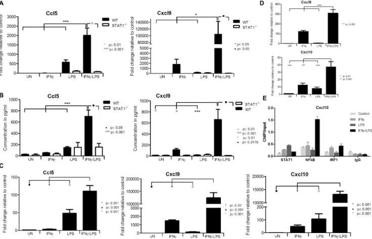
Raw.cel files downloaded from GEO were normalized using RMA algorithm, signals were log-transformed and probes were combined to genes using ‘‘Combine Figure 1. CXCL10 amplified by IFNcand LPS in VSMCs is STAT1 dependent.A,WTandSTAT12/2
VSMCs were treated with 10 ng/ml IFNcfor 8 h or with 1 ug/ml of LPS for 4 h or with IFNcfor 4 h followed by LPS for additional 4 h. RNA was isolated and qRT-PCR forCxcl10usingGapdhas internal control was performed. B, Cells were treated as in A. On the medium remained after treatment ELISA for CXCL10 was performed. Data represent means of at least 3 independent biological experiments¡SEM and p,0.05 was considered as significant. Data were tested for significance by one-way ANOVA followed by post-hoc Tukey or unpaired two-tailed student T-test when appropriate.
doi:10.1371/journal.pone.0113318.g001
Pro-Atherogenic Role of STAT1 in the Vasculature
probes to genes’’ tool (Chipster software [29]). Fold change and corresponding p-values were calculated using ‘‘calculate fold change’’ tool (Chipster [29]). Genes up-regulated at least 1.5 times in both datasets were compared with a list of 30 genes amplified by IFNcand LPS treatment in VSMCs.
The list of STAT1 target genes up-regulated by IFNcand LPS in murine VSMCs was used as the starting point for promoter analysis. First, that list was fed to pSCAN online promoter analysis tool in order to look for GAS, interferon stimulated response element ISRE (recognized by IRFs) and NFkB binding sites. The software was set to analyse 950 bp upstream and 50 bp downstream of the transcription start site. PSCAN produced a list of over-represented transcription factor binding sites together with occurrences of each site and a matrix similarity score. Occurrences having the score of at least 0.8 were fed into MatDefine (Genomatix software package) to create a highly conserved matrix for each transcription factor binding site. The settings were as follows: tuple size – 8; no. of sequences containing tuple – 60%; matrix similarity score for sequence inclusion – 0.9. Matrices for GAS, ISRE and NFkB binding sites were then used in pSCAN as «user supplied matrices» to search for occurrences in genes two-fold upregulated in the atherosclerotic plaque datasets.
ChIP-qPCR
ChIP was performed as previously described [30] with minor modifications. Briefly, VSMCs were stimulated as depicted inFig. 1and next crosslinked with 1% formaldehyde for 10 minutes. After fixation chromatin was sonicated with a Diagenode Bioraptor to generate 200–1000 bp fragments. Chromatin was immunoprecipitated with pre-immune IgG (Millipore, 12–371B), or with an antibody against STAT1 (Santa Cruz, sc-346) or IRF1 (Santa Cruz, sc-13041X) or NFkB (Santa Cruz, sc-398442X). Chromatin-antibody complexes were precipi-tated with agarose beads according to the EZ ChIP protocol (Millipore, 17-371). After DNA fragments were column purified DNA concentration was measured with a Qubit fluorometer. Immunoprecipitated DNA was quantified by qPCR (primers for Cxcl10: 59-CCTGTAAACCGAGGGCATTG-39, 59
-CACGCTTTGGAAAGTGAAAC-39) and normalized to values obtained after amplification of unprecipitated (input) DNA.
Western blot analysis
with primary and horseradish peroxidase-conjugated secondary antibodies (Santa Cruz), immunoreactivity was detected by adding Luminata Forte Western Substrate (EMD Millipore) and measured by INTAS imaging system (Intas, Germany).
Cytokine detection ELISA
Expression of murine Cxcl10, Ccl5 (Peprotech) as well as Cxcl9 (Sigma) was performed on medium remained after treatment of VSMCs using sandwich ELISA tests according to the manufacturer’s instructions.
Measurement of nitric oxide (NO)
VSMCs were treated as depicted in cell experiment section. After treatment medium was refreshed and cells were cultivated for further 24 h. Subsequently medium was collected and 100 ul was used to measure amount of NO by Griess diazotization reaction [31]. Medium was incubated with freshly prepared solution containing 1% sulfanilamide 5% HCl, 0.1% aqueous solution of
2-(1-Naphthylamino)ethylamine dihydrochloride (Sigma). After 10 min incubation OD at 560 mm was measured and compared to the standard curve.
Migration assay
Migration assay was performed according to Guo et al [32]. Briefly, 106of isolated red blood cells depleted splenocytes isolated from WT mice, were loaded into the upper chamber of Transwell 24-well plates (Corning, 3421). The bottom chamber was filled with 600 ul of the medium collected after treatment of VSMCs with LPS, IFNcor IFNcand LPS. After incubation for 3 h at 37
˚
C, migrated cells were stained with CD45FITC and CD3APC antibody (Miltenyi Biotec 130091609, 130092977) and analyzed by flow cytometer (Miltenyi Biotec).Ex vivo
contractility studies
Isolated aortas were cleaned from perivascular fat, cut into 2 mm long rings (for myograph) and placed in a DMEM medium containing 2% FBS (Sigma). Next, aortas were treated with 10 ng/ml of IFNc and/or 1mg/ml of LPS. Vascular contractility studies were performed according to the technique described by Mulvany et al. [33]. After treatment, 2 mm long rings were mounted in a 4-channel myograph (620M, Danish Myo Technology, Aarhus, Denmark) in the organ chamber filled with physiological saline solution (PSS) containing 118.99 mM NaCl, 4,69 mM KCl, 1.17 mM MgSO4*7H2O, 1.18 mM KH2PO4, 2.5 mM CaCl2*2H2O, 25 mM NaHCO3, 0.03 mM EDTA, 5.5 mM Glucose. During the experiment PSS buffer was aerated with carbogen (95% O2+5%CO2). After calibration, vessels were pre-streched to obtain optimal passive tension. Next, vascular functions were analyzed. Contractility was evaluated by substitu-tion of PSS buffer for high potassium physiological saline solusubstitu-tion (KPSS;
Pro-Atherogenic Role of STAT1 in the Vasculature
74.7 mM NaCl, 60 mM KCl, 1.17 mM MgSO4*7H2O, 1.18 mM KH2PO4, 1.6 mM CaCl2, 14.9 mM NaHCO3, 0.026 mM EDTA, 5.5 mM Glucose). For testing viability, vessels were subjected to noradrenaline-induced constriction followed by acetylcholine dilation (Sigma). After washing out with PSS buffer and resting for 15 minutes, noradrenaline dose-response curves was performed. Noradrenaline was used in stepwise increased, cumulative concentration ranging from 10211 to 1026 mol/L. To study vasodilatation, sodium nitroprusside (Sigma) was used in concentrations from 10210 to 1025 mol/L.
Histology and immunohistochemistry
Histological analyses and immunohistochemistry were performed on represen-tative sections (2–3 mm) of formalin fixed in paraffin embedded tissue samples from six human carotid atherosclerotic lesions and four healthy controls. The human tissue samples used in our study were procured from Biobank of
Department of Vascular and Endovascular Surgery (Klinikum rechts der Isar der Technischen Universitaet Muenchen). Collecting of specimens for the mentioned Biobank was approved by the local ethics committee (Ethikkommission der Fakultaet fuer Medizin der Technischen Universitaet Muenchen) and written informed consent was given by all patients. Haemalaun-Eosin (HE) and Elastica-van-Gieson (EvG) staining were performed in order to assess sample morphology. For characterisation of the cells within atherosclerotic plaques, specimens were treated with antibodies against vascular smooth muscle cells (smooth muscle myosin heavy chain 1 and 2 (SM-M10), rabbit polyclonal, dilution 1:4.000 (Abcam, ab81031) and endothelial cells (anti-CD31, mouse monoclonal, clone JC70A, dilution 1:100; Dako).
For the detection of specific cytokines, CXCL9 (MIG) and CXCL10 (IP10), as well as the phosphorylated transcription factor STAT1, following primary antibodies were used: rabbit polyclonal anti-MIG (Abcam, ab9720; dilution 1:500), rabbit polyclonal anti-IP10 (Abcam, ab47045; dilution 1:200), and rabbit monoclonal phospho-Stat1 (Cell Signaling, 9171l; dilution 1:400). All antibodies were first optimised on tonsil using different dilutions, staining conditions and with or without blocking. Optimal results were achieved by blocking anti-MIG and anti-phospho-Stat1 with goat serum, anti-IP10 without the blocking procedure.
Following incubation with primary antibody visualisation was performed by peroxidase/DAB ChemMate Detection Kit according to the manufacturer’s instruction (biotinylated goat anti-mouse/anti-rabbit secondary Ab; Dako).
Statistical Analysis
Data are presented as mean ¡SEM. For comparisons between more than two groups one-way ANOVA with Tukey post-hoc test was used. In all other
contractility studies, two-way ANOVA test with Bonferroni post hoc test was used.
Results
IFNc
and LPS synergistically induce CXCL10 expression in
VSMCs, depending on STAT1
Recently, we showed that in ECs cross-talk between IFNcand TLR4 resulted in augmented STAT1 phosphorylation and increased expression of the chemokine CXCL10 [17]. To study if a similar mechanism affected the expression of Cxcl10 in VSMCs, these cells were isolated from WTandSTAT12/2mice and treated as depicted in Fig. 1. In WT-VSMCs, treatment with IFNc or LPS alone induced expression of Cxcl10at the mRNA (Fig. 1A) as well as at the protein level (
Fig. 1B). Furthermore, pre-treatment with IFNcfor 4 h followed by LPS for another 4 h led to synergistic amplification of Cxcl10expression compared with both factors alone (Fig. 1A and 1B). In contrast, this IFNc and LPS-induced synergistic amplification inCxcl10gene expression was dramatically abrogated in STAT12/2-VSMCs (Fig. 1A), which coincided with Cxcl10 protein levels in the medium (Fig. 1B) and correlated with a predominant STAT1-dependent mechanism.
Transcriptional responses in IFNc
and LPS treated VSMCs predict
dependence on STAT1, NFkB and IRF
Next, we compared genome-wide transcriptional responses ofWT-VSMCsto LPS (4 h) or IFNc(8 h) alone, or after combined treatment (IFNc8 h, LPS 4 h). IFNc
changed the expression of 297 and LPS of 553 genes under these conditions (
Fig. 2A). The interactions between IFNcand LPS (Fig. 2A) increased the number of up-regulated genes to 990. While 128 of the IFNc-regulated genes were modulated by LPS (Fig. 2A), 118 were also commonly regulated by IFNc+LPS. Likewise, we compared transcriptional responses of STAT12/2-VSMCsto LPS or IFNcalone, or after combined treatment. Only 16 genes were found to be up-regulated by IFNcinSTAT12/2-VSMCs, highlighting the importance of STAT1 in this response pathway. In contrast, LPS treatment of STAT12/2-VSMCs was similar to WT-VSMCs, with a total of 470 genes being modulated. However, in general the potency of the response was lower as compared to WT-VSMCs. Consequently, the additive or synergistic effect of IFNc and LPS as seen in WT-VSMCs, was no longer present inSTAT12/2-VSMCs. Only 493 genes were upregulated by IFNc+LPS, of which 323 were in common with LPS alone. The complete list of up and down-regulated genes in response to IFNcor LPS alone, or after combined treatment is shown in Table S1, S2 and S3, respectively.
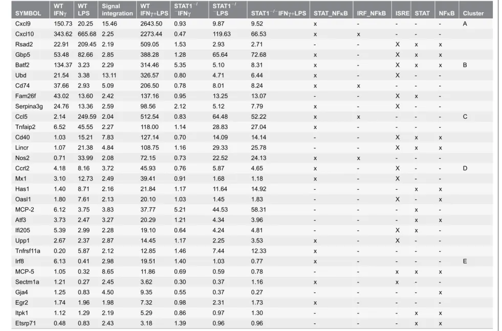
Subsequently, we aimed at identifying the genes, that similar to Cxcl10were synergistically affected by the interactions between IFNc and LPS, and their dependency on STAT1.Table 1shows the top 30 synergistically amplified genes of
Pro-Atherogenic Role of STAT1 in the Vasculature
which the expression was at least 2-fold higher upon stimulation with IFNc+LPS
as compared to the sum of the treatments with both factors alone. For example, expression of Cxcl9was .15-fold higher after combined treatment [2643.5-fold Figure 2. Identification of genes prone to synergistic amplification upon treatment with IFNcand LPS.WTandSTAT12/2VSMCs were treated as
described inFig. 1. On RNA isolated from untreated or IFNc, LPS or IFNc+LPS treated VSMCs genome-wide expression profiling was performed. A, Venn diagrams revealing number of differentially expressed genes upon stimulation. B, Heat map of the expression of synergistically amplified genes inWTand STAT12/2VSMCs. C, Clustering of the synergistically upregulated genes according to their expression profile. AVG, average expression in the group. For details see text.
Table 1.Genes prone to synergistic amplification upon stimulation with IFNcand/or LPS and their promoter analysis. SYMBOL WT IFNc WT LPS Signal integration WT IFNc+LPS
STAT12/
2IFN
c
STAT12/
2LPS STAT12/2IFN
c+LPS STAT_NFkB IRF_NFkB ISRE STAT NFkB Cluster
Cxcl9 150.73 20.25 15.46 2643.50 0.93 9.87 9.52 x - - - - A
Cxcl10 343.62 665.68 2.25 2273.44 0.47 119.63 66.53 x x - -
-Rsad2 22.91 209.45 2.19 509.05 1.53 2.93 2.71 - - X x x
Gbp5 53.48 82.66 2.85 388.28 1.28 65.64 72.68 x - X x x
Batf2 134.37 3.23 2.29 314.46 5.35 5.10 8.31 x - X x x B
Ubd 21.54 3.38 13.11 326.57 0.80 4.71 6.44 x - X -
-Cd74 37.66 2.93 5.09 206.50 0.78 8.01 8.24 x x - -
-Fam26f 43.02 13.60 2.42 137.16 0.95 13.25 13.07 - - X x
-Serpina3g 24.76 13.36 2.59 98.56 2.12 5.12 7.79 x - X -
-Ccl5 2.14 249.59 2.04 512.54 0.83 64.48 52.22 x x - - - C
Tnfaip2 6.52 45.55 2.27 118.00 1.14 28.83 27.04 x - - -
-Cd40 1.03 15.21 7.83 127.14 0.70 14.09 14.14 - - X x x
Lincr 1.07 21.38 4.84 108.75 1.16 29.33 25.78 - - X x x
Nos2 0.71 33.99 2.08 72.15 0.73 22.52 24.13 x x - -
-Ccrl2 4.18 8.16 3.72 45.93 0.76 5.87 4.65 x - X - - D
Mx1 3.10 12.73 2.49 39.41 0.91 1.68 1.18 x - X -
-Has1 1.40 8.71 2.16 21.84 1.17 11.64 14.92 - - - x x
Oasl1 1.80 7.61 2.13 20.10 1.03 1.45 1.83 - - X - x
MCP-2 6.12 3.75 3.83 37.77 5.21 44.53 58.31 - - - x
-Atf3 3.73 2.47 3.27 20.29 1.21 4.34 3.96 - - - x x
Ifi205 5.39 2.99 2.28 19.10 0.64 4.24 4.81 - - X x
-Upp1 2.67 2.37 2.87 14.45 1.17 2.25 3.53 x - X -
-Tnfrsf11a 0.20 5.87 2.12 12.85 1.46 7.44 12.33 x - - -
-Irf8 6.13 0.41 2.98 19.51 1.40 1.03 0.77 x - - - - E
MCP-5 1.05 0.32 8.65 11.86 0.69 0.59 0.78 - - x x x
Sectm1a 1.21 0.27 2.45 3.62 0.30 0.37 1.16 x - x -
-Gja4 1.25 0.83 4.50 9.35 0.55 0.37 0.27 - - - - x
Egr2 1.74 1.96 1.98 7.32 0.98 2.31 1.73 x - - -
-Itpk1 1.12 1.29 2.19 5.29 0.86 0.97 1.30 - - - x x
Etsrp71 0.48 0.83 2.43 3.18 1.39 0.96 0.96 - - - x x
The table introduces genes that expression is at least 2-fold higher upon stimulation with IFNc+LPS as compared to the sum of the treatments with both factors alone (see column ‘‘Signal integration’’). Other numbers represent fold changes compared to control. Cross indicates presence of specific sequence in the promoter regions.
increased by IFNc+LPS, divided by 171 times as the sum of IFNc (150.73) and
LPS (20.25) alone] as compared to the single treatments.
Subsequently, by grouping these 30 genes based on their response pattern in WT-VSMCs to IFNcor LPS (Fig. 2B), we could distinguish five groups of genes (
Fig. 2C). The first group contained genes with high response to both IFNc and LPS that were highly amplified after combined treatment (Cluster A in Table 1
and Fig. 2C). These include Cxcl9, Cxcl10, Rsad2 and Gbp5. The expression of a second group of genes, including Batf2, Ubd, Cd74, Fam26f and Serpina3g, showed high response to IFNc, mild response to LPS, and high amplification after combined treatment (Cluster B inTable 1andFig. 2C). In contrast, a third group of genes showed a mild or no response to IFNc, high response to LPS, and again high amplification after combined treatment (Cluster C in Table 1 andFig. 2C). This group was exemplified by Ccl5,Tnfaip2,Cd40, Lincrand Nos2 (iNOS). The fourth group of genes consisted of Ccrl2, Mx1,Has1, Oasl1, MCP-2,Atf3, Ifi205, Upp1andTnfrsf11aand displayed mild or no response to IFNc, mild response to LPS, and mild amplification after combined treatment (Cluster D inTable 1and
Fig. 2C). Finally, we could also identify genes which showed minor or no response to IFNcand LPS alone, but were highly amplified in expression after combined treatment [e.g.,Irf8, MCP-5, Sectm1a, Gja4, Egr2, Itpk1andEtsrp71] (Cluster E in
Table 1 and Fig. 2C).
In general, the absence of STAT1 severely abrogated the IFNc-induced
expression of all of these 30 genes. On the other hand, the LPS response of 50% of genes listed inTable 1, was decreased in the absence of STAT1. Consequently, the synergistic effect of IFNcand LPS as seen inWT-VSMCs, was no longer present in STAT12/2-VSMCs. As the only exception, the IFNc-induced expression of MCP-2 appeared STAT1-independent, with a similar fold induction in WT and STAT12/2VSMCs (Table 1, 6.12 vs. 5.12). In contrast, absence of STAT1 increased its response to LPS (Table 1, 3.75 vs. 44.53).
Successive promoter analysis of the genes listed inTable 1, predicted the presence of STAT-NFkB and IRF-NFkB modules or combinations of separate ISRE, STAT or NFkB binding sites, strongly implicating the cooperative
involvement of NFkB, STAT1 and/or IRFs in the transcriptional regulation of all of these genes in response to IFNcand LPS.
Transcriptional responses in IFNc
and LPS treated VSMCs and
ECs predict a pro-atherogenic phenotype
Gene ontology (GO) functional analysis of the top 30 genes listed in Table 1, revealed significant enrichment in biological functions involved in host defense, immune response, inflammatory response, cytokine response, response to stress and to wound healing (Table 2). All these categories generally recognize a similar group of genes, including the chemokines Cxcl9, Ccl12, Ccl8, Ccl5, Cxcl10and Ccrl2, adhesion molecules (Cd40, Cd74), and the antiviral and antibacterial response genes Irf8, Rsad2, Mx1, Oasl1, Gbp5, Nos2, Batf2and Tnfrsf11a.
profile that is mediated by interactions between IFNcand LPS in VSMCs and strongly depends on STAT1.
The expression of the chemokines Ccl5, Cxcl9, Ccl12and chemokine receptor Ccrl2 (not shown) was additionally examined by qPCR and ELISA (Fig. 3), and confirmed the microarray data. In agreement with Table 1, the response of this selected group of genes was severely abolished in STAT12/2-VSMCs, confirming the importance of STAT1 in the signal integration between IFNcand LPS. Because we were not able to isolate a homogeneous population of mouse aortic endothelial cells (data not shown), we instead used the human microvascular endothelial cell-line, HMEC [20]. Pre-treatment of HMECs with IFNcfor 4 h followed by LPS for another 4 h resulted in a similar amplification pattern ofCcl5, Cxcl9 andCxcl10 (Fig. 3C) as in WT-VSMCs, providing evidence for a universal STAT1-dependent mechanism in vascular cells triggered by IFNcand LPS. Similarly, we were able to observe a synergistic amplification after IFNcand LPS treatment of Cxcl9and Cxcl10in ex vivotreated aortic rings of WT animals as compared to IFNcor LPS alone (Fig. 3D)
Finally, Chromatin-immunoprecipitation (ChIP)-qPCR of untreated WT-VSMCs or treated with IFNc, LPS or IFNc+LPS and using antibodies against
STAT1 NFkB, IRF1 or IgG, clearly showed enhanced binding of these different transcription factors to the ISRE and NFkB binding elements of the Cxcl10gene, as compared to IgG controls (Fig. 3E). In a representative experiment, STAT1 binding to the ISRE increased after IFNcas well as LPS treatment, but not after IFNc+LPS stimulation. IRF1 binding was enriched upon treatment with IFNc
alone and after subsequent stimulation with LPS. LPS alone, on the other hand did not affect IRF1 binding. Finally, NFkB binding dramatically increased when cells were first treated with IFNcand then by LPS, but not in the presence of IFNc
or LPS alone (Fig. 3E). This confirms the cooperative involvement of STAT1, NFkB and IRF1 in the transcriptional regulation of Cxcl10 in response to IFNc
and LPS as predicted in Table 1. Table 2.Gene ontology classification of synergistically amplified genes.
Term ID Description frequency log10p-value Uniqueness Dispensability
GO:0051707 response to other organism 0.01 210.10 0.56 0.00
GO:0009607 response to biotic stimulus 0.01 29.62 0.66 0.40
GO:0006952 defense response 0.01 28.91 0.63 0.41
GO:0002376 immune system process 0.01 27.61 0.97 0.00
GO:0071345 cellular response to cytokine sti-mulus
0.00 26.87 0.52 0.32
GO:0006950 response to stress 0.04 26.59 0.61 0.50
GO:0006955 immune response 0.01 26.26 0.41 0.39
GO:0006954 inflammatory response 0.00 25.91 0.68 0.49
GO:0045071 negative regulation of viral genome replication
0.00 25.04 0.76 0.47
GO:0009611 response to wounding 0.00 24.93 0.68 0.53
doi:10.1371/journal.pone.0113318.t002
Pro-Atherogenic Role of STAT1 in the Vasculature
IRF8 mediates IFNc
and LPS induced
Ccl5
expression in vascular
cells
Notably, the transcription factor IRF8, which was thought to be restricted to lymphoid-cell lineages such as B-, T-, dendritic cells and macrophages, was identified among the amplified genes. Indeed, gene (Fig. 4Aleft panel) and protein expression (Fig. 4B left panel) of IRF8 in WT and STAT12/2 VSMCs in response to IFNc, LPS or IFNc+LPS, confirmed the microarray data. Interestingly,
pre-treatment of ECs with IFNcfor 4 h followed by LPS for another 4 h resulted in a similar amplification pattern of IRF8 RNA (Fig. 4A right panel) and protein expression (Fig. 4Bright panel) as inWT-VSMCs. These results provide evidence for STAT1-dependent expression of IRF8 in VSMCs and ECs upon treatment with Figure 3. Effect of STAT1 dependent signal integration on chemokine expression.WTandSTAT12/2VSMCs, HMECs orWTaortic ring segments were
treated as described inFig. 1. A, RNA from VSMCs was isolated and qRT-PCR forCcl5,Cxcl9usingGapdhas internal control was performed. B, On the medium remained after treatment of VSMCs ELISA for Ccl5 and Cxcl9 was performed. C, Expression ofCXCL10, CXCL9 and CCL5upon stimulation in ECs. D, RNA from incubated aortic rings was isolated and qRT-PCR forCxcl10,Cxcl9usingGapdhas internal control was performed. Data represent means of at least 3 independent biological experiments¡SEM and p,0.05 was considered as significant. Data were tested for significance by one-way ANOVA followed by post-hoc Tukey or unpaired two-tailed student T-test when appropriate. E, ChIP-qPCR analysis of theCxcl10promoter region containing NFkB and ISRE binding sites show the enrichment with STAT1, NFkB and IRF1 antibodies compared with IgG control in an IFNc, LPS or IFNy+LPS-dependent manner inWT VSMCs. Immunoprecipitated DNA was quantified by qPCR and normalized to values obtained after amplification of unprecipitated (input) DNA. A representative experiment is shown.
Pro-Atherogenic Role of STAT1 in the Vasculature
IFNc, and confirm amplification of IRF8 upon stimulation with IFNcand LPS in vascular cells.
Next the IRF8 dependent regulation ofCcl5 (a known IRF8 target in immune cells [34]) was examined. The amplified expression of Ccl5RNA (Fig. 4C, left panel) and protein (Fig. 4C, right panel) in response to IFNcand LPS, as seen in WT VSMCs, was highly attenuated in IRF82/2and STAT12/2-VSMC. In contrast, the expression of Cxcl10andCxcl9in response to IFNcand LPS inWT VSMCs was similar to that in IRF82/2-VSMCs (Fig. 4D).
Signal integration between IFNc
and LPS in VSMCs leads to
increased migration of T-lymphocytes
Since many of the chemokines characterized above are involved in chemotaxis of T-lymphocytes [35], we examined the effect of IFNcand LPS cross-talk on T-cell migration towards conditioned medium from treated VSMCs. Amplification of chemokines in WT-VSMCsupon stimulation with both stimuli indeed led to an increased migration of CD3+/CD45+ spleenocytes (Fig. 4E). Migration of CD3+/ CD45+cells towards medium ofWT-VSMCstreated with both IFNcand LPS was significantly higher (234%) as compared to both factors alone (125% and 175%, respectively). As expected, the chemotactic response of splenocytes towards the conditioned medium obtained after treatment of STAT12/2-VSMCs was highly attenuated (Fig. 4E).
Signal integration between IFNc
and LPS in aortic rings leads to
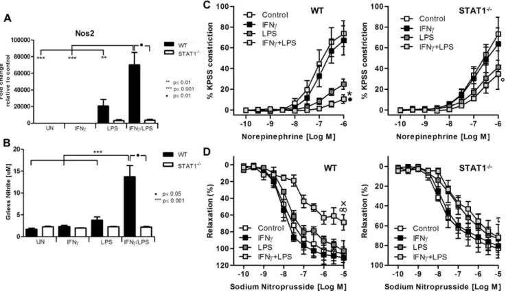
abolished response to norepinephrine and sodium nitroprusside
Among the genes that were highly amplified upon treatment with IFNcand LPS was inducible nitric oxide synthase (iNOS, Nos2). Indeed, treatment of WT-VSMCs but not STAT12/2with IFNcand LPS caused amplified expression of Nos2 as compared to stimulation with both factors alone (Fig. 5A). The RNA levels reflected nitrite accumulation in the medium (Fig. 5B). Since dysregulation of Nos2 expression and its activity affects vessel function, we evaluated the physiological ramifications of these experimental conditions using a wire myograph/organ chamber setting. Stimulation of the aortic rings isolated from WT animals with IFNcand LPS resulted in drastic impairment of contractility after subjection to norepinephrine treatment (Fig. 5C, left panel). WT vessels treated with both IFNcand LPS manifested also high loss of the sensitivity to sodium nitroprusside (Fig. 5D, left panel). In contrast to WT, aortic rings from Figure 4. IRF8 mediated cross-talk and functional activity of synergistically amplified chemokines.WT, STAT12/2and IRF82/2VSMCs and HMECswere treated as described inFig. 1. A, RNA was isolated and qRT-PCR forIRF8usingGAPDHas internal control was performed in VSMCs (left panel) and ECs (right panel). B, Protein extracts were analyzed for IRF8, tyrosine-phosphorylated STAT1, total STAT1 and GAPDH. C,CCL5mRNA expression (left panel) and protein presence in the medium (right panel) was measured. D, Expression profiles ofCxcl9(left panel) andCxcl10(right panel) between VSMCs WT, andIRF82/2were compared. E, Migration assay of CD45+/CD3+performed on conditioned medium remained after treatment of VSMCsWTand
STAT12/2. Data represent means of at least 3 independent biological experiments¡SEM and p,0.05 was considered as significant. Data were tested for
significance by one-way ANOVA followed by post-hoc Tukey or unpaired two-tailed student T-test when appropriate.
STAT1-deficient mice did not reveal ameliorated response to noradrenaline and sodium nitroprusside as compared to LPS stimulated vessel (Fig. 5C, Fig. 5D, right panel).
STAT1 activation and CXCL9 and CXCL10 expression in ECs and
VSMCs from human carotid atherosclerotic plaques
We performed immunohistochemistry staining for phosphorylated STAT1, CXCL9 and CXCL10 in human advanced atherosclerotic plaques of carotid arteries in comparison to healthy vessels. As can clearly be observed inFigure 6A, VSMCs in the lesion highly expressed phosphorylated STAT1 and both
chemokines CXCL9 and CXCL10. In contrast, healthy vessels were negative for all three markers (Figure 6A). Moreover, ECs covering the plaque likewise showed predominant staining for phosphorylated STAT1 and CXCL9, and to a lesser Figure 5. STAT1-mediated abolished response to norepinephrine and sodium nitroprusside is associated with disturbed NO production.A,WT andSTAT12/2VSMCswere treated as described inFig. 1. RNA was isolated and qRT-PCR forNos2usingGapdhas internal control was performed (upper
panel) B, After stimulation as described inFig. 1, medium was refreshed and left for 24 h. Next, 100ml of the medium was taken and the product of Nos2-nitrite was measured. Data represent means of at least 3 independent biological experiments¡SEM and p,0.05 was considered as significant. Data were tested for significance by one-way ANOVA followed by post-hoc Tukey or unpaired two-tailed student T-test when appropriate. C, D Isolated aortic rings from WT and STAT2/2mice were incubated with 10 ng/ml IFNcfor 8 h or with 1 ug/ml of LPS for 4 h or with IFNcfor 4 h followed by LPS for additional 4 h. Next,
response to norepinephrine and sodium nitroprusside was tested on the wire myograph. C, Response to noradrenaline in WT and STAT1-deficient aortic rings presented as a percentage of maximal constriction to KPSS.*p,0.001 vs. WT control;Np,0.001 vs. WT LPS;#p,0.001 vs. STAT12/2control. D, Response to stepwise increased concentration of sodium nitroprusside. xp,0.05 vs. WT control;‘p,0.01 vs. WT LPS;%p,0.05 STAT12/2control. Aortas isolated from 3–4 animals per group were taken. Two-way ANOVA test with Bonferroni post hoc test was used. Statistical significance for the highest concentration is given.
doi:10.1371/journal.pone.0113318.g005
Pro-Atherogenic Role of STAT1 in the Vasculature
extent for CXCL10 (Figure 6B). Again, healthy endothelium was negative. Staining for IRF8 was more difficult to interpret, but seemed present at low levels in SMCs (not shown).
STAT1-dependent pro-atherogenic target gene expression in
human atherosclerotic plaques
To obtain potential evidence for STAT1-mediated target gene expression in the human atherosclerotic plaque, we performed different types of experiments. First, we analyzed two independent microarray datasets obtained from human coronary plaques and human carotid plaques. These datasets are available in GEO NCBI database (acc. no. GSE40231 and GSE21545, respectively) [26,27]. In coronary and carotid plaques respectively we identified 1146 and 949 genes upregulated at least 1.5 times as compared to the healthy arterial tissue (Figure 7A). 201 of those genes are commonly expressed between the different plaque tissues, highly implying that there are common features between coronary and carotid plaques (Sikorski et al. [36]). Detailed promoter analysis of the differentially expressed genes in carotid and coronary plaques identified 128 (Figure 7B) and 362 (Figure 7C) genes, respectively containing GAS, ISRE or NFkB sites, either alone or in different combinations. This strongly suggests also the cooperative
involvement of NFkB, STAT1 and/or IRF in the transcriptional regulation of genes in the plaque tissue.
Next we compared the 30 IFNcand LPS amplified STAT1-target genes listed in
Table 1 to the genes upregulated in carotid and coronary plaques. Indeed, 12 out Figure 6. Expression of pSTAT1, CXCL9, CXCL10 in human atherosclerotic lesionsin situ.Staining of the sections prepared from normal human artery exhibited no presence of pSTAT1, CXCL9, CXCL10 (A, upper panel). In contrast, all three proteins could be detected in SM-M10 positive cells in atherosclerotic plaques (A, middle panel) as well as in the endothelial cells at the lumen side (B). A representative analysis is shown of 6 human carotid atherosclerotic lesions and 4 healthy controls. Arrows represent examples of positive staining. In B arrows with asterix indicate examples of positively stained VSMCs. Scale bar5100mm.
of the 30 genes were expressed in carotid plaques and 6 out of 30 in coronary plaques, including CXCL9, CXCL10, CCL5, CCL8, CRCL2, Cd74, GBP5, UBD, SECTM1, IFI16(homologueIfi-205),UPP1,FAM26Fand the transcription factor IRF8 as the above identified STAT1 targets (Fig. 7A).
Together, this points to a pro-atherogenic role of STAT1 in vascular cells of atherosclerotic plaques, and suggests the potential of a selection of STAT1-target genes as biomarkers to monitor plaque phenotype in human atherosclerosis.
Discussion
The involvement of STAT1 in experimental atherosclerosis has recently been appreciated, especially in immune cells. It is additionally accepted that in immune cells STAT1 is a unique point of convergence for the antimicrobial and
inflammatory synergism between IFNc and TLRs. Recently, we showed that also in ECs cross-talk between IFNc and TLR4 resulted in augmented STAT1
phosphorylation and increased expression of the chemokine CXCL10 [17]. Here, a similar STAT1-dependent mechanism for CXCL10 expression in response to IFNcand LPS was observed in VSMCs at the RNA and protein level (Fig. 1). To date, no information is available on the genome-wide induced changes modulated by IFNcand TLR4 in ECs and VSMCs and how this affects vascular function. Figure 7. Expression of synergistically amplified genes in atherosclerotic vessels.A, Venn diagram with analysis of microarray datasets obtained from human coronary plaques and human carotid plaques. B, Promoter analysis of the differentially expressed genes in carotid (left panel) and coronary plaques (right panel). For details see text.
doi:10.1371/journal.pone.0113318.g007
Pro-Atherogenic Role of STAT1 in the Vasculature
Therefore, we decided to further characterize the role of STAT1 in the
transcriptional response pathways involved in the interaction between IFNcand TLR4 signaling in VSMCs. Thus, we identified a specific set of STAT1-dependent genes that were synergistically affected by IFNcand LPS in VSMCsin vitro. These included the chemokines Cxcl9, Ccl12, Ccl8, Ccl5, Cxcl10and Ccrl2, adhesion molecules (Cd40, Cd74), and the antiviral and antibacterial response genes Irf8, Rsad2, Mx1, Oasl, Gbp5, Nos2, Batf2 andTnfrsf11a. Based on their response pattern to IFNc, LPS and IFNc+LPS inWT-VSMCs (Fig. 2B), we could
distinguish five clusters of genes (Fig. 2C). In general, the absence of STAT1 severely abrogated the IFNc-induced expression of all of these genes. Moreover, the LPS response of 50% of genes listed inTable 1was decreased in the absence of STAT1. Consequently, the synergistic effect of IFNc and LPS, as seen in WT-VSMCs, could no longer be detected inSTAT12/2-VSMCs. This strongly suggests the involvement of STAT1 in the signal integration between JAK/STAT and TLR4 pathways. The IFNc-induced expression of Ccl8appeared STAT1-independent, with a similar fold induction in WT and STAT12/2VSMCs. In contrast, absence of STAT1 increased its response to LPS. As Ccl8 is a known STAT3 target gene [37], it is possible that it’s IFNc and LPS inducibility in WT and STAT12/2 VSMCs, is regulated by this transcription factor. The increased LPS-mediatedCcl8 expression in STAT12/2VSMCs as compared to WT cells, could potentially be explained by the absence of a STAT1-dependent inhibitory mechanism of STAT3 activity mediated by the STAT1-target gene SOCS1.
The expression of a selection of these genes, includingCcl5,Cxcl9, Nos2, Irf8 andCcl12,Ccrl2(not shown) was additionally determined at the RNA and protein level, and confirmed the microarray data. Moreover, relative quantification as fold change compared to control, resulted in a similar range of induction values for RNA and protein expression. A similar expression pattern of some of these genes could also be identified in ECs and aortic ring segments, providing evidence for a universal STAT1-dependent mechanism in vascular cells triggered by IFNc and LPS.
Integration of IFNcand TLR signaling pathways occurs, for instance, through synergy between TLR- and IFNc-induced transcription factors. Promoter analysis of the genes listed in Table 1 indeed predicted the presence of STAT-NFkB and IRF-NFkB modules or combinations of separate ISRE, STAT or NFkB binding sites in their promoters. Indeed, ChIP-qPCR confirmed binding of STAT1, NFkB and IRF1 to theCxcl10gene, in an IFNcand LPS-dependent manner (Figure 3E). This strongly suggested that cooperation between NFkB, STAT1 and/or IRFs is involved in the transcriptional regulation of all of these genes in response to IFNc
between NFkB and STAT1 or NFkB and IRF1 [7]. They ultimately promote type I immune actions, which are associated with host-defense mechanisms against viral and bacterial infections and excessive immune responses [46]. Our data provides strong evidence that a similar mechanism of signal integration exists in vascular cells. The difference in expression pattern of these 30 genes did not correlate with the presence of a specific binding site or combination of binding sites. This implies that the affinity of the different transcription factors is most likely determining the transcriptional response of a particular gene.
Among the amplified genes we also identified the transcription factor IRF8, which expression is thought to be restricted to lymphoid-cell lineages such as B-, T- and macrophages. Thus, IRF8 may in part account for ‘‘immune cell-specific’’ STAT1-dependent functions of IFNc. IRF8 is also directly connected to TLR action, regulating the production of type I IFNs and other inflammatory mediators. For example, co-administration of IFNcand LPS to macrophages caused super-induction of IRF8 and IRF8 target genes [47]. As a consequence, synergistic induction of the pro-inflammatory genesIL1,IL6,IL12andTNFawas observed in an IRF8 dependent manner. In addition to its known immune cell functions, our results now uncover a novel ‘‘inflammation-dependent’’ role of IRF8 in cells from the vasculature. In ECs as well as VSMCs, combined treatment of IFNc and LPS resulted in a synergistic increase in IRF8 gene and protein expression as compared to both factors alone (Figure 4A and B). The presence of a potential STAT1-NFkB module in the IRF8 promoter (Table 1) highly suggests that the cooperation of these two transcription factors is at the basis of its synergistic expression. Consequently, this revealed the possible existence of IRF8-dependent cross-talk between IFNcand LPS in vascular cells. Indeed, we
subsequently identifiedCcl5(but notCxcl9andCxcl10) as a novel IRF8 target in VSMCs and ECs (Fig. 4C and D). The transcriptional regulation of theCcl5gene in macrophages in response to IFNcand LPS, was recently shown to involve IRF8 in combination with IRF1 and NFkB [34]. Therefore, the IRF8-dependent expression ofCcl5in IFNc-primed vascular cells that are subsequently stimulated by LPS is likely to comprise a similar mechanism. These results correlates with the predicted presence of an IRF-NFkB module in the Ccl5promoter (Table 1). On the other hand, the same promoter contains also a potential STAT1-NFkB module (Table 1) which suggests the additional involvement of STAT1 as well.
Together, our detailed investigation of STAT1-dependent transcriptional synergism between IFNcand LPS in cells from the vasculature predicts the existence of different regulatory mechanisms. It particularly involves cooperation between STAT1, IRF1, IRF8 and NFkB, with the novel role of IRF8 providing an additional layer to the overall complexity.
Functional analysis of the STAT1-dependent genes that were synergistically affected by interactions between IFNcand LPS in VSMCs (Table 1), revealed significant enrichment in biological functions connected to host defense, immune response, inflammatory response, cytokine response, response to stress and to wound healing (Table 2). All these categories generally represent a similar group
Pro-Atherogenic Role of STAT1 in the Vasculature
of genes, which together reflect an enhanced inflammatory and pro-atherogenic profile.
The fact that synergistic interactions between IFNcand LPS in VSMCs resulted in increased expression of multiple chemokines, prompted us to investigate T-cell migration. Indeed, a significant increase in migration of CD3+/CD45+splenocytes could be detected towards conditioned medium from IFNcand LPS treated WT-VSMCs as compared to that from cells treated with single factors. Importantly, splenocytes migration occurred in a STAT1-dependent manner, which correlated with decreased chemokine expression in STAT12/2-VSMCs under these
conditions (Fig. 4E). Interestingly a subset of these chemokines, including CXCL9, CXCL10, CCL5, CCL8 and CCRL2, has been reported to be increased in cells from the vasculature, which is in agreement with our results. Moreover, evidence exists that chemokines cooperate in leukocyte recruitment to the injured artery during vascular remodeling [35,48,49] and as such are involved in the pathogenesis of atherosclerosis.
To further elucidate the functional role of a cross-talk in the vessel, we performed contractility studies. The signal integration between IFNcand LPS in aortic ring segments resulted in impaired aortic contractility (Fig. 5) and
coincided with a dramatic increase in expression of Nos2. Nos2 participates in vascular dysfunction and is associated with progression of atherosclerosis [50,51].
More important, we were able to detect phosphorylated STAT1 in VSMCs and ECs of human atherosclerotic plaques (Fig. 6), which correlated with elevated expression of the chemokines CXCL9and CXCL10. Recently, Agrawal et al. [10] showed that STAT1 deficiency reduced foam cell formation in an intraperitoneal inflammation model and reduced atherosclerosis in an atherosclerosis-susceptible bone marrow transplantation mouse model. In combination with our results, this highlights the pro-atherogenic role of STAT1 in cells from the vasculature in human vascular disease.
Using data mining of human plaque transcriptomes, we were able to show that expression of a selection of the above identified STAT1-dependent
pro-atherogenic genes was significantly increased in human plaques from carotid and coronary arteries (Fig. 7A). Of these,CXCL9, CXCL10,CCL5,CCL8, CRCL2, Cd74andIRF8have previously been implicated in atherosclerosis [52,53]. This is not the case forGBP5,Ubd,SectM1,Ifi16,Upp1andFam26F, and could therefore represent potential novel biomarkers of atherosclerosis. Moreover, CCL5
both STAT1 and IRF8 in the regulation of gene expression in different cell types present in human atherosclerotic plaques.
In summary, our findings provide additional evidence to suggest that in ECs and VSMCs STAT1, in cooperation with IRF1, IRF8 and NFkB, coordinates a platform for cross-talk between IFNcand TLR4. This results in an increased pro-inflammatory phenotype and leads to amplified pro-atherogenic responses in the vasculature. As a consequence, in the presence of IFNcand LPS (or any other exogenous or endogenous TLR4 ligands), multiple chemokines, adhesion
molecules and antiviral and antibacterial response proteins can be over-produced in ECs and VSMCs. This may in turn modulate leukocyte attraction, adhesion and VSMC proliferation and migration, which are important characteristics of vascular dysfunction and early triggers of atherosclerosis. As such, a predefined STAT1-target gene signature could be developed as a novel diagnostic tool to monitor and diagnose plaque phenotype in human atherosclerosis. In addition, STAT1 represents an interesting novel target of therapeutic intervention that has a crucial role in mediating the interplay between damaged vessels and host
immunity during the process of atherosclerosis.
Supporting Information
Table S1. List of up and down-regulated genes in response to LPS in VSMCs
WT andSTAT12/2. Fold change compared to control. doi:10.1371/journal.pone.0113318.s001 (DOCX)
Table S2. List of up and down-regulated genes in response to IFNcin VSMCs
WT andSTAT12/2. Fold change compared to control. doi:10.1371/journal.pone.0113318.s002 (DOCX)
Table S3. List of up and down-regulated genes in response to IFNcand LPS in
VSMCs WT andSTAT12/2. Fold change compared to control.
doi:10.1371/journal.pone.0113318.s003 (DOCX)
Table S4. Primer sequences used in experimental procedures.
doi:10.1371/journal.pone.0113318.s004 (DOCX)
Acknowledgments
The authors would like to acknowledge Lilianna Schyschka from Klinik und Poliklinik fu¨r Unfallchirurgie, Klinikum rechts der Isar, Technical University Munich for valuable help with flow cytometer analysis as well as Alina Schmidt and Sandra Haderer for excellent technical assistance.
Author Contributions
Conceived and designed the experiments: SC MB HB. Performed the experiments: SC AO JP HN KB. Analyzed the data: SC AO KS JW MB CA HB. Contributed
Pro-Atherogenic Role of STAT1 in the Vasculature
reagents/materials/analysis tools: JP AZ. Wrote the paper: SC HB. Provided the funding and overall scientific oversight for the study: MB UH JW HB.
References
1. Orr AW, Hastings NE, Blackman BR, Wamhoff BR(2010) Complex regulation and function of the inflammatory smooth muscle cell phenotype in atherosclerosis. J Vasc Res 47: 168–180.
2. Hansson GK, Libby P(2006) The immune response in atherosclerosis: a double-edged sword. Nat Rev Immunol 6: 508–519.
3. Russell PS, Chase CM, Winn HJ, Colvin RB(1994) Coronary atherosclerosis in transplanted mouse hearts. III. Effects of recipient treatment with a monoclonal antibody to interferon-gamma.
Transplantation 57: 1367–1371.
4. Gupta S, Pablo AM, Jiang X, Wang N, Tall AR, et al.(1997) IFN-gamma potentiates atherosclerosis in ApoE knock-out mice. J Clin Invest 99: 2752–2761.
5. Nagano H, Mitchell RN, Taylor MK, Hasegawa S, Tilney NL, et al.(1997) Interferon-gamma deficiency prevents coronary arteriosclerosis but not myocardial rejection in transplanted mouse hearts. J Clin Invest 100: 550–557.
6. Tellides G, Tereb DA, Kirkiles-Smith NC, Kim RW, Wilson JH, et al.(2000) Interferon-gamma elicits arteriosclerosis in the absence of leukocytes. Nature 403: 207–211.
7. Sikorski K, Chmielewski S, Olejnik A, Wesoly JZ, Heemann U, et al. (2012) STAT1 as a central mediator of IFNgamma and TLR4 signal integration in vascular dysfunction. JAKSTAT 1: 241–249.
8. Tamura T, Yanai H, Savitsky D, Taniguchi T(2008) The IRF family transcription factors in immunity and oncogenesis. Annu Rev Immunol 26: 535–584.
9. Gough DJ, Levy DE, Johnstone RW, Clarke CJ(2008) IFNgamma signaling-does it mean JAK-STAT? Cytokine Growth Factor Rev 19: 383–394.
10. Agrawal S, Febbraio M, Podrez E, Cathcart MK, Stark GR, et al. (2007) Signal transducer and activator of transcription 1 is required for optimal foam cell formation and atherosclerotic lesion development. Circulation 115: 2939–2947.
11. Bjorkbacka H(2006) Multiple roles of Toll-like receptor signaling in atherosclerosis. Curr Opin Lipidol 17: 527–533.
12. Akira S, Uematsu S, Takeuchi O(2006) Pathogen recognition and innate immunity. Cell 124: 783–801.
13. Schroder K, Sweet MJ, Hume DA(2006) Signal integration between IFNgamma and TLR signalling pathways in macrophages. Immunobiology 211: 511–524.
14. Hu X, Chen J, Wang L, Ivashkiv LB(2007) Crosstalk among Jak-STAT, Toll-like receptor, and ITAM-dependent pathways in macrophage activation. J Leukoc Biol 82: 237–243.
15. Hu X, Chakravarty SD, Ivashkiv LB(2008) Regulation of interferon and Toll-like receptor signaling during macrophage activation by opposing feedforward and feedback inhibition mechanisms. Immunol Rev 226: 41–56.
16. Hu X, Ivashkiv LB(2009) Cross-regulation of signaling pathways by interferon-gamma: implications for immune responses and autoimmune diseases. Immunity 31: 539–550.
17. Sikorski K, Chmielewski S, Przybyl L, Heemann U, Wesoly J, et al.(2011) STAT1-mediated signal integration between IFNgamma and LPS leads to increased EC and SMC activation and monocyte adhesion. Am J Physiol Cell Physiol 300: C1337–1344.
18. Holtschke T, Lohler J, Kanno Y, Fehr T, Giese N, et al. (1996) Immunodeficiency and chronic myelogenous leukemia-like syndrome in mice with a targeted mutation of the ICSBP gene. Cell 87: 307– 317.
19. Geisterfer AA, Peach MJ, Owens GK(1988) Angiotensin II induces hypertrophy, not hyperplasia, of cultured rat aortic smooth muscle cells. Circ Res 62: 749–756.
21. Livak KJ, Schmittgen TD(2001) Analysis of relative gene expression data using real-time quantitative PCR and the 2(2Delta Delta C(T)) Method. Methods 25: 402–408.
22. Chen H, Boutros PC(2011) VennDiagram: a package for the generation of highly-customizable Venn and Euler diagrams in R. BMC Bioinformatics 12: 35.
23. Cartharius K, Frech K, Grote K, Klocke B, Haltmeier M, et al. (2005) MatInspector and beyond: promoter analysis based on transcription factor binding sites. Bioinformatics 21: 2933–2942.
24. Eden E, Navon R, Steinfeld I, Lipson D, Yakhini Z(2009) GOrilla: a tool for discovery and visualization of enriched GO terms in ranked gene lists. BMC Bioinformatics 10: 48.
25. Supek F, Bosnjak M, Skunca N, Smuc T(2011) REVIGO summarizes and visualizes long lists of gene ontology terms. PLoS One 6: e21800.
26. Folkersen L, Persson J, Ekstrand J, Agardh HE, Hansson GK, et al.(2012) Prediction of ischemic events on the basis of transcriptomic and genomic profiling in patients undergoing carotid
endarterectomy. Mol Med 18: 669–675.
27. Hagg S, Skogsberg J, Lundstrom J, Noori P, Nilsson R, et al.(2009) Multi-organ expression profiling uncovers a gene module in coronary artery disease involving transendothelial migration of leukocytes and LIM domain binding 2: the Stockholm Atherosclerosis Gene Expression (STAGE) study. PLoS Genet 5: e1000754.
28. Johnson WE, Li C, Rabinovic A(2007) Adjusting batch effects in microarray expression data using empirical Bayes methods. Biostatistics 8: 118–127.
29. Kallio MA, Tuimala JT, Hupponen T, Klemela P, Gentile M, et al. (2011) Chipster: user-friendly analysis software for microarray and other high-throughput data. BMC Genomics 12: 507.
30. Barish GD, Yu RT, Karunasiri M, Ocampo CB, Dixon J, et al.(2010) Bcl-6 and NF-kappaB cistromes mediate opposing regulation of the innate immune response. Genes Dev 24: 2760–2765.
31. Stuehr DJ, Nathan CF (1989) Nitric oxide. A macrophage product responsible for cytostasis and respiratory inhibition in tumor target cells. J Exp Med 169: 1543–1555.
32. Guo F, Weih D, Meier E, Weih F(2007) Constitutive alternative NF-kappaB signaling promotes marginal zone B-cell development but disrupts the marginal sinus and induces HEV-like structures in the spleen. Blood 110: 2381–2389.
33. Mulvany MJ, Halpern W (1977) Contractile properties of small arterial resistance vessels in spontaneously hypertensive and normotensive rats. Circ Res 41: 19–26.
34. Liu J, Ma X(2006) Interferon regulatory factor 8 regulates RANTES gene transcription in cooperation with interferon regulatory factor-1, NF-kappaB, and PU.1. J Biol Chem 281: 19188–19195.
35. Zernecke A, Weber C(2010) Chemokines in the vascular inflammatory response of atherosclerosis. Cardiovasc Res 86: 192–201.
36. Sikorski K, Wesoly J, Bluyssen H (2014) Data Mining of Atherosclerotic Plaque Transcriptomes Predicts STAT1-Dependent Inflammatory Signal Integration in Vascular Disease. International Journal of Molecular Sciences 15: 14313–14331.
37. Hiwatashi K, Tamiya T, Hasegawa E, Fukaya T, Hashimoto M, et al.(2011) Suppression of SOCS3 in macrophages prevents cancer metastasis by modifying macrophage phase and MCP2/CCL8 induction. Cancer Lett 308: 172–180.
38. Lowenstein CJ, Alley EW, Raval P, Snowman AM, Snyder SH, et al.(1993) Macrophage nitric oxide synthase gene: two upstream regions mediate induction by interferon gamma and lipopolysaccharide. Proc Natl Acad Sci U S A 90: 9730–9734.
39. Ohmori Y, Hamilton TA (2001) Requirement for STAT1 in LPS-induced gene expression in macrophages. J Leukoc Biol 69: 598–604.
40. Jahnke A, Johnson JP(1994) Synergistic activation of intercellular adhesion molecule 1 (ICAM-1) by TNF-alpha and IFN-gamma is mediated by p65/p50 and p65/c-Rel and interferon-responsive factor Stat1 alpha (p91) that can be activated by both IFN-gamma and IFN-alpha. FEBS Lett 354: 220–226.
41. Ohmori Y, Hamilton TA(1995) The interferon-stimulated response element and a kappa B site mediate synergistic induction of murine IP-10 gene transcription by IFN-gamma and TNF-alpha. J Immunol 154: 5235–5244.
Pro-Atherogenic Role of STAT1 in the Vasculature
42. Ohmori Y, Schreiber RD, Hamilton TA(1997) Synergy between interferon-gamma and tumor necrosis factor-alpha in transcriptional activation is mediated by cooperation between signal transducer and activator of transcription 1 and nuclear factor kappaB. J Biol Chem 272: 14899–14907.
43. Pine R (1997) Convergence of TNFalpha and IFNgamma signalling pathways through synergistic induction of IRF-1/ISGF-2 is mediated by a composite GAS/kappaB promoter element. Nucleic Acids Res 25: 4346–4354.
44. Naschberger E, Werner T, Vicente AB, Guenzi E, Topolt K, et al.(2004) Nuclear factor-kappaB motif and interferon-alpha-stimulated response element co-operate in the activation of guanylate-binding protein-1 expression by inflammatory cytokines in endothelial cells. Biochem J 379: 409–420.
45. Voraberger G, Schafer R, Stratowa C (1991) Cloning of the human gene for intercellular adhesion molecule 1 and analysis of its 59-regulatory region. Induction by cytokines and phorbol ester. J Immunol 147: 2777–2786.
46. O’Shea JJ, Ma A, Lipsky P(2002) Cytokines and autoimmunity. Nat Rev Immunol 2: 37–45.
47. Zhao J, Kong HJ, Li H, Huang B, Yang M, et al.(2006) IRF-8/interferon (IFN) consensus sequence-binding protein is involved in Toll-like receptor (TLR) signaling and contributes to the cross-talk between TLR and IFN-gamma signaling pathways. J Biol Chem 281: 10073–10080.
48. Braunersreuther V, Mach F, Steffens S(2007) The specific role of chemokines in atherosclerosis. Thromb Haemost 97: 714–721.
49. Seo D, Wang T, Dressman H, Herderick EE, Iversen ES, et al.(2004) Gene expression phenotypes of atherosclerosis. Arterioscler Thromb Vasc Biol 24: 1922–1927.
50. Kuhlencordt PJ, Chen J, Han F, Astern J, Huang PL(2001) Genetic deficiency of inducible nitric oxide synthase reduces atherosclerosis and lowers plasma lipid peroxides in apolipoprotein E-knockout mice. Circulation 103: 3099–3104.
51. Niu XL, Yang X, Hoshiai K, Tanaka K, Sawamura S, et al. (2001) Inducible nitric oxide synthase deficiency does not affect the susceptibility of mice to atherosclerosis but increases collagen content in lesions. Circulation 103: 1115–1120.
52. Martin-Ventura JL, Madrigal-Matute J, Munoz-Garcia B, Blanco-Colio LM, Van Oostrom M, et al.
(2009) Increased CD74 expression in human atherosclerotic plaques: contribution to inflammatory responses in vascular cells. Cardiovasc Res 83: 586–594.
53. Doring Y, Soehnlein O, Drechsler M, Shagdarsuren E, Chaudhari SM, et al.(2012) Hematopoietic interferon regulatory factor 8-deficiency accelerates atherosclerosis in mice. Arterioscler Thromb Vasc Biol 32: 1613–1623.
54. Michelsen KS, Wong MH, Shah PK, Zhang W, Yano J, et al.(2004) Lack of Toll-like receptor 4 or myeloid differentiation factor 88 reduces atherosclerosis and alters plaque phenotype in mice deficient in apolipoprotein E. Proc Natl Acad Sci U S A 101: 10679–10684.