Failure To Detect Functional Neutrophil B Helper Cells in
the Human Spleen
Sietse Quirijn Nagelkerke1, Daan Jacob aan de Kerk2,3, Machiel Hugo Jansen2,3, Timo Kars van den
Berg1, Taco Willem Kuijpers1,2*
1Department of Blood Cell Research, Sanquin Research and Landsteiner Laboratory, Amsterdam, The Netherlands,2Department of Pediatric Hematology, Immunology and Infectious Diseases, Emma Children’s Hospital, Academic Medical Center, Amsterdam, The Netherlands,3Department of Experimental Immunology, Academic Medical Center, Amsterdam, The Netherlands
Abstract
A novel role for human neutrophilic granulocytes was recently described, showing that these cells, upon entering the spleen, can be reprogrammed into a distinct B cell-helper neutrophil phenotype that is capable of eliciting B cell responses such as immunoglobulin secretion, class switch recombination and somatic hypermutation. Using similar protocols, we detected a homogeneous population of CD15highCD16highneutrophils in fresh human spleen samples, which did not differ
in phenotype and function from blood neutrophils. No phenotypic characteristics of costimulatory nature were detected on splenic or circulating neutrophils, nor could we reproduce the immunoglobulin production of splenic B cells in the presence of splenic neutrophils, although B cell function and neutrophil activity were normal. Independent confirmation of a role for NBHcells is required.
Citation:Nagelkerke SQ, aan de Kerk DJ, Jansen MH, van den Berg TK, Kuijpers TW (2014) Failure To Detect Functional Neutrophil B Helper Cells in the Human Spleen. PLoS ONE 9(2): e88377. doi:10.1371/journal.pone.0088377
Editor:Charaf Benarafa, University of Bern, Switzerland
ReceivedDecember 10, 2013;AcceptedJanuary 5, 2014;PublishedFebruary 10, 2014
Copyright:ß2014 Nagelkerke et al. This is an open-access article distributed under the terms of the Creative Commons Attribution License, which permits unrestricted use, distribution, and reproduction in any medium, provided the original author and source are credited.
Funding:This work was supported by a grant (LSBR-0916) from the Landsteiner Foundation for Bloodtransfusion Research (www.lsbr.nl). The funders had no role in study design, data collection and analysis, decision to publish, or preparation of the manuscript.
Competing Interests:The authors have declared that no competing interests exist.
* E-mail: [email protected]
Introduction
The marginal zone (MZ) in the spleen has a well defined structure and function [1]. It contains a specialized subset of B cells, the marginal zone B (MZ B) cells. A large proportion of the MZ B cells express B-cell receptors that recognize thymus-independent antigens (antigens) [2]. MZ B cells reactive to TI-antigens are able to undergo somatic hypermutation (SHM) [2–4] and class switch recombination (CSR) [2], but the co-stimulatory triggers that drive these events are not as clear as for TD-antigens. TLRs on the B cells themselves are known to be involved [5,6] and mice data show a role for dendritic cells [7] and monocytes [8], but not much is known about the human MZ B cells, which differ from rodents in many aspects [1,2,9]. Recently, Puga et al described a novel specialized subset of neutrophils in the human spleen capable of stimulating B-cell responses against TI-antigens [10]. These splenic neutrophils or ‘B cell-helper neutrophils’ (NBH
cells) were shown to induce IgM production, CSR and SHM in MZ B cells. This capacity was indicated to be specific for splenic neutrophils, as circulating or ‘conventional’ neutrophils (NCcells)
were not able to induce such reactions. NBHcells were reported to
express B-cell-stimulating molecules, such as CD40L, BAFF, APRIL and IL-21, to induce MZ B cell responses. These neutrophils were divided into 2 distinct subsets: NBH1(CD15int
C-D16int) and N
BH2 (CD15lowCD16low) cells. NBH2 cells were most
effective in eliciting MZ B cell responses. Since our laboratory has a longstanding interest in neutrophils, combined with the availability of fresh human spleen samples, we tried to characterize these neutrophil subsets further. Our findings indicated that the
phenotype of human splenic neutrophils is not different from circulating neutrophils, and their role in MZ B cell activation is limited, if present at all.
Materials and Methods
Human Subjects
Spleens were from organ transplant donors (Table S1 in File S1) without clinical signs of infection or inflammation. Written informed consent for organ donation was obtained according to national regulations regarding organ donation. Splenic tissue of the organ donor was obtained during transplantation surgery, as part of the standard diagnostic procedure for HLA-typing, and was transported in University of Wisconsin Fluid at 4uC. In case there was an excess of splenic tissue for diagnostic procedures, this excess of splenic tissue was used in an anonymous fashion for research in the present study, in accordance with the Dutch law regarding the use of rest material for research purposes. Blood samples were rest material from blood samples of organ donors drawn at the time of surgery as a standard diagnostic procedure, or from age matched healthy volunteers. Written informed consent was obtained from all age matched healthy volunteers. The study was approved by the Medical Ethics Committee of the Academic Medical Center and Sanquin in Amsterdam, and was performed in accordance with the Declaration of Helsinki.
Preparation of cells
Connective tissue was removed and the tissue was subsequently incubated in the collagenase buffer for 30 minutes at 37uC. Tissue was then filtered using a 100mm filter. Subsequently, erythrocytes were lysed with an isotonic ammoniumchloride buffer for 5 minutes at 4uC, after which lysis buffer was washed away. Blood leukocytes were isolated essentially the same way. In a selected set of experiments, spleen tissue was injected with PBS instead of collagenase buffer, and was immediately filtered afterwards.
The NIH3T3 mouse fibroblasts expressing human CD40L have been described previously [11].
Isolation of neutrophils
Neutrophils were isolated directly from splenocytes or blood leukocytes with EasySep-Human Neutrophil Enrichment Kit (StemCell Technologies), according to the manufacturer’s proto-col. Isolation was performed at 4uC.
In a selected set of experiments, neutrophils were separated from splenocytes with a Histopaque-1077 gradient (Sigma),
followed by purification with the Human Neutrophil Enrichment kit.
Flowcytometry
Sorting of neutrophils and different B cell subsets was performed on a FACS Aria II machine (BD). Flowcytometric analysis was performed on a FACS Canto II machine (BD). For a list of antibodies see Table S3 in File S1.
B cell cultures & immunoglobulin determination Essentially as described in [12,13]. In brief, MZ B cells were cultured for 7 days at a 1:1 ratio with stimulating cells, or with 1mg/ml CpG and 50 U/ml IL-2. Supernatants were tested for secreted IgM and IgG by ELISA using polyclonal rabbit-anti-human IgG and IgM and a serum protein calibrator (Dako) [12,13].
Measurement of reactive oxygen species production NADPH-oxidase activity of neutrophils was measured by hydrogen peroxide (H2O2) production for 30 minutes in an
Figure 1. Sorting strategy for sorting Neutrophils and Naı¨ve and Marginal Zone B Cells from splenocytes.Cells were sorted directly from splenocytes stained for CD19, IgD and CD27. Percentages indicate percentage of that population compared to the parent population. FN B Cells: follicular naı¨ve B cells, MZ B cells: marginal zone B cells.
Amplex Red assay (Invitrogen, Carlsbad, CA, USA), essentially as described before [14].
Results
Using fresh spleen sample from healthy organ donors, we isolated MZ B cells (CD19+
IgD+ CD27+
) and Follicular Naı¨ve (FN) B cells (CD19+
IgD+
CD272) by FACS-sorting (Figure 1). We isolated splenic neutrophils in two ways: by the EasySep-Neutrophil-Enrichment-Kit as reported by Pugaet al.[10], or by FACS-sorting from splenocytes as based on their FSC/SSC profile (Figure 1). When co-culturing these B cells with neutrophils, neither of the two splenic neutrophil isolates induced any IgM, IgG or IgA production after 7 days (Figure 2), nor did these splenic neutrophils induce differentiation of B cells to plasmablasts (Figure S1 in File S1) [12]. The MZ B cells were viable and able to produce significant amounts of IgM, IgG and IgA upon stimulation with CpG (Figure 2), concomitant with plasmablast formation (Figure S1 in File S1). Similarly, viability and function of neutrophils was normal, as indicated by a normal production of reactive oxygen species in response to various stimuli (Figure S2 in File S1).
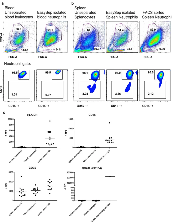
Whereas the purity of FACS-sorted splenic neutrophils and EasySep-isolated circulating neutrophils was excellent (,0.5% lymphocytes), the EasySep-isolated splenic neutrophils consistently contained a large population (ranging from 10 to 25%) of lymphoid cells (Figure 3a and Figure S3a in File S1). These contaminating cells were mainly CD20+
/CD27+
B cells, contain-ing both IgMposand IgGposcells (Figure S3b in File S1). A pre-enrichment step by density gradient centrifugation did not remove this population of B cells (Figure S4 in File S1).
Upon further analysis of these freshly isolated splenocytes, we tested EasySep-isolated and FACS-sorted neutrophils as well as unseparated splenocytes for expression of various surface antigens. In contrast to the findings of Pugaet al, the majority (.96%) of these cells showed high expression levels of CD15 and CD16, both in spleen and blood (Figure 3a,b and Figure S5e in File S1). We
did not detect any surface expression of CD40L, CD86, HLA-DR or increased levels of CD95 on these splenic neutrophils (Figure 3c and Figure S5a–d in File S1).
Discussion
We could not reproduce the stimulation of immunoglobulin production in human MZ B cells by splenic neutrophils, despite the fact that both the MZ B cells and the neutrophils we isolated were fully viable and functional. Possible explanations for the discrepancy with the findings of Pugaet al.[10] include differences in the protocols for obtaining spleen tissue and isolating spleen cells, discussed further below. Of particular concern is our finding of a consistent contamination with B cells in EasySep-isolated splenic neutrophils. The notion that NBHcells can induce CSR in
MZ B cells inin vitrocultures of 1 week is solely based on cultures with EasySep-isolated splenic neutrophils [10]. Our data show that splenic neutrophil samples isolated by the Easysep-Neutrophil-Enrichment-Kit, as opposed to blood samples, consistently contained a contaminating B-cell population that was partly IgGpos. Such a population will contain germline Ic1-Cc1 or Ic 2-Cc2 transcripts, and Ic-Cmswitch circles, in itself. Presence of these transcripts in cultures of MZ B cells stimulated with B-cell contaminated neutrophils therefore does not prove the induction of CSR. The Easysep-Neutrophil-Enrichment-Kit can be used for isolation of blood neutrophils, but the isolation of neutrophils from other sources is not mentioned by the manufacturer. Purity of EasySep-isolated splenic neutrophil fractions was not shown by Pugaet al.
Furthermore, we could not find the characteristic phenotypes of NBH1and NBH2 cells as described [10]. Slight differences in the
protocol for obtaining splenocytes may form an obvious explana-tion. Regarding the isolation protocol, the only difference between the protocols consisted of an incubation step with collagenase and DNAse. However, when we isolated splenocytes by perfusion with PBS only [10], expression of HLA-DR, CD40L, CD86 or CD95 on splenic neutrophils was still absent (Figure S6 in File S1), showing that the treatment with collagenase did not influence expression of these molecules. Treatment of splenic tissue with collagenase and DNAse is a widely used method [15] that is used to obtain also cells that are embedded in or have infiltrated tissue matrix, as NBHcells supposedly do, which is the reason we have
chosen to include it in our protocol.
Instead, differences in the protocol for obtaining splenic tissue may have been critical. We used only fresh spleen samples that were obtained from heart-beating organ donors and were transported at 4uC in a specialized medium for organ conserva-tion. Neutrophils are known to be easily activated during transport and isolation, resulting in the cleavage of CD16 from the cell surface [16–18]. Such events may have led to the reported phenotypes of NBH cells. In fact, the finding of Pugaet al. that
splenic samples contained only NBHcells in the absence of NCcells
is surprising in itself, as the spleen is highly vascularized and must contain lots of circulating blood cells. Further evidence for the fact that handling of splenic tissue prior to isolation can influence the phenotype of neutrophils comes from the intriguing finding of Puga et al. that inflamed splenic tissue only contained the (less active) NBH1cells, as these were isolated from frozen or
paraffin-embedded tissue [10]. In our case, we regard it unlikely that unfavorable circumstances during isolation of our neutrophils have prevented us from finding the phenotype of specialized NBH1and
NBH2 cells in 11 spleens, which consistently revealed neutrophil
populations very similar to circulating neutrophils. Figure 2. Splenic B cells do not produce immunoglobulin in
response to splenic neutrophils.ELISA of IgM, IgG and IgA from splenic marginal zone B cells (MZ B) or follicular naı¨ve B cells (FN), sorted as shown in Figure 1, after co-culture for 7 days with circulating neutrophils (N circ), FACS-sorted spleen neutrophils (N spl sorted), EasySep-isolated spleen neutrophils (N spl EasySep) or CpG/IL-2. Data summarize three independent experiments (error bars, SEM.) doi:10.1371/journal.pone.0088377.g002
Apart from technical issues, the findings of specific splenic neutrophil functions led to a lot of confusion and debate, because patients with neutropenia or chronic granulomatous disease are not known to present with clinical infections associated with B cell deficiencies [19,20]. Concluding, we did not find evidence for the reprogramming of circulating neutrophils into distinct NBH1and
NBH2cells in the human spleenin vivo, nor did we find evidence for
the major role of neutrophils in MZ B cell stimulation that was proposed by Puga et al. We encourage others to attempt to reproduce these experiments in order to establish whether splenic NBHcells exist and play a role in B-cell activation. In our opinion,
further research defining the triggers that drive B cell responses against TI-antigens in the human situation should not be solely focused on neutrophils, but should also consider other potentially involved cell populations such as monocytes or dendritic cells.
Supporting Information
File S1 Includes Figures S1–S4 and Tables S1–S3.Figure S1. Splenic marginal zone B cells are able to differentiate into plasmablasts in response to CpG/IL2, but not in response to blood or spleen neutrophils. FACS plot of CD27/CD38 double staining of CD20pos B cells cultured for 7 days with indicated stimuli. Numbers indicate percentage of the total B cell population. Figure S2. Measurement of neutrophil reactive oxygen species in response to different stimuli. Production of reactive oxygen species by neutrophils from spleen and blood. RFU: relative fluorescence units, which are a derivative of H2O2production. Zymosan 1 mg/
ml; STZ: serum-treated zymosan 1 mg/ml; PMA: Phorbol 12-Myristate 13-Acetate 100 ng/ml; fMetLeuPhe 1mM; PAF: platelet-activating factor 1mM. Spleen n = 2, blood n = 3. Error bars represent standard deviation. Figure S3. Purity analysis of different neutrophil isolates. a. FSC/SSC plot and May-Gru¨n-wald/Giemsa stained cytospins of different neutrophil isolates. Neutrophils (upper gate) and lymfocytes (lower gate) are gated according to canonical FSC/SSC pattern. Numbers indicate percentages of total events. Original magnification of cytospins 540x. Data are representative of four independent experiments. b. Characterisation of the lymfocyte population contaminating the EasySep-isolated spleen neutrophils. Numbers indicate percentag-es of the lymfocyte population. Figure S4. Ficoll density gradient centrifugation prior to EasySep isolation does not remove the contaminating B cell population from EasySep-isolated splenic neutrophils. FSC/SSC pattern of splenic neutrophils separated either directly from splenocytes with the Human Neutrophil Enrichment kit (left), or separated from splenocytes with a Histopaque-1077 gradient followed by purification with the Human Neutrophil Enrichment kit (right). Numbers indicate percentage of total events. Data are representative of 2 independent experiments. Figure S5. Expression patterns of
splenic neutrophils do not differ from their circulating counterpart. a.Gating strategy for neutrophils (upper gate) and monocytes (lower gate) by canonical FSC/SSC pattern in blood and spleen. The monocyte gate may include small percentages of other cells, especially in spleen, and serves only to show a positive control for the antibodies used. b.Staining of HLA-DR, CD86, CD95 and CD40L of splenic neutrophils as gated in Figure 2 and splenic monocytes as gated in Figure S5a. Data are representative of 11 independent experiments. Black lines: Staining with monoclonal antibody. Gray shading: isotype control. c. Staining of HLA-DR, CD86, CD95 and CD40L of blood neutrophils as gated in Figure 2 and blood monocytes as gated in Figure S5a. Data are representative of 11 independent experiments. Black lines: Staining with monoclonal antibody. Gray shading: isotype control. d.Staining of CD40L in human CD40L expressing fibroblast cell line. Black lines: Staining with monoclonal antibody. Gray shading: isotype control. e. CD15/CD16 double staining of blood and splenic neutrophils, as gated in Figure 2. Antibodies used: CD15: clone HI98, FITC. CD16: clone 3G8, APC. Data shown are of blood and splenic neutrophils from a single donor, and representative of two other experiments with unpaired samples. Figure S6. Incubation with collagenase buffer does not influence expression profile of neutrophils. Expression levels of HLA-DR, CD40L, CD86 and CD95 in neutrophils and monocytes from unseparated splenocyte suspensions, gated as in Figure S5a. Splenocytes suspensions were obtained either by perfusion with ‘collagenase buffer’ followed by 30 minute incubation at 37uC, or by perfusion with PBS alone with direct further processing. Data summarize 2 independent experiments, open triangles represent 1 donor, closed circles represent the other donor.D MFI: median fluorescence intensity minus median fluorescence intensity of appropiate isotype control. Table S1. Origin and characteristics of tissue samples. Table S2. Contents of collagenase buffer. Table S3. Antibodies used in flow cytometry.
(PDF)
Acknowledgments
The authors would like to thank mr Robin van Bruggen and mrs Paulien Kuin-Valkenhoff (Sanquin, Amsterdam, the Netherlands) for help in providing tissue samples, and would like to thank Prof. Dirk Roos for critical reading of the manuscript. The CD154 expressing NIH3T3 cells were a kind gift from mr Peter-Paul Unger (Sanquin, Amsterdam, the Netherlands).
Author Contributions
Conceived and designed the experiments: SQN TWK. Performed the experiments: SQN DJK MHJ. Analyzed the data: SQN DJK MHJ. Contributed reagents/materials/analysis tools: SQN DJK MHJ. Wrote the paper: SQN TWK. Discussed data: TKB TWK.
References
1. Mebius RE, Kraal G (2005) Structure and function of the spleen. Nat Rev Immunol 5: 606–616. nri1669 [pii];10.1038/nri1669 [doi].
2. Weill JC, Weller S, Reynaud CA (2009) Human marginal zone B cells. Annu Rev Immunol 27: 267–285. 10.1146/annurev.immunol.021908.132607 [doi].
3. Scheeren FA, Nagasawa M, Weijer K, Cupedo T, Kirberg J, et al (2008) T cell-independent development and induction of somatic hypermutation in human IgM+ IgD+ CD27+ B cells. J Exp Med 205: 2033–2042. jem.20070447 [pii];10.1084/jem.20070447 [doi].
spleen donor and one blood donor, and are representative of eight (unseparated), four (EasySep) or three (FACS sorted) independent experiments.c. Expression levels of HLA-DR, CD40L, CD86 and CD95 in neutrophils from unseparated leukocyte/splenocyte suspensions, as gated in a and b. Monocytes or a CD40L expressing cell line serve as positive control, gating strategy as in Figure S5a. Data summarize 11 independent experiments. In three of these experiments, the blood and spleen tissue were of the same donor, as indicated by the open symbols (n = 3). The MoAbs used are all commercially available, quality-tested antibodies (Table S3), with the same CD15 and CD16 clones as used by Pugaet al[10]. DMFI: median fluorescence intensity minus median fluorescence intensity of appropiate isotype control.
doi:10.1371/journal.pone.0088377.g003
4. Weller S, Braun MC, Tan BK, Rosenwald A, Cordier C, et al. (2004) Human blood IgM ‘‘memory’’ B cells are circulating splenic marginal zone B cells harboring a prediversified immunoglobulin repertoire. Blood 104: 3647–3654. 10.1182/blood-2004-01-0346 [doi];2004-01-0346 [pii].
5. Bernasconi NL, Onai N, Lanzavecchia A (2003) A role for Toll-like receptors in acquired immunity: up-regulation of TLR9 by BCR triggering in naive B cells and constitutive expression in memory B cells. Blood 101: 4500–4504. 10.1182/ blood-2002-11-3569 [doi];2002-11-3569 [pii].
6. Pone EJ, Zan H, Zhang J, Al-Qahtani A, Xu Z, et al. (2010) Toll-like receptors and B-cell receptors synergize to induce immunoglobulin class-switch DNA recombination: relevance to microbial antibody responses. Crit Rev Immunol 30: 1–29. 42750210589e65a5,553e1f3a5548cfc2 [pii].
7. Balazs M, Martin F, Zhou T, Kearney J (2002) Blood dendritic cells interact with splenic marginal zone B cells to initiate T-independent immune responses. Immunity 17: 341–352. S1074761302003898 [pii].
8. Chen Q, Snapper CM (2013) Inflammatory monocytes are critical for induction of a polysaccharide-specific antibody response to an intact bacterium. J Immunol 190: 1048–1055. jimmunol.1202455 [pii];10.4049/jimmunol.1202455 [doi]. 9. Steiniger B, Timphus EM, Barth PJ (2006) The splenic marginal zone in humans
and rodents: an enigmatic compartment and its inhabitants. Histochem Cell Biol 126: 641–648. 10.1007/s00418-006-0210-5 [doi].
10. Puga I, Cols M, Barra CM, He B, Cassis L, et al. (2012) B cell-helper neutrophils stimulate the diversification and production of immunoglobulin in the marginal zone of the spleen. Nat Immunol 13: 170–180. ni.2194 [pii];10.1038/ni.2194 [doi].
11. Urashima M, Chauhan D, Uchiyama H, Freeman GJ, Anderson KC (1995) CD40 ligand triggered interleukin-6 secretion in multiple myeloma. Blood 85: 1903–1912.
12. Aan de Kerk DJ, Jansen MH, Ten Berge IJ, van Leeuwen EM, Kuijpers TW (2013) Identification of B Cell Defects Using Age-Defined Reference Ranges for
In Vivo and In Vitro B Cell Differentiation. J Immunol 190: 5012–5019. jimmunol.1201807 [pii];10.4049/jimmunol.1201807 [doi].
13. Kuijpers TW, Bende RJ, Baars PA, Grummels A, Derks IA, et al. (2010) CD20 deficiency in humans results in impaired T cell-independent antibody responses. J Clin Invest 120: 214–222. 40231 [pii];10.1172/JCI40231 [doi].
14. van Bruggen R, Drewniak A, Jansen M, van Houdt M, Roos D, et al. (2009) Complement receptor 3, not Dectin-1, is the major receptor on human neutrophils for beta-glucan-bearing particles. Mol Immunol 47: 575–581. S0161-5890(09)00720-2 [pii];10.1016/j.molimm.2009.09.018 [doi].
15. Swirski FK, Nahrendorf M, Etzrodt M, Wildgruber M, Cortez-Retamozo V, et al (2009) Identification of splenic reservoir monocytes and their deployment to inflammatory sites. Science 325: 612–616. 325/5940/612 [pii];10.1126/ science.1175202 [doi].
16. Huizinga TW, van der Schoot CE, Jost C, Klaassen R, Kleijer M, et al. (1988) The PI-linked receptor FcRIII is released on stimulation of neutrophils. Nature 333: 667–669. 10.1038/333667a0 [doi].
17. Kuijpers TW, Tool AT, van der Schoot CE, Ginsel LA, Onderwater JJ, et al. (1991) Membrane surface antigen expression on neutrophils: a reappraisal of the use of surface markers for neutrophil activation. Blood 78: 1105–1111. 18. Wang Y, Wu J, Newton R, Bahaie NS, Long C, et al. (2012) ADAM17 cleaves
CD16b (FcgammaRIIIb) in human neutrophils. Biochim Biophys Acta 1833: 680–685. S0167-4889(12)00361-8 [pii];10.1016/j.bbamcr.2012.11.027 [doi]. 19. Moir S, De Ravin SS, Santich BH, Kim JY, Posada JG, et al. (2012) Humans
with chronic granulomatous disease maintain humoral immunologic memory despite low frequencies of circulating memory B cells. Blood 120: 4850–4858. blood-2012-05-430959 [pii];10.1182/blood-2012-05-430959 [doi].