Splenic difuse red-pulp small B-cell lymphoma associated
with hepatitis B virus: a report of two cases
Linfoma esplênico difuso da polpa vermelha, de linfócitos B pequenos, associado ao
vírus da hepatite B: relato de dois casos
Mariana Nassif Kerbauy
I, Carolina Melo Fernandes
II, Evandro Dantas Bezerra
II, Luis Alberto de Padua Covas Lage
II,
Sheila Aparecida Coelho Siqueira
III, Juliana Pereira
IVFaculdade de Medicina da Universidade de São Paulo (FMUSP), São Paulo, SP, Brazil
ABSTRACT
CONTEXT: Splenic difuse red-pulp small B-cell lymphoma is a rare disease, representing less than 1% of all non-Hodgkin lymphomas (NHL). This entity is characterized by involvement of bone marrow sinusoids and peripheral blood. The majority of cases are at an advanced stage when diagnosed. Its pathogenesis is still poorly understood.
CASE REPORTS: We report on two patients with chronic non-replicating hepatitis B virus (HBV) who de-veloped splenic difuse red-pulp small B-cell lymphoma. Both of them were in stage IV at diagnosis and evolved with aggressive disease. Both of them achieved a complete response through chemotherapy, but one of them died due to infectious complications during bone marrow transplantation. The other decided not to undergo transplantation and continues not to show any evidence of disease today (three years after treatment). Some studies have shown a possible association between B-cell NHL and HBV. Nonetheless, the mechanism through which this oncogenic virus interacts with B-cell NHL is still poorly understood. HBV is lymphotropic and may insert into the host’s genome, thus causing overexpression of oncogenes and downregulation of tumor suppressor genes. Therefore, chronic stimulation by HBV can increase B-cell proliferation, which promotes monoclonal expansion of these cells and results in malignancy.
CONCLUSION: HBV may be implicated in the pathogenesis of this lymphoma, although no direct associa-tion between these two entities could be proved in the present study. Further investigaassocia-tions are necessary.
RESUMO
CONTEXTO: Linfoma esplênico difuso da polpa vermelha, de linfócitos B pequenos, é uma doença rara, representando menos do que 1% de todos os linfomas não Hodgkin. Essa entidade é caracterizada por envolvimento de sinusoides da medula óssea e sangue periférico. A maioria dos casos está em estádio avançado ao diagnóstico. Sua patogênese ainda é pouco compreendida.
RELATOS DE CASOS: Reportamos dois pacientes com vírus da hepatite B (HBV) crônica não replicante que desenvolveram linfoma esplênico difuso da polpa vermelha, de linfócitos B pequenos. Ambos esta-vam em estádio IV ao diagnóstico e evoluíram com doença agressiva. Ambos alcançaram resposta com-pleta com a quimioterapia, porém um deles evoluiu a óbito por intercorrências infecciosas durante o transplante de medula óssea e o outro optou por não realizar o transplante e encontra-se sem evidência de doença até os dias atuais (três anos após tratamento). Alguns estudos demonstraram a possível asso-ciação entre linfomas não Hodgkin B e HBV. Entretanto, o mecanismo pelo qual esse vírus oncogênico interage com linfoma não Hodgkin B ainda é pouco compreendido. HBV é linfotrópico e pode se inserir no genoma do receptor, causando superexpressão de oncogenes e downregulation de genes supressores tumorais. Portanto, o estímulo crônico pelo HBV pode aumentar a proliferação de células B, promovendo expansão monoclonal dessas células, resultando em malignidade.
CONCLUSÃO: HBV pode estar implicado na patogênese desse linfoma, entretanto, uma associação direta entre essas duas entidades não pôde ser provada no presente estudo e investigações adicionais são necessárias.
IMD. Resident Physician, Department of Hematology and Hemotherapy, Faculdade de Medicina da Universidade de São Paulo (FMUSP), São Paulo, SP, Brazil.
IIMD. Hematologist, Department of Hematology and Hemotherapy, Faculdade de Medicina da Universidade de São Paulo (FMUSP), São Paulo, SP, Brazil.
IIIMD, PhD. Professor in the Department of Pathology, Faculdade de Medicina da Universidade de São Paulo (FMUSP), São Paulo, SP, Brazil.
IVMD, PhD. Professor in the Department of Hematology and Hemotherapy, Faculdade de Medicina da Universidade de São Paulo (FMUSP - University of São Paulo), São Paulo, SP, Brazil.
KEY WORDS: Lymphoma.
Lymphoma, non-Hodgkin. Lymphoma, B-cell. Hepatitis B. Hepatitis B virus.
PALAVRAS-CHAVE: Linfoma.
INTRODUCTION
Splenic difuse red-pulp small B-cell lymphoma was recognized as a provisional entity in the 2008 update of the World Health Organization (WHO) classiication.1 his new WHO update rec-ognized two categories of primary splenic lymphomas; splenic marginal-zone lymphoma (SMZL) and the provisional group of splenic B-cell lymphoma/leukemia unclassiiable (SLLU), that includes splenic difuse red-pulp small B-cell lymphoma and hairy-cell leukemia variant (HCL-v).1 he median age at which patients are diagnosed with splenic difuse red-pulp small B-cell lymphoma is 72 years (range: 69-74 years),2 and the disease is seen predominantly among males, with a male/female ratio of 2.4:1.
Splenic difuse red-pulp small B-cell lymphoma is a rare dis-ease and represents less than 1% of all non-Hodgkin lymphomas (NHL) and 10% of B-cell lymphomas that are described in post-splenectomy series.1 his malignancy is characterized by difuse iniltrate of monomorphic small to medium-sized B-cells with cytoplasmic villi, going into the red pulp of the spleen. he bone marrow sinusoids and peripheral blood are frequently involved. Patients usually present with an advanced stage of disease when they are diagnosed. Peripheral lymph node involvement and B-symptoms are rarely reported and massive splenomegaly is common.1 Splenic difuse red-pulp small B-cell lymphomas have an indolent clinical course and there is no known standard treat-ment approach. So far, most of these patients have been treated with splenectomy.3
he pathogenesis of this lymphoma is still poorly understood and no risk factors associated with its development have been described. Recent studies have shown an association between B-cell NHL and hepatitis B virus (HBV), but the mechanism through which this oncogenic virus results in chronic lymphop-roliferative B-cell disorders is not clearly understood. To the best of our knowledge, there is no case report in the literature corre-lating HBV with splenic difuse red-pulp small B-cell lymphoma.
Here, we describe two cases of splenic difuse red-pulp small B-cell lymphoma associated with chronic HBV infection.
CASE REPORTS
Case 1
A 62-year-old woman presented at our clinic complaining of weight loss, fever, night sweats and increasing abdominal size over the preceding two months. In her medical history, she reported having had rheumatoid arthritis, which was previously treated with chloroquine and methotrexate, and an untreated chronic non-replicating HBV infection (AntiHbC total +, AgHbe −, AntiHbe +, AgHbs +, AntiHbsAg -) with quantitative polimer-ase chain reaction (PCR) for HBV of 104 IU/ml (normal range of values: 20–170,000,000 IU/ml). Other viral serological tests
for human immunodeiciency virus (HIV) and hepatitis C virus (HCV) were negative. In her family history, her daughter had had a central nervous system (CNS) neoplasm, a 60-year-old brother had had prostate cancer and another 50-year-old brother had died of acute leukemia.
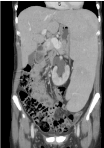
In her physical examination, she presented as emaciated and pale, with massive splenomegaly and no palpable hepatomegaly or peripheral lymphadenopathy. Computed tomography (CT) scans showed subtle hepatomegaly and greatly increased spleen size, with no lymphadenopathy (Figure 1).
Laboratory tests showed normocytic and normochro-mic anemia (hemoglobin 8.8 g/dl, platelet count of 124 x 109/l and total leukocyte count of 6.18 x 10⁹/l); 21% of the lymphoid cells had a high nuclear-to-cytoplasmic ratio, with loose chro-matin, small clear nucleolus and thin cytoplasmic projections. Immunophenotyping of peripheral blood cells showed that they were positive for CD19, CD20, FMC7, CD22, and CD23, with strong expression of CD79b and CD25, weak surface expres-sion of immunoglobulin (Ig) M, and partial expresexpres-sion of CD5. No expression of CD200, CD10, CD11c or CD103 was seen. Monoclonality was demonstrated through restriction of
cytoplasmic light kappa chain Ig. Bone marrow biopsy revealed intrasinusoidal iniltration by small B lymphoid cells.
Splenectomy was performed as a therapeutic procedure and the histological analysis showed that the spleen weighed 1.818 kg, measured 27 x 14.3 x 8.3 cm and presented difuse iniltrate in the red pulp of the spleen, with both cord and sinusoid iniltra-tion by small B-cell lymphocytes (Figure 2) expressing CD20 and DBA-44 antigens. hese cells had a low Ki67 proliferative index and were negative for CD3, CD5, CD10, CD23 and cyclin-D1. A hepatic biopsy revealed mixed inlammatory iniltration and stage 1 steatohepatitis, with an activity index of 5.
he patient improved clinically and her laboratory parame-ters normalized ater splenectomy. However, 10 months later, the patient presented with weight loss, fever, night sweats, asymmet-rical eyelid edema and pleural efusion. At that time, laboratory examinations showed leukocytosis of 30.18 x 10⁹/l; 22% of the cells were of moderate size and presented loose chromatin and evident peripheral nucleoli, which were morphologically sug-gestive of centroblasts. Her laboratory values showed hypercal-cemia and elevated lactate dehydrogenase (LDH), of more than 3,000 U/l (normal values range from 240 to 480 U/l). Pleural luid showed iniltration by neoplastic cells. New immunophe-notyping of peripheral blood cells revealed aberrant lymphoid cells expressing B antigens, including CD19, surface IgM (sIgM), strong CD20 expression, heterogeneous partial CD79b expres-sion and FMC7 with a lack of CD5 antigen. Bone marrow karyo-typing revealed 46,XX [20]. he bone marrow biopsy was hyper-cellular due to iniltration by B cells.
Brain and orbit magnetic resonance imaging (MRI) showed iniltration of the optic nerve, bilateral lacrimal glands and CNS. Restaging CT scans revealed renal iniltration and axillary and abdominal lymph node enlargement. It was concluded that transformation to high-grade B-cell lymphoma had occurred,
and treatment with R-CHOP was started, with the following given on day 1 of each cycle: intravenous rituximab, 375 mg/m2; intravenous cyclophosphamide, 750 mg/m2; intravenous doxoru-bicin, 50 mg/m2; and intravenous vincristine, 1.4 mg/m2. In addi-tion, oral prednisone, 100 mg once daily, was given on days 1–5. R-CHOP was administered in combination with intrathecal che-motherapy containing methotrexate (12 mg), cytarabine (60 mg) and dexamethasone (2 mg) (MADIT) and two cycles of metho-trexate (3 g/m2 intravenously) ater the last cycle of R-CHOP and radiotherapy to the orbits (24 Gy). Tenofovir (300 mg/day) was started along with the chemotherapy to avoid replication of HBV. he patient achieved a complete response, and autologous bone marrow transplantation was performed as a consolidation strat-egy. However, unfortunately, the patient died due to infectious complications during the transplantation.
Case 2
A 29-year-old female patient with a seven-year history of spleno-megaly, without portal hypertension and with chronic non-rep-licating HBV infection (AntiHbC total +, AntiHbe +, AgHbe −, AgHbs +, AntiHbs −) showing quantitative PCR for HBV of less than 20 IU/ml (normal range of values: 20-170,000,000 IU/ ml) presented to our service complaining of progressive spleen enlargement and B symptoms (fever, night sweats and weight loss) for the past two months. Laboratory tests showed hemoglo-bin 8.1 g/dl, platelets 80×109/l and white blood cells 12 × 109/l, with 85% atypical lymphoid cells. Immunophenotyping demon-strated strong expression of CD45, positivity for the B-cell anti-gens CD20 and CD19, weak expression of CD25, partial expres-sion of CD23 and CD200 and negativity for CD11c and CD103. Monoclonality for kappa light chain Ig was demonstrated in both the peripheral blood and bone marrow.
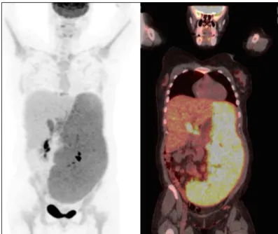
he karyotype was complex, with structural abnormalities in the 14q32 region, deletions on chromosomes 6 and 7, isochro-mosomes 8 and 17 and trisomy 18. Fluorescence in situ hybrid-ization analysis revealed an extra copy of the MYC gene and a p53 deletion. 18FDG-PET-CT showed splenomegaly (standard uptake value [SUV]: 6.1), hepatomegaly (SUV: 3.2), abdominal lymph node enlargement (SUV: 6.3) and cervical and mediasti-nal lymphadenopathy (Figure 3).
Splenectomy was indicated and the anatomopathological analysis showed that the spleen dimensions were 33.0 x 20.6 x 7.5 cm. he spleen showed iniltration by small to intermediate-sized mature lymphoid cells, with large cells among them, primarily in the red pulp and sinusoidal space of the spleen and secondarily in the white pulp of the spleen (Figure 4). Immunohistochemical analysis showed expression of CD20 and DBA44, with Ki67 of 40%. he cells were negative for CD5, CD10, CD23, CD3 and cyclin D1. A hepatic biopsy demonstrated nodular lymphoid Figure 2. Difuse iniltrate of small B-cell lymphocytes in the
iniltrate involvement, with suspected lymphoid neoplasia. A hilum splenic lymph node (measuring 2.6 x 1.1 x 1.0 cm) showed lymphoid neoplasia of mature cells with predominance of intermediate to large cells with a nodular pattern.
he diagnosis of splenic difuse red-pulp small B-cell lym-phoma was made and treatment was then instituted, consisting of eight cycles of R-CHOEP: intravenous rituximab (375 mg/m2), intravenous cyclophosphamide (750 mg/m2), intravenous doxo-rubicin (50 mg/m2), and intravenous vincristine (1.4 mg/m2) on
day 1; intravenous etoposide (100 mg/m2) on days 1–3; and oral prednisone (100 mg orally once daily) on days 1–5. It was pro-posed that this would be followed by autologous stem cell trans-plantation. Ater eight cycles of R-CHOEP, the patient achieved complete response, but she became pregnant and chose not to undergo the transplantation. Nonetheless, she continues not to present any evidence of disease today (three years ater the treatment). Administration of tenofovir (300 mg/day) was started along with the chemotherapy.
DISCUSSION
We have reported on two patients with chronic non-replicating HBV who developed splenic difuse red-pulp small B-cell lym-phoma. he diagnosis of primary chronic lymphoproliferative disorders of the spleen is established based on analysis on sple-nectomy tissue, or on integration of clinical data, assessment of lymphocyte morphology in peripheral blood, immunophenotyp-ing of these lymphocytes and evaluation of bone marrow iniltra-tion pattern from biopsy. For our patients, we used both criteria in order to be sure of the diagnosis. In both cases, the splenectomy product demonstrated difuse neoplastic iniltration consisting of small mature lymphocytes with primary immunophenotype B of the red pulp of the spleen. With this presentation, the diag-nosis of splenic marginal-zone lymphoma was ruled out, since this entity is characteristically a primary neoplasm of the splenic white pulp, usually with nodular architectural arrangement.
Other entities that might have formed diferential diagno-ses were hairy-cell leukemia and hairy-cell leukemia variant. However, the clinical data in association with morphological and immunophenotyping data made it possible to safely rule out diagnoses of hairy-cell leukemia (absence of pancytopenia, no cells with the classical morphology of “hairy cell” and immuno-phenotyping not characterized by strong expression of CD11c, CD25, CD103 and CD123 antigens) or hairy-cell leukemia vari-ant (no lymphocytes with morphology characterized by the pres-ence of central vesicular nucleolus, cytoplasm similar to hairy cell and absence of strong expression of CD11c and CD20 antigens). hus, integration of all these indings, i.e. lymphoprolifera-tive malignancy of small B cells, difuse iniltration of the spleen and primary from splenic red pulp, with exclusion criteria for “hairy cell” and its variant form, enabled the deinitive accurate diagnosis of splenic difuse red-pulp small B-cell lymphoma in both cases reported here.
We conducted a systematic search in the main electronic databases (PubMed, Google Scholar and Lilacs Library), to ind articles relating to splenic difuse red-pulp small B-cell lym-phoma in association with hepatitis B infection. In order to make the search as wide as possible, no limits were applied regard-ing the date of publication, the language or the research design Figure 3. 18FDG-PET-CT (ludeoxyglucose F18 position emission
computed tomography) demonstrating major splenomegaly (standard uptake value [SUV]: 6.1) and hepatomegaly (SUV: 3.2)
(Table 1). here were fewer than 100 search results and none of them described any association between splenic difuse red-pulp small B-cell lymphoma and hepatitis B.
Mollejo et al. irst proposed this lymphoma as a new sub-type in 2002,4 consequent to reviewing 85 cases of SMZL. hese authors identiied four cases with predominance of monomor-phic iniltration in the red pulp of the spleen instead of the typi-cal micronodular component in the white pulp commonly seen in SMZL. Later on, two series of cases of splenic difuse red-pulp small B-cell lymphoma were published by Traverse-Glehen et al.5 and Kanellis et al.2
Similarly to our two patients, all cases previously reported were also in stage IV at diagnosis. Difuse iniltration of the red pulp of the spleen, afecting both cords and sinuses, along with marked DBA-44 positivity, was frequently found by Kanellis et al.2 (88.2% of their cases), as well as in our cases. Additionally, previous studies described situations of lymph node replacement by difuse neoplastic iniltrate, with preserved sinuses.2,4
he most common inding in the bone marrow biopsies was predominance of intrasinusoidal lymphoid iniltration, some-times associated with interstitial and nodular involvement, along with hematopoietic tissue and absent or mild ibrosis. he malig-nant cells presented with round to slightly irregular nuclei, vesic-ular chromatin and moderate amounts of pale cytoplasm and cytoplasmic projections similar to SMZL villous cells. hese cells expressed CD20, DBA44 and IgG. Presence of annexin A1, CD25, CD3, CD5, CD23, CD103, CD123, CD11c, CD38, CD10, Bcl6, bcl2, cyclin D1 and IgD was uncommon.1,4 Our cases are consistent with those presented in the literature regarding the pathological and immunophenotypic indings of splenic dif-fuse red-pulp small B-cell lymphoma. he majority of the cases reported in the literature had a normal karyotype and the most
frequent chromosomal abnormalities were 7q and 3q deletions and trisomy 18.1,6 One of our cases showed a complex karyotype.
Kanellis et al.2 found p53 inactivation in all cases of splenic difuse red-pulp small B-cell lymphoma, comprising either p53 mutation (2/4 cases) or anomalous p53 staining. Mollejo et al.4 reported that TP53 abnormality was present in 2 out of 13 cases (15.3%): one of these patients showed disease progression and ultimately died of the disease. In the same way as described by these authors, one of our patients also had a p53 deletion and evolved to aggressive disease. Splenic difuse red-pulp small B-cell lymphoma is an indolent lymphoma and patients can be maintained using a watchful waiting approach or may undergo splenectomy.
Compared with SMZL, splenic difuse red-pulp small B-cell lymphoma has demonstrated better disease-free survival, but overall survival is not statistically diferent. At diagnosis, our patients presented with anemia, which is associated with worse prognosis according to Kanellis et al.,2 and both of them had HBV infection as a comorbidity. HBV infection may be asso-ciated with worse prognosis, since both patients evolved with aggressive disease.
Virus-induced carcinogenesis is known to occur in several lymphoid malignancies in which the virus has a pathogenic role, such as in Burkitt’s lymphoma with Epstein-Barr virus (EBV), adult T-cell leukemia with human T-cell lymphotropic virus-1, and primary efusion B-cell lymphoma with human herpes virus 8. Some malignancies may be caused by chronic stimulation such as in mucosa-associated lymphoid tissue lymphoma, which is associated with Helicobacter pylori, or through an indirect mech-anism. While EBV has a direct carcinogenic efect on Burkitt’s lymphoma through activating the c-Myc oncoprotein, HIV acts on the immune system to reduce immune surveillance.7,8
HBV is a small DNA virus that is a member of the Hepadnaviridae family. It replicates through a RNA intermedi-ary and can integrate into the host genome. HBV infection is highly prevalent worldwide, with around 350 million chronically infected individuals. It is endemic in Asia, Africa, the Middle East, Eastern Europe and South America.9 here are 500,000 to 1.2 million deaths due to chronic hepatitis B annually, out of a total of 350 million cases worldwide,8 and about 340,000 cases of liver cancer relating to HBV.10
HBV and HCV are known to induce acute and chronic hep-atitis and are strongly associated with hepatocellular carcinoma (HCC). HBV carriers have a 200 times higher risk of HCC, and this is one of the highest risks for a human malignancy.11 Its pathogenesis involves multiple pathways, including oxidative stress, hepatic inlammation leading to genetic damage and inte-gration of HBV DNA into the host genome, thereby leading to genetic alterations such as chromosomal and gene translocations Table 1. Systematic search of the literature performed on
April 4, 2016
Electronic
databases Search strategy
Results
Found Paper used
Medline (via PubMed)
“Lymphoma, B-Cell”[Mesh] AND “Hepatitis B”[Mesh] AND Case
Reports[ptyp]
62 0
Lilacs (via Bireme)
(“Linfoma de Células B OR Lymphoma, B-Cell OR Linfoma de Células B “ [Decs] AND Hepatite B OR
Hepatitis B OR Hepatitis B [Decs])
0 0
Embase (via Ovid)
(Lymphoma, B-Cell) and (Hepatitis B)). All ields.
Search terms cross-referenced: b; b-cell; hepatitis; hepatitis b; lymphoma; lymphoma, b-cell
and deletions or generation of fusion transcripts. hese alter-ations may change oncogenes, tumor-suppressor genes and expression of small non-coding RNA molecules (miRNAs).12
Non-structural HBV X protein is a key regulatory protein that modulates viral replication and pathogenesis. It is tran-scribed in human HCC tumor cells, even when HBV replica-tion is absent, due to viral DNA integrareplica-tion. his protein modu-lates gene expression, since it interacts with transcription factors, activates mitogenic signaling cascades and interacts with cellular proteins, including p53. Consequently, DNA repair, tumor sup-pressor genes and the cell cycle could become deregulated.13
he relationship between NHL and HCV has already been proven, with evidence demonstrating a causal relationship.11,14-17 here is a known multicausal process involving direct lympho-cytic activation mediated by viral proteins and chronic antigenic stimulation, for promoting B-cell transformation.18 A recent prospective cohort study conducted in South Korea evaluated 603,585 participants, among whom 53,045 (9%) tested positive for HBsAg. he HBsAg-positive patients were at higher risk of NHL than those who were HBsAg-negative (hazard ratio: 1.74; 95% CI: 1.45–2.09), especially for difuse large B-cell lymphoma and immunoproliferative diseases. In this study, HBsAg was not associated with follicular lymphoma, T-cell NHL, Hodgkin’s lym-phoma, multiple myeloma or leukemia.19 A meta-analysis on 17 case-control and ive cohort studies found that HBV-infected individuals had an odds ratio (OR) of 2.24 (95% CI: 1.80-2.78; P ≤ 0.001) for developing NHL.20 Another meta-analysis on 12 case-control studies, comprising 11 studies evaluating HBV infection in NHL and one study that had investigated NHL in HBV infection, reported that the OR of detecting HBV infection in lymphoma patients was 2.5 times higher than in controls.21 hese studies suggest a possible relationship between HBV and NHL. he pathogenic association is less well studied than rela-tionships with HCV, but it appears to result from diferent HBV-driven events.22
HBV is a hepatotropic virus and replicates within hepato-cytes, but it can also cause lymphotropism for lymphocytes in the peripheral blood, bone marrow, spleen, lymph nodes, and thymus.23-26 his lymphotropism of HBV is a fundamental prop-erty favoring causality between HBV and B-cell malignancies. Additionally, a study evaluating CD20 cell expression in the liver of HBsAg-positive and HBeAg-negative patients found that all of them were CD20-positive.27
Wang et al.28 found higher prevalence of HBeAg and anti-HBe in NHL cases than in controls, thus suggesting that viral replication may be required to support neoplastic proliferation. HBV integrates into the host genome and can lead to overexpression of cellular oncogenes or downregulation of tumor suppressor gene expres-sion. Introduction of whole-genome sequencing identiied several
preferred sites for HBV integration, some of which have also been described in lymphomas, CCNE (cyclin E1)29 and the PDGF recep-tor.30 Also, the HBX protein transactivates cellular promoters and enhancers, including the binding site for NF-KB.12 he HBX pro-tein is involved in blocking p53-mediated apoptosis, which has been implicated in the pathogenesis of splenic difuse red-pulp small B-cell lymphoma.12,28,31,32 A second mechanism for lymphomagen-esis could relate to chronic stimulation by HBV antigens, thereby leading to incremental proliferation of B-lymphocytes and, conse-quently, monoclonal malignant expansion. he incremental B-cell proliferation may lead to predisposition towards genetic aberrations, thus promoting neoplastic transformation.28
CONCLUSION
We have described two cases of splenic difuse red-pulp small B-cell lymphoma associated with HBV. Although we hypothe-sized that HBV might be implicated in the pathogenesis of this lymphoma, no direct association between these two entities could be proved in this study, and further investigations are necessary.
REFERENCES
1. Piris MA, Foucar KM, Campo E, Falini B. Splenic lymphoma/leukemia,
unclassiiable. In: Swerdlow SH, Campo E, Harris NL, et al. WHO
classiication of tumours of haematopoietic and lymphoid tissues. 4th
ed. Lyon: IARC Press; 2008. p. 191-3.
2. Kanellis G, Mollejo M, Montes-Moreno S, et al. Splenic difuse red pulp
small B-cell lymphoma: revision of a series of cases reveals characteristic
clinico-pathological features. Haematologica. 2010;95(7):1122-9.
3. Jarrett RF. Viruses and lymphoma/leukaemia. J Pathol. 2006;208(2):
176-86.
4. Mollejo M, Algara P, Mateo MS, et al. Splenic small B-cell lymphoma
with predominant red pulp involvement: a difuse variant of splenic
marginal zone lymphoma? Histopathology. 2002;40(1):22-30.
5. Traverse-Glehen A, Baseggio L, Bauchu EC, et al. Splenic red pulp
lymphoma with numerous basophilic villous lymphocytes: a distinct
clinicopathologic and molecular entity? Blood. 2008;111(4):2253-60.
6. Traweek ST, Sheibani K. Monocytoid B-cell lymphoma. The biologic
and clinical implications of peripheral blood involvement. Am J Clin
Pathol. 1992;97(4):591-8.
7. Pinato DJ, Rossi D, Minh MT, et al. Hepatitis B virus and
lymphomagenesis: novel insights into an occult relationship. Dig
Liver Dis. 2012;44(3):235-8.
8. Liaw YF, Chu CM. Hepatitis B virus infection. Lancet. 2009;373(9663):
582-92.
9. Lim ST, Fei G, Quek R, et al. The relationship of hepatitis B virus
infection and non-Hodgkin’s lymphoma and its impact on clinical
characteristics and prognosis. Eur J Haematol. 2007;79(2):132-7.
10. Parkin DM. The global health burden of infection-associated cancers
11. Dal Maso L, Franceschi S. Hepatitis C virus and risk of lymphoma
and other lymphoid neoplasms: a meta-analysis of epidemiologic
studies. Cancer Epidemiol Biomarkers Prev. 2006;15(11):2078-85.
12. Tarocchi M, Polvani S, Marroncini G, Galli A. Molecular mechanism
of hepatitis B virus-induced hepatocarcinogenesis. World J
Gastroenterol. 2014;20(33):11630-40.
13. Knoll S, Fürst K, Thomas S, et al. Dissection of cell context-dependent
interactions between HBx and p53 family members in regulation of
apoptosis: a role for HBV-induced HCC. Cell Cycle. 2011;10(20):3554-65.
14. Feitelson MA, Duan LX. Hepatitis B virus X antigen in the pathogenesis
of chronic infections and the development of hepatocellular
carcinoma. Am J Pathol. 1997;150(4):1141-57.
15. Anderson LA, Engels EA. Hepatitis C virus infection and non-Hodgkin
lymphoma: interesting association or causal relationship? Int J
Cancer. 2008;122(8):x-xii.
16. Viswanatha DS, Dogan A. Hepatitis C virus and lymphoma. J Clin
Pathol. 2007;60(12):1378-83.
17. de Sanjose S, Benavente Y, Vajdic CM, et al. Hepatitis C and
non-Hodgkin lymphoma among 4784 cases and 6269 controls from
the International Lymphoma Epidemiology Consortium. Clin
Gastroenterol Hepatol. 2008;6(4):451-8.
18. Marcucci F, Mele A. Hepatitis viruses and non-Hodgkin lymphoma:
epidemiology, mechanisms of tumorigenesis, and therapeutic
opportunities. Blood. 2011;117(6):1792-8.
19. Engels EA, Cho ER, Jee SH. Hepatitis B virus infection and risk of
non-Hodgkin lymphoma in South Korea: a cohort study. Lancet Oncol.
2010;11(9):827-34.
20. Dalia S, Chavez J, Castillo JJ, Sokol L. Hepatitis B infection increases
the risk of non-Hodgkin lymphoma: a meta-analysis of observational
studies. Leuk Res. 2013;37(9):1107-15.
21. Nath A, Agarwal R, Malhotra P, Varma S. Prevalence of hepatitis B virus
infection in non-Hodgkin lymphoma: a systematic review and
meta-analysis. Intern Med J. 2010;40(9):633-41.
22. Marcucci F, Spada E, Mele A, Caserta CA, Pulsoni A. The association
of hepatitis B virus infection with B-cell non-Hodgkin lymphoma - a
review. Am J Blood Res. 2012;2(1):18-28.
23. Ciesek S, Helfritz FA, Lehmann U, et al. Persistence of occult hepatitis
B after removal of the hepatitis B virus-infected liver. J Infect Dis.
2008;197(3):355-60.
24. Michalak TI. Occult persistence and lymphotropism of hepadnaviral
infection: insights from the woodchuck viral hepatitis model.
Immunol Rev. 2000;174:98-111.
25. Becker N, Schnitzler P, Bofetta P, et al. Hepatitis B virus infection
and risk of lymphoma: results of a serological analysis within the
European case-control study Epilymph. J Cancer Res Clin Oncol.
2012;138(12):1993-2001.
26. Yofe B, Burns DK, Bhatt HS, Combes B. Extrahepatic hepatitis B
virus DNA sequences in patients with acute hepatitis B infection.
Hepatology. 1990;12(2):187-92.
27. Mohamadkhani A, Naderi E, Sotoudeh M, et al. Clinical feature
of intrahepatic B-lymphocytes in chronic hepatitis B. Int J Inlam.
2014;2014:896864.
28. Wang XW, Gibson MK, Vermeulen W, et al. Abrogation of
p53-induced apoptosis by the hepatitis B virus X gene. Cancer Res.
1995;55(24):6012-6.
29. Nagel I, Akasaka T, Klapper W, et al. Identiication of the gene
encoding cyclin E1 (CCNE1) as a novel IGH translocation partner in
t(14;19)(q32;q12) in difuse large B-cell lymphoma. Haematologica.
2009;94(7):1020-3.
30. Duşe AO, Ceauşu RA, Mezei T, et al. The characterization of
PDGFR-alpha and PDGFR-beta expression in malignant non-Hodgkin
lymphoma. Rom J Morphol Embryol. 2012;53(3 Suppl):749-53.
31. Pontisso P, Vidalino L, Quarta S, Gatta A. Biological and clinical
implications of HBV infection in peripheral blood mononuclear cells.
Autoimmun Rev. 2008;8(1):13-7.
32. Natoli G, Avantaggiati ML, Chirillo P, et al. Ras- and Raf-dependent
activation of c-jun transcriptional activity by the hepatitis B virus
transactivator pX. Oncogene. 1994;9(10):2837-43.
Sources of funding: None
Conlict of interest: No conlicts of interest to declare
Date of irst submission: February 7, 2016
Last received: April 4, 2016
Accepted: April 13, 2016
Address for correspondence:
Juliana Pereira
Faculdade de Medicina da Universidade de São Paulo (FMUSP)
Av. Dr. Enéas de Carvalho Aguiar, 155
São Paulo (SP) — Brasil
CEP 05403-000
Tel. (+55 11) 3061-5544
Fax. (+55 32) 3215-1523