Avian Leukosis Virus Subgroup J Contributes to Its
Replication
in Vitro
but Is Not Related to Its
Pathogenicity
in Vivo
Xiaolin Ji., Qi Wang., Xiaofei Li, Xiaole Qi, Yongqiang Wang, Honglei Gao, Yulong Gao*, Xiaomei Wang*
Division of Avian Infectious Diseases, State Key Laboratory of Veterinary Biotechnology, Harbin Veterinary Research Institute, Chinese Academy of Agricultural Sciences, Harbin, China
Abstract
Subgroup J avian leukosis virus (ALV-J) was first isolated from meat-type chickens that had developed myeloid leukosis and since 2008, ALV-J infections in chickens have become widespread in China. A comparison of the sequence of ALV-J epidemic isolates with HPRS-103, the ALV-J prototype virus, revealed several distinct features, one of which is a 19-nucleotide (nt) insertion in the leader sequence. To determine the role of the 19-nt insertion in ALV-J pathogenicity, a pair of viruses were constructed and rescued. The first virus was an ALV-J Chinese isolate (designated rSD1009) containing the 19-nt insertion in its leader sequence. The second virus was a clone, in which the leader sequence had a deleted 19-19-nt sequence (designated rSD1009g19). Compared with rSD1009g19, rSD1009 displayed a moderate growth advantage in vitro. However, no differences were demonstrated in either viral replication or oncogenicity between the two rescued viruses in chickens. These results indicated that the 19-nt insertion contributed to ALV-J replicationin vitrobut was not related to its pathogenicityin vivo.
Citation:Ji X, Wang Q, Li X, Qi X, Wang Y, et al. (2014) A 19-Nucleotide Insertion in the Leader Sequence of Avian Leukosis Virus Subgroup J Contributes to Its Replicationin Vitrobut Is Not Related to Its Pathogenicityin Vivo. PLoS ONE 9(1): e84797. doi:10.1371/journal.pone.0084797
Editor:Zandrea Ambrose, University of Pittsburgh, United States of America
ReceivedSeptember 12, 2013;AcceptedNovember 25, 2013;PublishedJanuary 20, 2014
Copyright:ß2014 Ji et al. This is an open-access article distributed under the terms of the Creative Commons Attribution License, which permits unrestricted use, distribution, and reproduction in any medium, provided the original author and source are credited.
Funding:This study was funded by the National Natural Science Foundation of China (31072146), the National Natural Science Foundation of China 31201923) and the earmarked fund for the Modern Agro-industry Technology Research System (no. nycytx-42-G3-01). The funders had no role in study design, data collection and analysis, decision to publish, or preparation of the manuscript.
Competing Interests:The authors have declared that no competing interests exist.
* E-mail: [email protected]; [email protected]
.These authors contributed equally to this work.
Introduction
Avian leukosis viruses (ALVs) constitute a species belonging to the family Retroviridae, subfamily Orthoretrovirinae, and genus
Alpharetrovirus.ALVs that infect chickens can be classified into six subgroups (A, B, C, D, E, and J) based on their host ranges, viral cross-neutralization patterns, and viral envelope interference [1,2]. They can also be divided into exogenous (subgroups A, B, C, D, and J) and endogenous (subgroup E) groups according to the mode of transmission [1]. In China, ALV-J-associated myeloid leukosis in chickens was first reported in 1999 [3] and has since become widespread and responsible for the spread of disease among flocks in many parts of the country and subsequently has caused enormous economic losses to the poultry industry. The morbidity rates for some chicken flocks due to ALV-J infections have reportedly reached 60% and mortality rates for some flocks may be more than 20%. ALV-J infections can induce formation of various tumors and have caused production problems in commercial laying hens and local chicken breeds throughout China [4,5,6].
ALV-J has an overall structure of a typical slowly transforming replication-competent ALV: a 59 long terminal repeat (LTR) leadergag-pol-envsequence followed by a 39LTR [7]. The leader
sequence has two unique ATG codons upstream of the conserved ATG codon that introduces the large open reading frame encoding the Gag protein, which is composed of several subunits that form the internal structural components of the viral core [8]. Further along, the leader sequence contains cis-acting sequences and essential transcriptional control elements that are required for viral gene expression [9]. Thegag,pol, andenvgenes encode Gag (a group-specific antigen), a reverse transcriptase integrase, and the envelope glycoproteins, respectively [10,11].
Materials and Methods
Ethics Statement
All animal experiments were approved by the ethical review board of Harbin Veterinary Research Institute (HVRI) of the Chinese Academy of Agricultural Sciences (CAAS) and performed in accordance with approved animal care guidelines and protocols. The animal Ethics Committee approval number is Heilongjiang-SYXK-2006–032.
Cell culture and viral isolation
DF-1 cells (ATCC accession number: CRL-12203) were grown in Dulbecco’s modified Eagle’s medium (DMEM; Invitrogen, Shanghai, China) supplemented with 10% fetal bovine serum and 100mg/ml of penicillin and streptomycin. Tumors from chickens raised in Shandong province, China [13], were collected aseptically from clinical samples with spontaneous infections and identified as erythroblastosis. Tumor homogenate prepared in serum-free DMEM was lysed by freezing and thawing three times and then the supernatant was purified by low-speed centrifugation and 0.22-mm-pore-size cellulose–acetate filtration. The superna-tant was inoculated into a continuous line of chicken fibroblasts (DF-1 cells) and the viruses were then harvested from log-phase culture supernatant and stored at270uC for further analysis.
Construction of the rSD1009 and rSD1009g19 infectious
clones
Proviral DNA was extracted from SD1009-infected DF-1 cells for use as a template for polymerase chain reaction (PCR) amplification. The SD1009 genome was cleaved into three fragments using the XbaI and ClaI restriction enzymes, which were amplified separately. The primer sets used to amplify
fragment I were 59
-CCCGGCCGTGTAGTGTTATGCAA-TACTCTTATGTAACGATGAAAC-39 (upstream, position 1–
37)
and 59-GGCCATTTTCATGTCTAGATT-39 (downstream,
position 3600–3620). The primer sets used to amplify fragment II were 59-GGCGAGGGAATGGAATCT-39(upstream, position
3587–3608) and 59-GCGCCAGGAGTAAGAAATCGATG-39
(downstream, position 6633–6655). The primer sets used to
amplify fragment III were 59
-CGAGCAGCCATCGATTTCT-TACTC-39(upstream, position 6625–6648) and
59-CCCTCGAGTGAAGCCATCCGCTTCATG-CAGGTGCTCGTAGTTGTCAGG-39 (downstream, position
7802–7841). The amplicons were gel purified and subcloned into pMD-18T vectors (TaKaRa Biotechnology Co. Ltd., Dalian, China) and ligated in order into pBlueScript II KS(+) plasmids (TaKaRa Biotechnology Co. Ltd.) via restriction enzyme diges-tion. The recombinant plasmid was named pBlu-SD1009. To delete the 19-nt insertion in the leader sequence, fragment I was replaced with a fusion product of two PCR fragments containing overlapping sequences spanning the deleted region. The primer
sets were F1, 59-CGAGCAGCCATCGATTTCTTACTC-39and
R1, 59-CTGGTGCGGACCACTCAGT-39 and F2,
59-ACT-GAGTGGTCCGCACCAGGCGTGATTCTGGTCGCCCT-39
and R2,
59-CCCTCGAGTGAAGCCATCCGCTTCATG-CAGGTGCTCGTAGTTGTCAGG-39. Finally, the new frag-ments I, II, and III were ligated into the pBlueScript II KS(+) plasmid, in that order, to obtain the pBlu-SD1009g19 recombi-nant plasmid. The above construction for the two full-length cDNA clones is shown in Fig. 1.
Viral rescue and identification
Highly purified pBlu-SD1009 and pBlu-SD1009g19 DNA was obtained using Qiagen Plasmid Midi Kits (Qiagen, Hilden, Germany) according to the manufacturer’s instructions. The purified plasmid DNA (4mg) from SD1009 and
pBlu-SD1009g19 was introduced into DF-1 cells using Lipofectamine 2000 (Invitrogen, Carlsbad, CA, USA). The culture supernatant containing the viral stocks was harvested 48 h later and then blind-passaged into secondary DF-1 cells. The rescued viruses were named rSD1009 and rSD1009g19. To determine the specificity of the rescued viruses, the secondary DF-1 cell supernatants were analyzed using an enzyme-linked immunosorbent assay (ELISA; IDEXX, Inc., Westbrook, MA, USA) with the ALV group-specific antigen p27. An indirect immunofluorescence assay (IFA) was performed with virally infected DF-1 cells. Briefly, DF-1 cells were grown to 60% confluence in a tissue-culture plate and then infected with supernatants from the passage of rSD1009 and rSD1009g19. After incubation for 48 h at 37uC, the cells were washed once with phosphate-buffered saline/Tween 20 (PBST) and then fixed with ethanol for 20 min at room temperature. The fixed cells were incubated with monoclonal antibodies specific for ALV-J gp85 [14] in a humidified chamber for 60 min at 37uC and reacted with fluorescein-labeled goat anti-mouse immunoglobulin G (IgG) antibody (Sigma-Aldrich, St. Louis, MO, USA) in a humidified chamber for 60 min at 37uC. Next, the cells were washed with PBST and then examined by fluorescence micros-copy. Non-infected DF-1 cells were used as a negative control and identification by electron microscopy was performed as described previously [15]. The HPRS-103 prototype virus was a generous gift from Prof. Venugopal Nair (Avian Viral Diseases Programme, BBSRC Institute for Animal Health, Compton, UK).
Determination of the ALV-J titer
rSD1009, rSD1009g19 and HPRS-103 was inoculated into DF-1 cell culture bottles, freeze-thawed three times and then centrifuged at 70006gand 4uC for 5 min. The supernatant was collected and 10-fold serially diluted to 1028and then inoculated into 96-well plates along with DF-1 cells. Eight parallel well were created in each gradient. After incubation for 5 days, the cells were washed once with PBST and then fixed with ethanol for 20 min at room temperature. The fixed cells were incubated with monoclo-nal antibodies specific for ALV-J gp85 in a humidified chamber for 60 min at 37uC and reacted with fluorescein-labeled goat anti-mouse immunoglobulin G (IgG) antibody (Sigma-Aldrich, St. Louis, MO, USA) in a humidified chamber for 60 min at 37uC. Next, the cells were washed with PBST. The number of fluorescent cells per well were counted using a fluorescence microscope, and the TCID50 was calculated using the Reed -Muench method [16].
Reverse transcriptase (RT) activity and replication kinetics of the rescued viruses
RT activity and the replication kinetics in the culture supernatants of rSD1009, rSD1009g19, and HPRS-103 were measured. Each 60-mm-diameter plate of DF-1 cells (approxi-mately 106cells/plate) was infected with approximately 0.1 ml of 102.5/ml 50% tissue culture infectious doses (TCID
0.1 ml per plate of 102.5/ml TCID50of rSD1009, rSD1009g19, and HPRS-103, respectively. Infected cell cultures were harvested at various time points and the titers of infectious progeny were determined as TCID50 per milliliter using the Reed–Muench method as described above. Results are presented as mean values and standard deviations from three independent experiments.
Luciferase-reporter plasmid construction
The luciferase-expression vector pGL3-Promoter Vector (Pro-mega, Madison, WI, USA) was used as the parent vector for the leader sequences of rSD1009, rSD1009g19, and HPRS-103 reporter-analysis experiments. The leader sequences of rSD1009 (279 nts), rSD1009g19 (259 nts), and HPRS-103 (259 nts) were amplified by PCR. To facilitate cloning,KpnI andXhoI restriction sites were added to the 59 and 39 primers. The primers used to amplify all leader sequences were as follows: forward,
59-CCGGTACCCGTTAGGGAATAGTGGTCGGC-39 and
re-verse, 59-CCCTCGAGGCTTGATCCACAGGGCGACCA-39.
The PCR products were ligated to the pGL3-Promoter vector directly through gel retrieval (BioFlux, Hangzhou, China) and the constructs were named rSD1009-leader, rSD1009g19-leader, and HPRS103-leader after confirmation by sequencing.
Pathogenicity analyses of the rescued viruses
One-day-old chicks of specific-pathogen-free (SPF) layers were purchased from the Experimental Animal Center of the Harbin Veterinary Research Institute, CAAS, China, and housed in negative-pressure-filtered air isolators. The animal experiments were approved by the Animal Ethics Committee of the institute. The SPF layers were infected on the day of hatching by intra-abdominal inoculation with approximately 0.25 ml of 102.5/ml TCID50 of rSD1009 and rSD1009g19 viral stock per chicken. The titer used to inoculate the chickens in this study was based on
a previous report that showed that similar titers could induce tumors [12]. Control chickens were injected with 0.25 ml of uninfected tissue culture fluid. Serum samples, collected from post-hatching birds 7 weeks post-infection, were analyzed using a microneutralization test as described previously [9]. The chickens were observed for 238 days and examined postmortem for any gross or microscopic tumors.
Statistical analysis
The statistical significance was determined by Student’s t test using GraphPad Prism (version 5.0) software (GraphPad Inc., La Jolla, CA, USA).
Results
Viral isolation and identification
The ALV-J isolate SD1009, which was isolated from a layer chicken in the Shandong province in China [13], was used as the initial viral strain to infect DF-1 cells. Using primers H5 and H7, PCR amplification of DNA extracted from infected DF-1 cells produced an ALV-J-specific 545-bp fragment; however, the same DNA samples failed to produce specific fragments using primers specific to other viruses (data not shown). Further evidence of ALV-J in the samples was demonstrated by the positive results in the IFA using infected DF-1 cells with ALV-J-specific monoclonal antibodies (data not shown). These data indicated that ALV-J SD1009 was the pathogen associated with tumor growth in infected chickens.
The 19-nt insertion in the leader sequence of ALV-J was isolated in China
The sequence of SD1009 was analyzed, which identified a unique 19-nt insertion in the leader sequence that was absent in Figure 1. Schematic diagrams showing the construction of the rSD1009 and rSD1009g19 proviral DNA molecules.The structure of
the SD1009 proviral genome (including the LTR, leader,gag-pol,env[gp85 and gp37], rTM, DR-1, and E element) is shown at the top. (A) The full-length rSD1009 proviral genome was assembled into the pBlueScript II KS (+) plasmid from subgenomic DNA fragments (I–III) that were generated by
high-fidelity PCR (not to scale) to construct the rSD1009 proviral DNA molecules. The black arrows indicate the PCR primers. (B) The 19-nt deletion in the leader sequence was constructed as the rSD1009g19 proviral DNA.
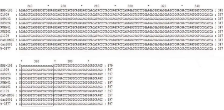
the prototype ALV-J strain HPRS-103. To determine whether the 19-nt insertion in the ALV-J leader sequence was a common and new molecular characteristic of the ALV-J genome, the sequences of other ALV-J isolates were collected and analyzed. As shown in Fig. 2, a comparison of the leader sequences revealed the emergence of a unique, naturally occurring 19-nt insertion in the ALV-J genome from China (Fig. 2). A 19-nt insertion (59 -GCGCGGTTCCGGTTGCTCT-39), located between nt 573 and 574 according to the HPRS-103 sequence, was present in the leader sequence. The leader sequence from 10 ALV-J isolates (including one classical ALV-J strain and nine Chinese isolates) were analyzed (Table 1).
Two viable viruses were rescued
Two viruses, rSD1009 (with the 19-nt insertion in the leader sequence) and rSD1009g19 (without the 19-nt insertion), were rescued using reverse genetics. An IFA was performed to identify the rescued viruses. The DF-1 cells infected with the rescued viruses were stained by monoclonal antibodies specific for ALV-J gp85. Primary antibody binding was detected using fluorescein isothiocyanate-labeled anti-mouse IgG, which exhibited green fluorescence (Fig. 3A and B). Electron microscopy revealed two viruses with diameters of approximately 90 nm in the ultrathin sections of rSD1009- and rSD1009g19-infected DF-1 cells (Fig. 3D and E). These data demonstrated that the rSD1009 and rSD1009g19 viruses were rescued.
The 19-nt insertion in the leader sequence enhanced leader enhancer activity and viral replication in DF-1 cells
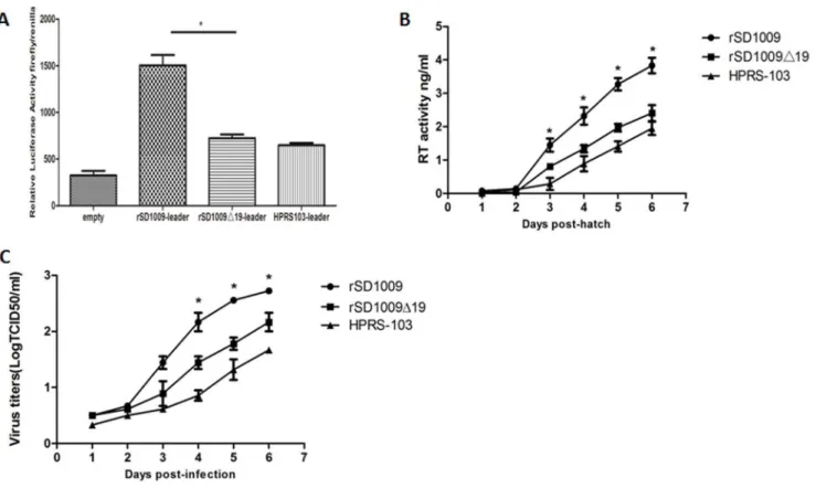
To explore whether the 19-nt insertion in the leader sequence influenced the enhancer activity of the leader sequence, we constructed a luciferase vector with a leader sequence with and without the 19-nt insertion. We also constructed a luciferase vector with the same leader sequence as HPRS-103. As shown in Fig. 4A, the results indicated a significant increase in the leader sequence constructs with the 19-nt insertion, which suggested that the 19-nt insertion in the leader region enhanced the leader enhancer activity.
To further explore whether the 19-nt insertion in the leader sequence conferred a growth advantage to the virus, DF-1 cells, which are commonly used for ALV-J proliferation [17], were used to evaluate in vitro replication of the rescued virus. DF-1 cell cultures were infected with equivalent amounts of rSD1009,
rSD1009D19, and HPRS-103. The culture supernatants were harvested periodically thereafter for reverse transcriptase (RT) activity assays. For the first 2 days, rSD1009 displayed growth curves similar to rSD1009D19 before the RT activity could be detected. Subsequently, rSD1009-infected cells produced more viral particles than those infected with rSD1009g19. By day 6, the number of viral particles of both strains peaked. The results revealed that rSD1009 had a moderate growth advantage over rSD1009D19 (Fig. 4B), suggesting that the 19-nt insertion enhanced ALV-J replication in DF-1 cells. Although RT activity can be used to determine retroviral replication [18], direct titration was performed to confirm replication of rSD1009, rSD1009D19 and HPRS-103. As shown in Fig. 4C, the direct titration of rSD1009 and rSD1009D19 was similar to the RT activity results and the 19-nt insertion-enhanced viral replication in DF-1 cells.
rSD1009 and rSD1009D19 have similar replication and pathogenicityin vivo
To evaluatein vivoreplication and pathogenicity of the rescued viruses, 1-day-old SPF layer chicks were intra-abdominally inoculated with rSD1009 and rSD1009. Compared to controls, most of the infected chickens gradually became emaciated as they grew and some became paralyzed by 3 months post-infection, which is a classical phenomenon in ALV-J-infected chickens [19]. To compare the pathogenic effects of the rescued viruses, infected chickens were observed for 238 days. Evidence of viral replication was apparent in these chickens via examination of serum samples 7 weeks post-infection [9], which confirmed neutralizing antibodies in 4/12 (33.33%) of the rSD1009-infected and 4/13 (30.76%) of the rSD1009g19-infected chickens. No significant differences in viral replication were observed between the rSD1009 and rSD1009g19 infection groups. These data demonstrated that the 19-nt insertion did not influence ALV-J replicationin vivo.
Levels of oncogenicity were determined from the incidence of histological tumors. In chickens infected with either of the viruses, there was only one chicken with an induced tumor. There was no difference in the incidence of tumors between chickens infected with rSD1009 (8.3%) and rSD1009g19 (7.6%). Details regarding the types of identified tumors are shown in Table 2. The clinical manifestations of the rSD1009- and rSD1009g19-induced tumors and the histological findings of those tumors (diagnosed as erythroblastosis) are shown in Fig. 5. PCR detection and sequencing also confirmed that all chickens with tumors had infections with the inoculated viruses (data not shown). Uninfected control chickens displayed no tumors.
Discussion
Since 2008, ALV-J infections in chickens have become widespread in China [6,20]. The genomic sequences of ALV-J epidemic isolates compared with HPRS-103, the ALV-J prototype virus, exhibits several distinct features. In this study, a unique 19-nt insertion in the leader sequence was identified. Thus, we investigated the function of the 19-nt sequence bothin vitro and
in vivoby construction and rescue of a pair of viruses (rSD1009 and rSD1009g19). The results indicated that the 19-nt insertion in the leader sequence of ALV subgroup J contributed to its replicationin vitro, but was not related to its pathogenicityin vivo.
It is important to monitor sequence changes in the leader sequence of ALV-J because the 5’ leader sequences of avian retroviruses contain functional elements involved in key steps of the retroviral life cycle [21]. A previous report indicated that ALV-J was prone to mutations that facilitate the relatively rapid
Table 1.ALV-J strains used in sequence analysis.
Isolate Origin Yr Accession no. Hosta
HPRS-103 UK 1988 Z46390 M
SD1009 China 2010 KF768000 CL
JS09GY3 Jiangsu, China 2009 GU982308 CL JS09GY6 Jiangsu, China 2009 GU982310 CL
CAUHM01 China 2009 JF932000 C
CAUXT01 China 2009 JF932003 C
GD1109 China 2012 JX254901 C
SCAU-HN06 China 2007 HQ900844 CL
sdau1001 China 2010 JN389517 CL
TW-3577 Taiwan 2013 HM582657 C
chicken; C, chicken.
doi:10.1371/journal.pone.0084797.t001
19nt Contributes to Replication of ALV-Jin Vitro
evolution of the virus [12]. A 19-nt insertion, observed in the leader sequence of ALV-J in this study, was naturally occurring, suggesting that it was the result of the natural evolution of the ALV-J genome.
We employed DF-1 cells, which are commonly used for propagating ALV-J [22], to evaluate in vitro replication of the rescued viruses. Our results indicated that rSD1009 replicated more rapidly than rSD1009g19 in DF-1 cells (Fig. 4B and C). In Figure 2. Sequence comparisons of the reference strains with the 19-nt insertion in the leader sequence. The 19-nt insertion in the leader sequence is enclosed in a black box.
doi:10.1371/journal.pone.0084797.g002
Figure 3. The identification of the rescued viruses by IFA and electron microscopy.(A) The DF-1 cells infected with rSD1009 using IFA mediated by monoclonal antibodies specific for ALV-J gp85 produced ALV-J-specific green fluorescence. (B) The DF-1 cells infected with rSD1009g19
using IFA mediated by monoclonal antibodies specific for ALV-J produced ALV-J-specific green fluorescence. (C) Uninoculated DF-1 cells served as a negative control. (D) Electron microscopy of the ultrathin sections of rSD1009-infected DF-1 cells revealed the virion. (E) Electron microscopy of ultrathin sections of rSD1009g19-infected DF-1 cells revealed the virion. The red arrows point to virion. Scale bar = 1mm.
addition, we also confirmed that the leader sequence with the 19-nt insertion enhanced protein expression using a luciferase assay (Fig. 4A). The ALV-J leader sequence has three unique open reading frames, which may be positioned to influence viral activities, such as expression of viral proteins [23]. The 19-nt insertion was found to influence viral replication in vitro, which may be the reason that the insertion changed the length of the leader sequence, thereby re-adjusting its structure and ultimately influencing the cis-acting characteristics of the leader sequence in the ALV-J strain.
Although the 19-nt insertion enhancedin vitroreplication, it did not appear to affect viral replicationin vivo, as demonstrated by the neutralizing-antibody response of infected chickens at 7 weeks of age (Table 2). This discrepancy between viral replicationin vitro
andin vivohas also been observed in other viruses, such as a single amino acid mutation in very virulent infectious bursal disease virus that could attenuate replicationin vivobut increased replicationin vitro[24]. The mechanism explaining why the 19-nt insertion had different effects on ALV-J replicationin vitro and in vivorequires further investigation.
Figure 4. The 19-nt insertion in the leader sequence enhanced activity of the leader sequence and viral replication in DF-1 cells.(A) Comparison of enhancer activity of the leader sequence of rSD1009, rSD1009g19, and HPRS-103 in constructs expressing a firefly reporter gene. The
Renilla luciferase gene was used as a reference gene for normalization of gene expression in transiently transfected cells. (B) The results of the RT assays performed on the virus particles isolated from the culture supernatants of DF-1 cells infected with each virus are shown. The growth curves were drawn by RT assays. (C) One-step growth curves for rSD1009, rSD1009g19, and HPRS-103 in DF-1 cells. The growth curves were drawn by
assaying the viral titers. The viral titers harvested at different intervals were calculated and expressed as TCID50per milliliter. The standard deviations
(error bars) from three independent experiments are shown. Significant differences between the rSD1009 and rSD1009g19 groups are indicated by
asterisks (*,p,0.05). Statistical analysis was performed by Student’s t test. doi:10.1371/journal.pone.0084797.g004
Table 2.In vivo infection and tumorigenesis in rSD1009- and rSD1009g19-infected groups
Virus Age at infection Infection status*(%) Details of tumors
SPF layer chicken SPF layer chicken
Tumor incidence(%) Tumor of type(n)
rSD1009 1-day-old chick 4/12(33.33) 1/12(8.33) EB(1)
rSD1009g19 1-day-old chick 4/13(30.76) 1/13(7.69) EB(1)
Uninfected control 1-day-old chick 0/13(0) 0/13(0) NA
Infection status is based on the results of neutralizing antibodies of serum samples 7 weeks after infection. NA, not applicable; erythroblastosis (EB). doi:10.1371/journal.pone.0084797.t002
19nt Contributes to Replication of ALV-Jin Vitro
There was no significant difference in the prevalence of tumors in chickens following infection with either rSD1009D19 or rSD1009. In addition, the oncogenic spectrum of the two viruses was very similar (Table 2). The oncogenicity of ALV-J is an important index of its pathogenicity [9,12]. These data strongly suggest that the 19-nt insertion in the leader sequence was not related to the pathogenicity of ALV-J in SPF layers, which is in agreement with the similar replication levels of rSD1009 and rSD1009g19 in vivo. A previous hypothesis suggested a link between in vivoreplication and pathogenicity and proposed that viral replication and pathogenicity were positively correlated [25]. Here, the similarin vivoreplication of rSD1009 and rSD1009g19 led to similar pathogenicity to the SPF layers, supporting the proposed hypothesis. Although a correlation between pathogenic-ity and effective viral replication has also been observed in other reports, including a study that linked the replication of Newcastle disease virus with its pathogenicity [26] and a second that revealed increased pathogenicity of avian influenza viruses with efficient viral replication [27], the molecular mechanisms mediating the relationship between the viral replication and pathogenicity remain unclear.
ALV-J-induced disease among chicken flocks in China has been surprising and generated a large amount of attention worldwide [5,6,28,29,30]. In the present study, molecular epidemiological research revealed that the 19-nt insertion, naturally occurring in ALV-J isolates from China, was a newly added molecular characteristic to the ALV-J genome. Using reverse genetics and animal experiments, here, we report the first evidence that the 19-nt insertion in the leader sequence of ALV-J co19-ntributed to the increased replication of ALV-Jin vitro, but was not related to its pathogenicityin vivo. This study not only demonstrated a unique molecular characteristic of ALV-J, but also generated data that may benefit the understanding of the relationships between variations in genomic sequences and the oncogenicity of ALV-J.
Author Contributions
Conceived and designed the experiments: QW XJ XW. Performed the experiments: XJ QW XL. Analyzed the data: QW XJ. Contributed reagents/materials/analysis tools: YW XQ HG. Wrote the paper: XJ QW YG XW.
References
1. Payne LN, Brown SR, Bumstead N, Howes K, Frazier JA, et al. (1991) A novel subgroup of exogenous avian leukosis virus in chickens. J Gen Virol 72 ( Pt 4): 8012807.
2. Payne LN, Howes K, Gillespie AM, Smith LM (1992) Host range of Rous sarcoma virus pseudotype RSV(HPRS-103) in 12 avian species: support for a new avian retrovirus envelope subgroup, designated J. J Gen Virol 73 ( Pt 11): 299522997.
3. Du Y, Cui Z, Qin A (1999) Subgroup J of avian leukosis viruses in China. China Poult Sci 3: 124.
4. Cheng ZQ, Zhang L, Liu SD, Zhang LJ, Cui ZZ (2005) Emerging of avian leukosis virus subgroup J in a flock of Chinese local breed]. Wei Sheng Wu Xue Bao 45: 5842587.
Figure 5. Clinical manifestations and histological examinations of tumors from chickens infected with rSD1009 and rSD1009g19. (A) The liver of an SPF layer infected with rSD1009. (B) The liver of an SPF layer infected with rSD1009g19. The red arrows point to clinical lesions. (C)
Histological examination of (A) liver; a large, well-demarcated, non-encapsulated nodule consisting of numerous erythroblasts (as shown on the right side). (D) Histological examination of (B) liver; a large, well-demarcated, non-encapsulated nodule consisting of numerous erythroblasts. All histological examinations used hematoxylin and eosin stain. Scale bar = 50mm.
5. Cheng Z, Liu J, Cui Z, Zhang L (2010) Tumors associated with avian leukosis virus subgroup J in layer hens during 2007 to 2009 in China. J Vet Med Sci 72: 102721033.
6. Gao YL, Qin LT, Pan W, Wang YQ, Qi X, et al. (2010) Avian leukosis virus subgroup J in layer chickens, China. Emerg Infect Dis 16: 163721638. 7. Bai J, Howes K, Payne LN, Skinner MA (1995) Sequence of host-range
determinants in the env gene of a full-length, infectious proviral clone of exogenous avian leukosis virus HPRS-103 confirms that it represents a new subgroup (designated J). J Gen Virol 76 ( Pt 1): 1812187.
8. Venugopal K (1999) Avian leukosis virus subgroup J: a rapidly evolving group of oncogenic retroviruses. Res Vet Sci 67: 1132119.
9. Chesters PM, Smith LP, Nair V (2006) E (XSR) element contributes to the oncogenicity of Avian leukosis virus (subgroup J). J Gen Virol 87: 268522692. 10. Payne LN, Gillespie AM, Howes K (1993) Unsuitability of chicken sera for detection of exogenous ALV by the group-specific antigen ELISA. Vet Rec 132: 5552557.
11. Tsukamoto K, Kono Y, Arai K, Kitahara H, Takahashi K (1985) An enzyme-linked immunosorbent assay for detection of antibodies to exogenous avian leukosis virus. Avian Dis 29: 111821129.
12. Wang Q, Gao Y, Wang Y, Qin L, Qi X, et al. (2012) A 205-nucleotide deletion in the 3’ untranslated region of avian leukosis virus subgroup J, currently emergent in China, contributes to its pathogenicity. J Virol 86: 12849212860. 13. Wang Q, Wang Y, Gao Y, Qin L, Qi X, et al. (2011) Isolation and identification molecular characterization of a subgroup J avian leukosis virus isolate SD1009. Chinese Journal of Preventive Veterinary Medicine 33: 5932597.
14. Qin A, Lee LF, Fadly A, Hunt H, Cui Z (2001) Development and characterization of monoclonal antibodies to subgroup J avian leukosis virus. Avian Dis 45: 9382945.
15. Deng X, Qi X, Wu G, Gao Y, Qin L, et al. (2012) Construction and characterization of the infectious clone of Reticuloendotheliosis virus carrying a genetic marker. Virus Res 167: 1462151.
16. Reed LJ, Muench H (1938) A simple method of estimating fifty percent endpoints. Am J Hygiene 27: 4932497.
17. Sacco MA, Howes K, Smith LP, Nair VK (2004) Assessing the roles of endogenous retrovirus EAV-HP in avian leukosis virus subgroup J emergence and tolerance. J Virol 78: 10525210535.
18. Ogert RA, Lee LH, Beemon KL (1996) Avian retroviral RNA element promotes unspliced RNA accumulation in the cytoplasm. J Virol 70: 383423843. 19. Lai H, Zhang H, Ning Z, Chen R, Zhang W, et al. (2011) Isolation and
characterization of emerging subgroup J avian leukosis virus associated with hemangioma in egg-type chickens. Vet Microbiol 151: 2752283.
20. Gao Y, Yun B, Qin L, Pan W, Qu Y, et al. (2012) Molecular epidemiology of avian leukosis virus subgroup J in layer flocks in China. J Clin Microbiol 50: 9532960.
21. Donze O, Spahr PF (1992) Role of the open reading frames of Rous sarcoma virus leader RNA in translation and genome packaging. EMBO J 11: 37472 3757.
22. Maas R, van Zoelen D, Oei H, Claassen I (2006) Replacement of primary chicken embryonic fibroblasts (CEF) by the DF-1 cell line for detection of avian leucosis viruses. Biologicals 34: 1772181.
23. Hackett PB, Swanstrom R, Varmus HE, Bishop JM (1982) The leader sequence of the subgenomic mRNA’s of Rous sarcoma virus is approximately 390 nucleotides. J Virol 41: 5272534.
24. Yu F, Ren X, Wang Y, Qi X, Song J, et al. (2013) A single amino acid V4I substitution in VP1 attenuates virulence of very virulent infectious bursal disease virus (vvIBDV) in SPF chickens and increases replication in CEF cells. Virology 440: 2042209.
25. Ebert D, Bull JJ (2003) Challenging the trade-off model for the evolution of virulence: is virulence management feasible? Trends Microbiol 11: 15220. 26. Dortmans JC, Rottier PJ, Koch G, Peeters BP (2010) The viral replication
complex is associated with the virulence of Newcastle disease virus. J Virol 84: 10113210120.
27. Suzuki K, Okada H, Itoh T, Tada T, Mase M, et al. (2009) Association of increased pathogenicity of Asian H5N1 highly pathogenic avian influenza viruses in chickens with highly efficient viral replication accompanied by early destruction of innate immune responses. J Virol 83: 747527486.
28. Nair LNPV ( 2012) The long view: 40 years of avian leukosis research. Avian Pathology 41: 11219.
29. Gao Y, Yun B, Qin L, Pan W, Qu Y, et al. (2011) Molecular epidemiology of avian leukosis virus subgroup J in layer flocks in China. J Clin Microbiol. 30. Sun H, Qin M, Xiao Y, Yang F, Ni W, et al. (2010) Haemangiomas,
leiomyosarcoma and myeloma caused by subgroup J avian leukosis virus in a commercial layer flock. Acta Vet Hung 58: 4412451.