Alterations in Perivascular Sympathetic and
Nitrergic Innervation Function Induced by
Late Pregnancy in Rat Mesenteric Arteries
Esther Sastre1,2☯‡, Javier Blanco-Rivero1,2☯‡, Laura Caracuel1,2, María Callejo1, Gloria Balfagón1,2
*
1Departamento de Fisiología, Facultad de Medicina, Universidad Autónoma de Madrid, Madrid, Spain, 2Instituto de Investigación Sanitaria IdiPAZ, Madrid, Spain
☯These authors contributed equally to this work. ‡These authors are joint first authors on this work.
Abstract
Background and Purpose
We investigated whether pregnancy was associated with changed function in components of perivascular mesenteric innervation and the mechanism/s involved.
Experimental Approach
We used superior mesenteric arteries from female Sprague-Dawley rats divided into two groups: control rats (in oestrous phase) and pregnant rats (20 days of pregnancy). Modifica-tions in the vasoconstrictor response to electrical field stimulation (EFS) were analysed in the presence/absence of phentolamine (alpha-adrenoceptor antagonist) or L-NAME (nitric oxide synthase-NOS- non-specific inhibitor). Vasomotor responses to noradrenaline (NA), and to NO donor DEA-NO were studied, NA and NO release measured and neuronal NOS (nNOS) expression/activation analysed.
Key Results
EFS induced a lower frequency-dependent contraction in pregnant than in control rats. Phentolamine decreased EFS-induced vasoconstriction in segments from both experimen-tal groups, but to a greater extent in control rats. EFS-induced vasoconstriction was in-creased by L-NAME in arteries from both experimental groups. This increase was greater in segments from pregnant rats. Pregnancy decreased NA release while increasing NO re-lease. nNOS expression was not modified but nNOS activation was increased by pregnan-cy. Pregnancy decreased NA-induced vasoconstriction response and did not modify DEA-NO-induced vasodilation response.
OPEN ACCESS
Citation:Sastre E, Blanco-Rivero J, Caracuel L, Callejo M, Balfagón G (2015) Alterations in Perivascular Sympathetic and Nitrergic Innervation Function Induced by Late Pregnancy in Rat Mesenteric Arteries. PLoS ONE 10(5): e0126017. doi:10.1371/journal.pone.0126017
Academic Editor:Marcia B. Aguila, State University of Rio de Janeiro, Biomedical Center, Institute of Biology, BRAZIL
Received:June 16, 2014
Accepted:March 27, 2015
Published:May 7, 2015
Copyright:© 2015 Sastre et al. This is an open access article distributed under the terms of the
Creative Commons Attribution License, which permits unrestricted use, distribution, and reproduction in any medium, provided the original author and source are credited.
Data Availability Statement:All relevant data are within the paper.
Funding:This study was supported by Ministerio de Economía y Competitividad (SAF2012-38530), Red RIC (RD12/0042/0024), and Fundación MAPFRE. E. Sastre received a FPI-UAM fellowship. L. Caracuel received a fellowship from Alianza 4 Universidades Program.
Conclusions and Implications
Neural control of mesenteric vasomotor tone was altered by pregnancy. Diminished sympa-thetic and enhanced nitrergic components both contributed to the decreased vasoconstric-tion response to EFS during pregnancy. All these changes indicate the selective
participation of sympathetic and nitrergic innervations in vascular adaptations produced during pregnancy.
Introduction
Pregnancy is associated with a decrease in systemic vascular resistance that, despite the marked increase in blood volume and cardiac output, maintains or reduces maternal blood pressure, in both experimental animals and humans. Adaptations to pregnancy have been studied in several vascular beds, but the mechanisms underlying the altered vessel function are complex and only partially understood.
Vascular adaptations to pregnancy include both an endothelium-dependent pathway asso-ciated with increased production of vasodilators [1] and an endothelium-independent pathway associated with altered vasomotor smooth muscle cell responses to different vasoactive sub-stances [2,3,4], that decrease myogenic reactivity [5] and increase vascular compliance [6]. However, activation of additional endothelium-independent pathways has been strongly sug-gested [7]. Perivascular innervation has a significant influence on peripheral vascular resistance involving the sympathetic, cholinergic, nitrergic, peptidergic and/or sensory innervations, which are specific to the vascular bed under consideration.
The mesenteric artery plays a pivotal role in global peripheral resistance in rats, especially in pregnancy; during this physiological process, mesenteric perfusion is strongly increased. These arteries are innervated by sympathetic nerves, which mediate vasoconstriction mainly via nor-adrenaline (NA) release, but also by nitrergic innervation, which induces vasodilatation by ni-tric oxide (NO) release, and sensory innervation through release of the vasodilator calcitonin gene-related peptide neuropeptide (CGRP) [8,9,10]. Electric field stimulation (EFS) produces a vasomotor response that is the integrated result of the effect of these different neurotransmit-ters [10]. The alterations in the functional roles of these components have been associated with changes in synthesis, release, response and/or metabolism of the different neurotransmitters in several physiological and pathological circumstances [11,12,13,14].
Neuronal adaptation to pregnancy by mesenteric arteries it has been reported to be time-de-pendent. In late pregnancy diminished sympathetic nerve-mediated constriction has been as-sociated with a decreased vasoconstrictor response to NA [7], while possible changes in NA release have been suggested but not investigated [4]. No changes have been reported in sensory innervation [4] but there is an increased vasodilation to CGRP [7,15,16]. It is widely known that estrogens modulate vascular tone activating endothelial nitric oxide synthase (eNOS) and several studies have reported that vascular adaptation in pregnancy is associated with an in-crease in eNOS protein expression [17,18,19]. In previous studies we have observed that changes in levels of sex steroids are associated with changes in nitrergic innervation function [14,20]. However, to the best of our knowledge, the possible role of nitrergic innervation in vas-cular adaptations to pregnancy remains unexplored.
and sensory innervations in late pregnancy could be associated with the decreased vascular re-sistance observed in the mesenteric artery, as well as the mechanisms that may be implicated.
Materials and Methods
Animals
Female Sprague-Dawley rats (4–6 months old) were obtained from the Animal Quarters and housed in the Animal Facility of the Universidad Autónoma de Madrid (registration number EX-021U) in accordance with guidelines 609/86 of the E.E.C., R.D. 233/88 of the Ministerio de Agricultura, Pesca y Alimentación of Spain, and theGuide for the Care and Use of Laboratory Animalspublished by the United States National Institute of Health [NIH publication No. 85– 23, revised 1996]. All experimental procedures involving animal use were approved by the Eth-ics Committee of the Universidad Autónoma de Madrid. Rats were housed at a constant room temperature, humidity, and light cycle (12:12 h light-dark) with free access to tap water and fed with standard rat chowad libitum, and were divided into two groups: Control (virgin females in oestrous phase) and pregnant rats (20 days of pregnancy). The stage of the oestrous cycle was determined by examination of vaginal smears; oestrous females exhibited the presence of corni-fied cells. The day when a vaginal plug was found was considered to be day 1 of pregnancy.
Animals were sacrificed by CO2inhalation followed by decapitation; superior mesenteric
ar-tery was carefully dissected, cleaned of connective tissue and placed in Krebs-Henseleit solution (KHS, in mmol/L: NaCl 115, CaCl22.5, KCl 4.6, KH2PO41.2, MgSO4.7H2O 1.2, NaHCO325;
glucose 11.1; Na2EDTA 0.03) at 4°C. For protein expression analysis, some segments were
rap-idly frozen in liquid nitrogen and kept at -80°C until the day of analysis.
Vascular reactivity
The method used for isometric tension recording has been described in full elsewhere [10,21]. Two parallel stainless steel pins were introduced through the lumen of the vascular segment: one was fixed to the bath wall, and the other connected to a force transducer (Grass FTO3C; Quincy, MA, USA); this, in turn, was connected to a model 7D Grass polygraph. For EFS ex-periments, segments were mounted between two platinum electrodes 0.5 cm apart and con-nected to a stimulator (Grass, model S44) modified to supply adequate current strength. Segments were suspended in an organ bath containing 5 mL of KHS at 37°C and continuously bubbled with a 95% O2to 5% CO2mixture (pH of 7.4). Some experiments were performed in
endothelium-denuded segments to eliminate the main source of vasoactive substances, includ-ing endothelial NO. This avoided possible actions by different drugs on endothelial cells that could lead to misinterpretation of results. Endothelium was removed by gently rubbing the lu-minal surface of the segments with a thin wooden stick. The segments were subjected to a ten-sion of 0.5 g, which was readjusted every 15 min during a 90-min equilibration period before drug administration. After this, vessels were exposed to 75 mmol/L KCl to check their func-tional integrity. After a washout period, the presence/absence of vascular endothelium was test-ed by the ability/inability of 10μmol/L acetylcholine (ACh) to relax segments precontracted
with NA (1μmol/L).
Vasodilator response to ACh (0.1 nmol/L- 10μmol/L) was tested in endothelium-intact
ar-teries precontracted with NA (1μmol/L) from all experimental groups.
neural origin of the EFS-induced contractile response, the nerve impulse propagation blocker, tetrodotoxin (TTX, 0.1μmol/L) was added to the bath 30 min before the second
frequency-re-sponse curve was performed.
To determine the participation of the adrenergic component of sympathetic innervation in EFS-induced responses in endothelium-denuded segments from control and pregnant rats, 1μmol/L phentolamine, an alpha-adrenoceptor antagonist was added to the bath 30 min
be-fore performing the frequency-response curve. Additionally, the vasoconstrictor response to exogenous NA (1 nmol/L- 10μmol/L) was tested in segments from both experimental groups.
The method to deplete sympathetic innervation has been used previously by our group in this artery [22]. Briefly, endothelium-denuded mesenteric segments from control and pregnant rats were incubated at room temperature for 10 minutes in KHS (NaHCO3 and NaH2PO4 were omitted, unbuffered solution) containing 0.02 mmol/L glutathione and 1.46 mmol/L of the neurotoxin 6-hydroxydopamine (6-OHDA). The pH of this solution was adjusted to 4.9 with 0.05 mmol/L NaOH and then the solution was covered with paraffin oil. Subsequently, the arteries were immersed in normal KHS and EFS-induced contraction experiments were performed. After EFS-contraction experiments mesenteric segments from control and preg-nant rats were exposed to 75 mmol/L KCl to check that their functional integrity was not af-fected by 6-OHDA.
To study the possible participation of sensory innervation in EFS-induced responses in en-dothelium-denuded segments from control and pregnant rats, 0.5μmol/L CGRP (8–37), a
CGRP receptor antagonist was added to the bath 30 min before performing the second fre-quency-response curve.
To analyse the participation of NO in the EFS-induced response in endothelium-denuded segments from control and pregnant rats, 0.1 mmol/L Nω
-nitro-L-arginine methyl ester (L-NAME), a non-specific inhibitor of nitric oxide synthase (NOS), was added to the bath 30 min before performing the second frequency-response curve. The vasodilator response to the NO donor, diethylamine NONOate (DEA-NO, 0.1 nmol/L-0.1 mmol/L) was determined in NA-precontracted arteries from both experimental groups. To assess the participation of super-oxide anion in DEA-NO-induced vasodilation, 0.1 mmol/L tempol, a supersuper-oxide anion scaven-ger, was added to the bath before the frequency-response curve to DEA-NO was performed.
Noradrenaline release
Endothelium-denuded mesenteric segments from control and pregnant rats were preincubated for 30 min in 5 mL of KHS at 37°C and continuously gassed with a 95% O2-5% CO2mixture
(stabilisation period). This was followed by two washout periods of 10 min in a bath of 0.4 mL of KHS. Then, the medium was collected to measure basal release. Next, the organ bath was re-filled, and cumulative EFS periods of 30 s at 1, 2, 4 and 8 Hz were applied at 1 min intervals. Af-terwards, the medium was collected to measure EFS-induced neurotransmitter release. The EFS-induced NA release was calculated by subtracting basal NA release from that evoked by EFS. Mesenteric segments were weighed in order to normalise the results.
NA release was measured using the Noradrenaline Research EIA (Labor Diagnostica Nord, Gmbh and Co., KG, Nordhon, Germany). The assay was performed following the manufactur-er’s instructions. Results were expressed as ng NA/mL mg tissue.
Nitric Oxide release
NaCl 119; HEPES 20; CaCl21.2; KCl 4.6; MgSO41; KH2PO40.4; NaHCO35; glucose 5.5;
Na2HPO40.15; pH 7.4) at 37°C. Arteries were incubated with 2μmol/L DAF-2 for 30 min. The
medium was then collected to measure basal NO release. Once the organ bath was refilled, cu-mulative EFS periods of 30 s at 1, 2, 4 and 8 Hz were applied at 1 min intervals. Afterwards, the medium was collected to measure EFS-induced NO release. The fluorescence of the medium was measured at room temperature using a spectrofluorometer (LS50 Perkin Elmer Instru-ments, FL WINLAB Software, Whaltmann, MA, USA) with excitation wavelength set at 492 nm and emission wavelength at 515 nm. Mesenteric segments were weighed in order to nor-malize the results.
The EFS-induced NO release was calculated by subtracting basal NO release from that evoked by EFS. Also, blank samples were collected in the same way from segment-free medium in order to subtract the background emission. Some assays were performed in the presence of 0.1 mmol/L L-NAME, 0.1 mmol 7-nitroindazol (7-NI), the specific nNOS inhibitor, 1μmol/L
1400W, the specific iNOS inhibitor, or 0.1μmol/L TTX. The amount of NO released was
ex-pressed as arbitrary units/mg tissue.
Detection of O
2.-O2.-levels were measured using lucigenin chemiluminescence [24]. Endothelium-denuded
mesenteric segments from control and pregnant rats were rinsed in KHS for 30 min, equilibrat-ed for 30 min in HEPES buffer at 37°C, transferrequilibrat-ed to test tubes that containequilibrat-ed 1 mL HEPES buffer (pH 7.4) with lucigenin (5μmol/L) and then kept at 37°C. The luminometer was set to
report arbitrary units of emitted light; repeated measurements were collected for 5 min at 10 s intervals and averaged. 4,5-Dihydroxy-1,3-benzene-disulphonic acid“Tiron”(10 mmol/L), a cell-permeant, non-enzymatic O2.-scavenger, was added to quench the O2.-dependent
chemilu-minescence. Some segments were preincubated with 0.3 mmol/L apocinin, a NADPH oxidase inhibitor, or 0.1 mmol/L allopurinol, a xanthine oxidase inhibitor. Also, blank samples were collected in the same way without mesenteric segments to subtract background emission.
nNOS and P-nNOS Expression
Western blot analysis of nNOS and phosphorylated nNOS (P-nNOS) expression was per-formed in endothelium-denuded mesenteric segments from control and pregnant rats, as pre-viously described [23,24]. For these experiments, we used mouse monoclonal nNOS antibody (1:1000, Abcam, Cambridge, UK), rabbit polyclonal phospho1417-nNOS antibody (1:1000, Abcam, Cambridge, UK), and monoclonal anti-β-actin-peroxidase antibody (1:50000, Sigma-Aldrich, Spain). Rat brain homogenates were used as a positive control.
Drugs used
L-NA hydrochloride, ACh chloride, diethylamine NONOate diethylammonium salt, CGRP (8–37), TTX, L-NAME hydrochloride, 7-NI, 1400W, phentolamine, apocinin, allopurinol, luci-genin, tiron, tempol and DAF-2 (Sigma-Aldrich, Madrid, Spain) were used. Stock solutions (10 mmol/L) of drugs were made in distilled water, except for NA, which was dissolved in a NaCl (0.9%)-ascorbic acid (0.01% w/v) solution. These solutions were kept at -20°C and appropriate dilutions were made in KHS on the day of the experiment.
Data analysis
induced by ACh or DEA-NO was expressed as a percentage of the initial contraction elicited by NA (Control: 1373 ± 178.4 mg; pregnant: 1366 ± 126.5 mg; P>0.05). For
concentration-re-sponse curves, non-linear regression and Emax and—log EC50 were performed. Results are given as mean ± S.E.M. Statistical analysis was done by comparing the curve obtained in the presence of the different substances with the previous or control curve by means of repeated measure analysis of variance (ANOVA) followed by the Bonferronipost-hoctest using Graph-Pad Prism 5.0 software (CA, USA). Some results were expressed as differences of area under the curve (dAUC) for EFS obtained in segments from control and pregnant rats. AUC were cal-culated from the individual concentration-response plots. For dAUC, NO, O2.-and NA release
data, the statistical analysis was done using one-way ANOVA followed by Newman-Keuls’
post-hoctest. P<0.05 was considered significant.
Results
Vasomotor response to KCl
In endothelium-intact mesenteric segments, the vasoconstrictor response to 75 mmol/L KCl was similar in mesenteric segments from control and pregnant rats (Control: 1421 ± 114 mg; pregnant; 1538 ± 67.3 mg; P>0.05). Endothelium removal did not alter KCl-induced
vasocon-striction (Control: 1677 ± 63.5 mg; pregnant: 1788 ± 54 mg; P>0.05).
Vasodilator response to ACh
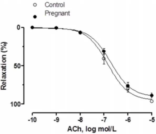
Vasodilator response to ACh was similar in NA-precontracted segments from both experimen-tal groups (Fig 1,Table 1).
Vascular responses to EFS
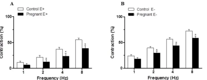
The application of EFS induced a frequency-dependent contractile response in both endotheli-um-intact and endothelium-denuded mesenteric segments from both experimental groups. This vasoconstriction was lower in segments from pregnant rats compared to control animals (Fig2Aand2B). Endothelium removal increased the EFS-induced contractile response to a similar extent in segments from control and pregnant rats (Fig2Aand2B,Table 2). EFS-in-duced contractions were practically abolished by preincubation with neurotoxin TTX (0.1μmol/L), indicating the neuronal origin of the factors inducing this response (Table 3).
Effect of pregnancy on the sympathetic component of vascular
responses to EFS in endothelium-denuded mesenteric segments
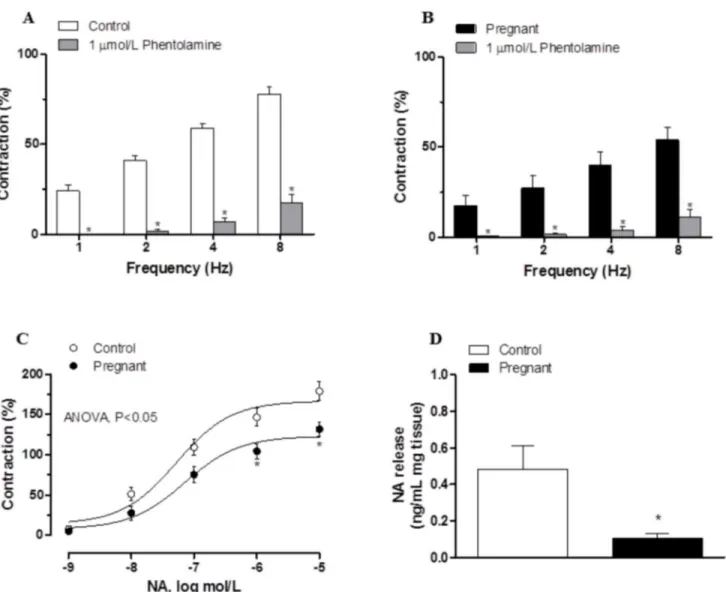
Preincubation with the alpha-adrenergic antagonist phentolamine (1μmol/L) decreased the
vasoconstrictor response induced by EFS in segments from both experimental groups (Fig3A
and3B). This decrease was lower in arteries from pregnant rats (Table 4). The phentolamine-resistant contractile response to EFS was lower in segments from pregnant rats (Table 5). Pre-incubation with 6-OHDA practically abolished the EFS-induced contraction in segments from control and pregnant rats (Table 3). After 6-OHDA preincubation, vasomotor response to KCl was similar in mesenteric segments from both experimental groups (Control: 1501 ± 95.4 mg; pregnant; 1622 ± 109 mg; P>0.05).
The contractile response elicited by exogenous NA (1 nmol/L-10μmol/L) was reduced in
Effect of pregnancy on the sensory component of vascular responses to
EFS in endothelium-denuded mesenteric segments
Preincubation with the CGRP receptor antagonist CGRP (8–37) (0.5μmol/L) did not alter the
EFS-induced contraction in any experimental group, indicating that sensory innervation did not contribute to the observed effects (Fig4Aand4B).
Fig 1. Vasodilator response to ACh.ACh-induced vasodilation in endothelium-intact mesenteric segments from control and pregnant rats. Results (mean
±SEM) were expressed as a percentage of the previous tone elicited by exogenous NA. n = 6 animals each group.
doi:10.1371/journal.pone.0126017.g001
Table 1. Emax(%) and log EC50(mmol/L) values of vasodilator responses to Ach and DEA-NO or vasoconstrictor responses to NA in mesenteric
ar-teries from control and pregnant rats.
Control Pregnant
Emax -logEC50 Emax -logEC50
ACh 95.09±3.07 6.84±0.08 90.58±2.76 6.72±0.07
NA 167.4±10.2 7.26±0.12 129.7±8.44* 7.23±0.2
DEA-NO 101.8±2.11 6.83±0.06 94.27±3.15 7.21±0.11
Results are expressed as mean±SEM.
*P<0.05 vs. Control. n = 7 animals each group.
Effect of pregnancy on the nitrergic component of vascular responses to
EFS in endothelium-denuded mesenteric segments
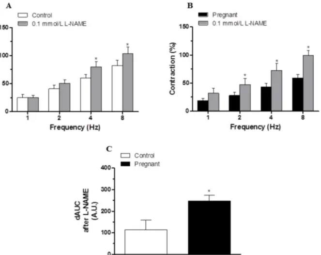
Preincubation with the unspecific NOS inhibitor L-NAME (0.1 mmol/L) significantly in-creased the EFS-contractile response at all frequencies in segments from control and pregnant rats (Fig5Aand5B). This increase was greater in arteries from pregnant animals (Fig 5C). Fig 2. Vasoconstrictor response to EFS.EFS-induced vasoconstriction in endothelium intact (A) and endothelium-denuded (B) mesenteric segments from control and pregnant rats. Results (mean±SEM) were expressed as a percentage of the initial contraction elicited by KCl. ANOVA P<0.05 Control vs. pregnant in both endothelium intact (A) and endothelium-denuded (B).*P<0.05 vs. control animals at each frequency (Bonferroni test). n = 10 animals each group.
doi:10.1371/journal.pone.0126017.g002
Table 2. EFS potentiation after endothelium removal in superior mesenteric artery from control and pregnant rats.
1 Hz 2 Hz 4 Hz 8 Hz
Control 12.51±2.61 18.19±2.79 19.38±3.05 17.23±3.32
Pregnant 11.55±2.92 17.39±3.85 20.26±4.37 19.79±4.26
Percentage (%) of potentiation in EFS-induced contraction after endothelium removal. Calculations are performed taking KCl-induced contraction as 100% of the contractile response. Results are expressed as mean±SEM. n = 10 animals each group.
doi:10.1371/journal.pone.0126017.t002
Table 3. Effect of preincubation with tetrodotoxin (TTX, 0.1μmol/L) or 6-hydroxydopamine (6-OHDA, 1.46 mmol/L) on the frequency—contraction curves performed in mesenteric segments of control and pregnant rats.
1 Hz 2 Hz 4 Hz 8 Hz
Control 21.31±2.23 34.10±2.56 52.41±2.22 69.28±3.91
TTX 0 0 0 0.5±0.06
6-OHDA 0 0 0.1±0.02 0.2±0.04
Pregnant 16.42±2.72 25.9±3.58 39.28±4.04 53.75±3.99
TTX 0 0 0 0.4±0.05
6-OHDA 0 0 0.1±0.01 0.3±0.05
Results (means±S.E.M.) are expressed as percentages of the response elicited by 75 mM KCl; zeros are used when contraction was not detected. n = 7 animals each group.
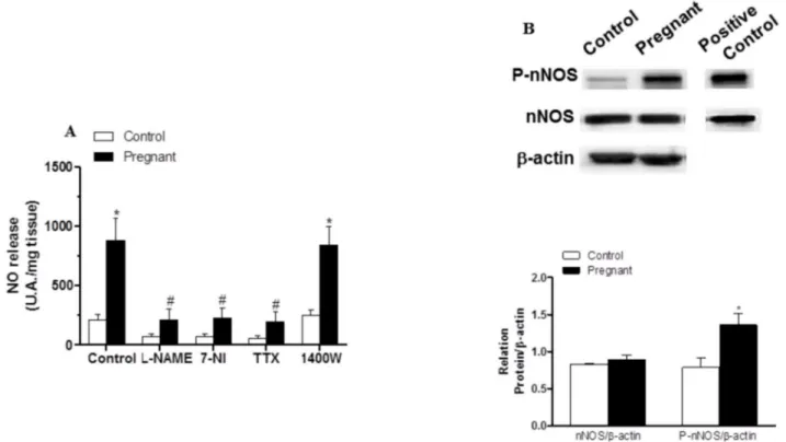
EFS-induced NO release was significantly greater in segments from pregnant rats (Fig 6A). Preincubation with 0.1 mmol L-NAME, 0.1 mmol 7-NI or 0.1μmol/L TTX abolished
EFS-in-duced NO release in both experimental groups (Fig 6A), while preincubation with 1μmol/L
1400W did not modify this release (Fig 6A). Pregnancy did not modify nNOS expression but did increase P-nNOS expression (Fig 6B).
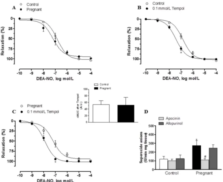
In segments precontracted with NA, vasodilator response elicited by the exogenous NO donor DEA-NO (0.1 nmol/L- 10μmol/L) was similar in mesenteric segments from both
exper-imental groups (Fig 7A,Table 2). Preincubation with 0.1 mmol/L tempol augmented DEA-NO-induced vasodilation similarly in segments from control and pregnant rats (Fig7B
and7C). On the other hand, after subtracting the lucigenin chemiluminescence obtained in the presence of tiron from that obtained in its absence, superoxide anion formation was greater in Fig 3. Effect of pregnancy on sympathetic innervation function.Effect of preincubation with 1μmol/L phentolamine on vasonstriction response induced by EFS in endothelium-denuded mesenteric segments from control (A) and pregnant rats (B). Results (mean±SEM) were expressed as a percentage of the
initial contraction elicited by KCl. ANOVA P<0.05 vs. conditions without phentolamine in both experimental groups.*P<0.05 vs. conditions without
phentolamine at each frequency (Bonferroni test). n = 8 animals per group. (C) Vasoconstriction response to NA in segments of control and pregnant rats. Results (mean±SEM) were expressed as a percentage of the initial contraction elicited by KCl. ANOVA P<0.05 Control vs. pregnant.*P<0.05 vs. control animals at each concentration (Bonferroni test). n = 8 animals per group. (D) EFS-induced NA release in mesenteric segments of control and pregnant rats. Results expressed as ng NA/mL mg tissue.*P<0.05 vs. Control. n = 6 animals per group.
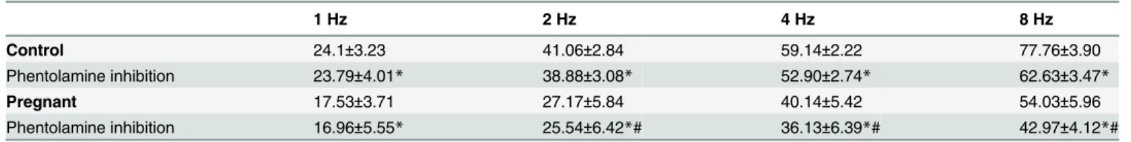
Table 4. Effect of preincubation with phentolamine (0.1μmol/L) on the frequency—contraction curves performed in mesenteric segments from control and pregnant rats.
1 Hz 2 Hz 4 Hz 8 Hz
Control 24.1±3.23 41.06±2.84 59.14±2.22 77.76±3.90
Phentolamine inhibition 23.79±4.01* 38.88±3.08* 52.90±2.74* 62.63±3.47*
Pregnant 17.53±3.71 27.17±5.84 40.14±5.42 54.03±5.96
Phentolamine inhibition 16.96±5.55* 25.54±6.42*# 36.13±6.39*# 42.97±4.12*# Percentage (%) of inhibition in EFS-induced contraction after preincubation with phentolamine. Calculations are performed taking KCl-induced contraction as 100% of the contractile response. Results are expressed as mean±SEM.
*P<0.05 vs. conditions without phentolamine at each frequency (Bonferroni test).
#P<0.05 vs. Control.
n = 8 animals per group.
doi:10.1371/journal.pone.0126017.t004
Table 5. Remnant vasoconstriction after preincubation with phentolamine (0.1μmol/L) on the frequency—contraction curves performed in mesen-teric segments of control and pregnant rats.
1 Hz 2 Hz 4 Hz 8 Hz
Control 24.1±3.23 41.06±2.84 59.14±2.22 77.76±3.90
Phentolamine Remnant 0.40±0.19* 2.17±0.88* 7.98±1.38* 18.35±5.54*
Pregnant 17.53±3.71 27.17±5.84 40.14±5.42 54.03±5.96
Phentolamine Remnant 0.61±0.4* 1.63±0.56* 3.72±2.06*# 9.97±3.63*#
Results (means±S.E.M.) are expressed as percentages of the response elicited by 75 mM KCl.
*P<0.05 vs. conditions without phentolamine at each frequency (Bonferroni test).
#P<0.05 vs. Control.
n = 8 animals per group.
doi:10.1371/journal.pone.0126017.t005
Fig 4. Effect of pregnancy on sensory innervation.Effect of preincubation with 0.5μmol/L CGRP (8–37) on the vasoconstrictor response induced by EFS in mesenteric segments from (A) control and (B) pregnant rats. Results (mean±SEM) are expressed as a percentage of the previous contraction elicited by
KCl. n = 8 animals per group.
arteries from pregnant rats (Fig 7D). Preincubation with apocinin decreased superoxide anion formation significantly in mesenteric segments of pregnant rats, while allopurinol did not modify this formation in any experimental group (Fig 7D)
Discussion
The present study shows that pregnancy diminished the vasoconstrictor response due to stimu-lation of perivascular innervation in mesenteric artery through an endothelium-independent mechanism. This effect is associated with a reduction in sympathetic innervation function me-diated by a decreased NA release and vasoconstrictor response and an increased nitrergic func-tion associated with increased neuronal NO release. Sensory innervafunc-tion did not participate in any experimental conditions.
When analyzing the vasoconstrictor response induced by EFS in endothelium-intact seg-ments, we observed that EFS produced a frequency-dependent contraction in segments from both control and pregnant rats that was lower in segments from pregnant rats, as previously re-ported [4,25,26]. The fact that in our experimental conditions the vasoconstrictor response to Fig 5. Effect of pregnancy on nitrergic innervation.Effect of preincubation with 0.1 mmol/L L-NAME on the vasoconstrictor response induced by EFS in mesenteric segments from (A) control and (B) pregnant rats. Results (mean±SEM) are expressed as a percentage of the previous contraction elicited by KCl.
ANOVA P<0.05 vs. conditions without L-NAME in both experimental groups.*P<0.05 vs. conditions without L-NAME for each frequency (Bonferroni test). n = 8 animals per group. (C) Differences of area under curve (dAUC) in the absence or presence of 0.1 mmol/L L-NAME, dAUC values are expressed as arbitrary units.*P<0.05 Control vs. pregnant rats.
KCl remained unmodified by pregnancy, as has been previously reported in this artery [4,27], indicates the maintenance of vasoconstrictor capacity, ruling out possible changes in the intrin-sic contractile machinery of smooth muscle cells.
It has been widely described that endothelium affects the response to several vasomotor sub-stances, including neurotransmitters [28], and since pregnancy could affect the production of endothelial factors [29] it would consequently affect these responses. In our experimental con-ditions ACh-induced vasodilation was not modified by pregnancy as also reported by Ralevic and Burnstock at late-term pregnancy [4]. Endothelium removal increased vasoconstrictor re-sponse to EFS similarly in both groups, indicating that the modulating role of endothelium in the EFS vasoconstrictor response is unaltered by pregnancy so the decreased contractile re-sponse would be due to modifications in perivascular innervation function produced by an en-dothelium-independent mechanism. Taking into account these results, we performed the following experiments only in endothelium-denuded mesenteric segments. The abolishment of EFS-induced vasoconstriction in the presence of TTX indicates that the response to EFS is due to neurotransmitter release. Sympathetic and nitrergic innervation involvement in the vasomo-tor response to EFS in rat superior mesenteric artery has been widely reported [9,10], while sen-sory innervation participation only appears in several pathological situations [13,30,31,32,33].
A decreased participation of sympathetic innervation in splanchnic blood flow as an adapta-tion to pregnancy has been demonstrated previously and associated with decreased vasocon-strictor response to NA [7]. However the possibility that the NA release was also affected by Fig 6. Effect of pregnancy on neuronal NO synthesis.(A) Effect of 0.1 mmo/L L-NAME or 0.1 mmol/L 7-NI or 0.1μmol/L TTX on EFS-induced NO release in segments from control and pregnant rats. Results (mean±SEM) were expressed as arbitrary (A.U.)/mg tissue.*P<0.05 vs. Control; #P<0.05 compared with conditions without specific inhibitor; n = 8 animals per group. (B) Effect of pregnancy on nNOS and P-nNOS expression. The blot is representative of four separate segments from each group. Rat brain homogenates were used as a positive control. Lower panel shows relation between P-nNOS or nNOS expression andβ-actin. Results (mean±SEM) are expressed as ratio of the signal obtained for each protein and the signal obtained forβ-actin.*P<0.05
Control vs. pregnant rats.
pregnancy has been also suggested but not measured [3,4]. Preincubation with the alpha-adre-noceptor antagonist phentolamine significantly diminished the vasoconstrictor response to EFS in mesenteric segments from both experimental groups. This decrease was less marked in mesenteric segments from pregnant rats, indicating a decreased participation by the adrenergic component from sympathetic innervation in the lower vasoconstrictor response to EFS in pregnant rats, similarly to results described by Ralevic and Burnstock [4]. The differences in the vasoconstrictor response to EFS could be associated with changes in neurotransmitter re-lease and/or vasomotor response to NA. The results obtained demonstrate that late pregnancy decreases the vasoconstrictor response to NA and simultaneously decreases NA release in mesenteric artery. It is well known that there is a decrease in vasomotor responses to contractile Fig 7. Effect of pregnancy on DEA-NO vasodilation and superoxide anion production.(A) Vasodilator response to NO donor DEA-NO in segments from control and pregnant rats. Effect of preincubation with tempol (0.1 mmol/L) on vasodilator response to NO donor DEA-NO in segments from (B) control and (C) pregnant rats. Results (mean±SEM) are expressed as a percentage of the previous tone elicited by exogenous NA. ANOVA P<0.05 vs. conditions
without tempol in both experimental groups.*P<0.05 vs. conditions without tempol for each concentration (Bonferroni test). n = 7 animals per group. Insert
graph shows differences of area under curve (dAUC) in the absence or presence of 0.1 mmol/L tempol; dAUC values are expressed as arbitrary units. (D) Effect of 0.3 mmol/L apocinin or 0.1 mmol/L allopurinol on superoxide anion release in mesenteric segments from control and pregnant rats. Results (mean
±SEM) are expressed as chemoluminiscence units (U)/min mg tissue.*P<0.05 vs. Control; #P<0.05 compared with conditions without specific inhibitor; n = 7
animals per group.
substances in pregnancy, although the mechanisms implicated are not well known. No changes in alpha-adrenoceptor affinity in pregnancy [34], decreased alpha(1)-adrenoceptor expression [35] and modifications in the regulation of Ca(2+) mobilization and Ca(2+) sensitivity in alpha (1)-adrenoceptor-mediated contractions [36] have all been described. While the decrease in NA release could be associated with changes in sympathetic nerve density due to estrogens, previous studies have report that pregnancy produces no change [37], increased [38] and de-creased [39] sympathetic nerve density, depending on the tissue studied. We have reported that the remnant vasoconstriction after preincubation with phentolamine is due to the release of sympathetic cotransmitter ATP in mesenteric segments from female rats [14]. Preincubation with the neurotoxine 6-OHDA practically abolished EFS-induced contraction in both experi-mental groups while the remnant vasoconstriction after preincubation with phentolamine was decreased in mesenteric segments of pregnant rats, suggesting a minor role for ATP in late pregnancy. The diminished sympathetic innervation function could, by itself, explain the de-creased EFS-induced vasoconstriction observed in mesenteric segments from pregnant rats. However, the participation of other neural components cannot be ruled out.
We have previously reported that sensory innervation does not participate in vasomotor re-sponse induced by EFS in control Sprague Dawley rats [14]. However, the participation of this innervation is increased in several pathological conditions such as hypertension, cirrhosis and diabetes [20,30,31,32]. Important variations in serum levels of the vasodilator CGRP have been reported in humans and rats during pregnancy [40,41]. Additionally, in rat mesenteric arteries, both early and late pregnancy are associated with a rise in the vascular sensitivity to CGRP in selected areas of the vascular bed without a concomitant increase in vascular CGRP production and release [15,16,42]. This observation led us to study the possible role of sensory innervation in the decreased EFS vasoconstriction observed in pregnant rats. Preincubation with the CGRP receptor antagonist CGRP (8–37) did not alter EFS-induced vasoconstriction in any experi-mental group, indicating that in late pregnancy sensory innervation does not participate in the vasoconstrictor response to EFS. This result contrasts with those of Ralevic and Burnstock [4], who observed a participation of sensory innervation in the EFS-mediated relaxation, and this response was not modified by pregnancy. Nevertheless, Lanluaet al. [43] reported no effect from CGRP (8–37) in the EFS-mediated relaxation of small mesenteric arteries in pregnancy.
TTX in both experimental groups, confirming that EFS induced NO release from nitrergic in-nervation. Additionally, in the present study, the specific iNOS inhibitor 1400W did not change EFS-induced NO release in segments from the two experimental groups, ruling out the partici-pation of this inducible isoform in our experimental approach, as previously reported [22,46].
The differences in NO release observed in our experimental conditions could be dependent on nNOS expression and/or activity. We found that nNOS protein expression was not modi-fied by pregnancy, whereas P-nNOS, the active form of nNOS was increased, suggesting in-creased nNOS activity. Estrogens are reported to regulate constitutive nitric oxide synthases and this regulation could be implicated in changes occurring in pregnancy [47]. Additionally Martiniet al. [48] have reported an increase in nNOS expression correlated with increased fe-male sex hormone levels, suggesting a complex action by fefe-male sex hormones in nNOS ex-pression; therefore the increased nNOS activity could be due to the estrogens as has been reported previously [49].
Thus, increased NO release participates in the diminished response to EFS in segments from pregnant rats. However, altered smooth muscle cell sensitivity to NO cannot be ruled out since measurements of oxidative stress markers in maternal blood and urine show that preg-nancy itself is a state of oxidative stress due to the high metabolic activity of the placenta and maternal metabolism [50], and this could alter NO bioavailability. First, we confirmed that su-peroxide anion production is increased in mesenteric segments from pregnant rats. Oxidative stress in pregnancy is regulated differently depending on the organ analysed [51]. It is well known that NADPH oxidase is the main source of superoxide anion in vessels [52]. Taking this into account, we have measured superoxide anion production in the presence of the NADPH oxidase inhibitor apocinin and the xanthine oxidase inhibitor allopurinol. Preincuba-tion with apocinin significantly decreased superoxide anion producPreincuba-tion only in mesenteric seg-ments of pregnant rats, but allopurinol did not modify superoxide anion production in any experimental group. These results confirm NADPH oxidase as the principal source of oxidative stress in superior mesenteric artery in late pregnancy. However, the vasodilator response to DEA-NO was similar in segments from both experimental groups. Additionally, preincubation with the superoxide anion scavenger tempol increased vasodilator response to DEA-NO to a similar extent in mesenteric segments from both control and pregnant rats. These results sug-gest that, despite the increased superoxide anion levels observed, the oxidation process is un-able to affect the NO metabolism enough to significantly affect vasodilator response.
In conclusion, our results show that neural control of mesenteric vasomotor tone was al-tered by pregnancy. Diminished sympathetic and enhanced nitrergic components both con-tributed to a decreased vasoconstriction response to EFS. The sensory innervation did not participate in these modifications. All these changes indicate the simultaneous implication of different components of perivascular innervation in vascular adaptations to pregnancy.
Author Contributions
Conceived and designed the experiments: ES JBR LC GB. Performed the experiments: ES JBR LC MC. Analyzed the data: ES JBR LC MC GB. Contributed reagents/materials/analysis tools: JBR GB. Wrote the paper: ES JBR LC GB.
References
1. Gerber RT, Anwar MA, Poston L. Enhanced acetylcholine induced relaxation in small mesenteric arter-ies from pregnant rats: an important role for endothelium-derived hyperpolarizing factor (EDHF). Br J Pharmacol 1998; 125: 455–460. PMID:9806327
3. Crandall ME, Keve TM, McLaughlin MK. Characterization of norepinephrine sensitivity in the maternal splanchnic circulation during pregnancy. Am J Obstet Gynecol. 1990; 162: 1296–1301. PMID:
2339731
4. Ralevic V, Burnstock G. Mesenteric arterial function in the rat in pregnancy: role of sympathetic and sensory-motor perivascular nerves, endothelium, smooth muscle, nitric oxide and prostaglandins. Br J Pharmacol 1996; 117: 1463–1470. PMID:8730740
5. Learmont JG, Cockell AP, Knock GA, Poston L. Myogenic and flow-mediated responses in isolated mesenteric small arteries from pregnant and nonpregnant rats. Am J Obstet Gynecol 1996; 174: 1631– 1636. PMID:9065143
6. Hohmann M, McLaughlin MK, Künzel W. Direct assessment of mesenteric vein compliance in the rat during pregnancy. Z Geburtshilfe Perinatol 1992; 196: 33–40. PMID:1549918
7. van Drongelen J, Hooijmans CR, Lotgering FK, Smits P, Spaanderman ME. Adaptive changes of mesen-teric arteries in pregnancy: a meta-analysis. Am J Physiol Heart Circ Physiol 2012; 303: 639–657. 8. Kawasaki H, Takasaki K, Saito A, Goto K. Calcitonin gene related peptide acts as a novel vasodilator
neurotransmitter in mesenteric resistance vessels of the rat. Nature 1988; 335: 164–167. PMID:
2901042
9. Li YJ, Duckles SP. Effect of endothelium on the actions of sympathetic and sensory nerves in the per-fused rat mesentery. Eur J Pharmacol 1992; 210: 23–30. PMID:1376271
10. Marín J, Balfagón G. Effect of clenbuterol on non-endothelial nitric oxide release in rat mesenteric arter-ies and the involvement of beta-adrenoceptors. Br J Pharmacol 1998; 124: 473–478. PMID:9647470
11. Loesch A. Perivascular nerves and vascular endothelium: recent advances. Histol Histopathol 2002; 17: 591–597. PMID:11962760
12. Ferrer M, Sánchez M, Minoves N, Salaices M, Balfagón G. Aging increases neuronal nitric oxide re-lease and superoxide anion generation in mesenteric arteries from spontaneously hypertensive rats. J Vasc Res 2003; 40: 509–519. PMID:14646371
13. Sastre E, Márquez-Rodas I, Blanco-Rivero J, Balfagón G. Perivascular innervation of the superior mesenteric artery: pathophysiological implications. Rev Neurol 2010; 50: 727–737. PMID:20533251
14. Blanco-Rivero J, Sastre E, Caracuel L, Granado M, Balfagón G. Breast feeding increases vasoconstric-tion induced by electrical field stimulavasoconstric-tion in rat mesenteric artery. Role of neuronal nitric oxide and ATP. PLoS One 2013; 8: e53802. doi:10.1371/journal.pone.0053802PMID:23342008
15. Yallampali C, Chauhan M, Thota CS, Kondapaka S, Wimalawansa SJ. Calcitonin gene-related peptide in pregnancy and its emerging receptor heterogeneity. Trends Endocrinol Metab 2002; 13: 263–269. PMID:12128288
16. Gangula PR, Lanlua P, Bukoski RD, Wimalawansa SJ, Yallampalli C. Mesenteric arterial relaxation to calcitonin gene-related peptide is increased during pregnancy and by sex steroid hormones. Biol Reprod 2004; 71:1739–1745. PMID:15286037
17. Goetz RM, Morano I, Calovini T, Studer R, Holtz J. Increased expression of endothelial constitutive ni-tric oxide synthase in rat aorta during pregnancy. Biochem Biophys Res Commun 1994; 205: 905– 910. PMID:7528018
18. Xu DL, Martin PY, St John J, Tsai P, Summer SN, Ohara M, et al. Upregulation of endothelial and neu-ronal constitutive nitric oxide synthase in pregnant rats. Am J Physiol 1996; 271: 1739–1745. 19. Zancheta D, Troiano JA, Potje SR, Cavalari P, Sumida DH, Antoniali C (2015) The PI3K-Akt-eNOS
pathway is involved in aortic hyporeactivity to Phenylephrine associated with late pregnancy in sponta-neously hypertensive rats. Life Sci 122: 78–86. doi:10.1016/j.lfs.2014.12.014PMID:25534440
20. del Campo L, Ferrer M, Balfagón G. Hypertension alters the function of nitrergic and sensory innerva-tion in mesenteric arteries from female rats. J Hypertens 2009; 27: 791–799. doi:10.1097/HJH. 0b013e32832531e6PMID:19516178
21. Nielsen KC, Owman C. Contractile response and amine receptor mechanism in isolated middle cere-bral artery of the cat. Brain Res 1971; 27: 33–42. PMID:4396591
22. Blanco-Rivero J, Roque FR, Sastre E, Caracuel L, Couto GK, Avendaño MS, et al. Aerobic exercise
training increases neuronal nitric oxide release and bioavailability and decreases noradrenaline release in mesenteric artery from spontaneously hypertensive rats. J Hypertens 2012; 31: 916–926.
23. Blanco-Rivero J, de las Heras N, Martín-Fernández B, Cachofeiro V, Lahera V, Balfagón G. Rosuvasta-tin restored adrenergic and nitrergic function in mesenteric arteries from obese rats. Br J Pharmacol 2011; 162: 271–285. doi:10.1111/j.1476-5381.2010.01030.xPMID:20840472
25. Coelho EB, Ballejo G, Salgado MC. Nitric oxide blunts sympathetic response of pregnant normotensive and hypertensive rat arteries. Hypertension 1997; 30: 585–588. PMID:9322986
26. Nelson SH, Steinsland OS, Johnson RL, Suresh MS, Gifford A, Ehardt JS. Pregnancy-induced alter-ations of neurogenic constriction and dilation of human uterine artery. Am J Physiol 1995; 268: 1694– 1701
27. D’Angelo G, Osol G. Regional variation in resistance artery diameter responses to alpha-adrenergic stimulation during pregnancy. Am J Physiol 1993; 264: 78–85.
28. Vanhoutte PM, Houston DS. Platelets, endothelium, and vasospasm. Circulation 1985; 72: 728–734. PMID:3861273
29. Carbillon L, Uzan M, Uzan S. Pregnancy, vascular tone, and maternal hemodynamics: a crucial adapta-tion. Obstet Gynecol Surv 2000; 55: 574–581. PMID:10975484
30. Balfagón G, Márquez-Rodas I, Alvarez Y, Alonso MJ, Cachofeiro V, Salaices M, et al. Aldosterone modulates neural vasomotor response in hypertension: role of calcitonin gene-related peptide. Regul Pept 2004; 120: 253–260. PMID:15177944
31. Blanco-Rivero J, Márquez-Rodas I, Sastre E, Cogolludo A, Pérez-Vizcaíno F, del Campo L, et al. Cir-rhosis decreases vasoconstrictor response to electrical field stimulation in rat mesenteric artery: role of calcitonin gene-related peptide. Exp Physiol 2011; 96: 275–286. doi:10.1113/expphysiol.2010. 055822PMID:21148625
32. del Campo L, Blanco-Rivero J, Balfagon G. Fenofibrate increases neuronal vasoconstrictor response in mesenteric arteries from diabetic rats: role of noradrenaline, neuronal nitric oxide and calcitonin gene-related peptide. Eur J Pharmacol 2011; 666: 142–149. doi:10.1016/j.ejphar.2011.03.056PMID:
21620828
33. Sastre E, Balfagón G, Revuelta-López E, Aller MA, Nava MP, Arias J, et al. Effect of short- and long-term portal hypertension on adrenergic, nitrergic and sensory functioning in rat mesenteric artery. Clin Sci (Lond) 2012; 122: 337–348. doi:10.1042/CS20110303PMID:21999248
34. Jansakul C, King RG. Effects of pregnancy and endothelial cell removal on alpha-adrenoceptor-mediat-ed responses of rat thoracic aortae. J Auton Pharmacol 1990; 10: 353–362. PMID:1982669
35. López-Sánchez P, Valdés IB, Godínez-Hernández D, Bobadilla-Lugo RA. Angiotensin-II-dependent changes in alpha-1 adrenoceptor vascular expression in pregnant rats. Pharmacology 2009; 84: 367– 376. doi:10.1159/000258655PMID:19923872
36. Zhang H, Xiao D, Longo LD, Zhang L. Regulation of alpha1-adrenoceptor-mediated contractions of uterine arteries by PKC: effect of pregnancy. Am J Physiol Heart Circ Physiol 2006; 291: 2282–2289. PMID:16699075
37. Aukes AM, Bishop N, Gosfrey J, Cipolla MJ. The influence of pregnancy and gender on perivascular in-nervation of rat posterior cerebral arteries. Reprod Sci 2008; 15: 411–419. doi:10.1177/
1933719107314067PMID:18497348
38. Smith PG, George M, Bradshaw S. Estrogen promotes sympathetic nerve regeneration in rat proximal urethra. Urology 2009; 73: 1392–1396. doi:10.1016/j.urology.2008.11.052PMID:19362354
39. Barcena de Arellano ML, Oldeweme J, Arnold J, Schneider A, Mechsner S. Remodelling of estrogen-dependent sympathetic nerve fibers seems to be disturbed in ademomyosis. Fertil Steril 2013; 100: 801–809. doi:10.1016/j.fertnstert.2013.05.013PMID:23755957
40. Saggese G, Bertelloni S, Baroncelli GI, Pelletti A, Benedetti U. Evaluation of a peptide family encoded by the calcitonin gene in selected healthy pregnant women. A longitudinal study. Horm Res 1990; 34: 240–244. PMID:2100282
41. Gangula PR, Lanlua P, Wimalawansa S, Supowit S, DiPette D, Yallampalli C. Regulation of calcitonin gene-related peptide expression in dorsal root ganglia of rats by female sex steroid hormones. Biol Reprod 2000; 62: 1033–1039. PMID:10727274
42. van Eijndhoven HW, van der Heijden OW, Fazzi GE, Aardenburg R, Spaanderman ME, Peeters LL, et al. Vasodilator reactivity to calcitonin gene-related peptide is increased in mesenteric arteries of rats during early pregnancy. J Vasc Res 2003; 40: 344–350. PMID:12891003
43. Lanlua P, Bukoski RD, Wimalawansa SJ, Yallampalli C. Effects of pregnancy and female sex steroid hormones on calcitonin gene-related peptide content of mesenteric artery in rats. Biol Reprod 2002; 67: 1430–1444. PMID:12390872
44. Xavier FE, Salaices M, Márquez-Rodas I, Alonso MJ, Rossoni LV, Vassallo DV, et al. Neurogenic nitric oxide release increases in mesenteric arteries from ouabain hypertensive rats. J Hypertens 2004; 22: 949–957. PMID:15097235
46. Sastre E, Caracuel L, Xavier FE, Balfagón G, Blanco-Rivero J. Opposite effect of mast cell stabilizers ketotifen and tranilast on the vasoconstrictor response to electrical field stimulation in rat mesenteric ar-tery. PLoS One 2012; 8: e73232.
47. Weiner C, Lizasoain I, Baylis SA, Knowle RG, Charles IG, Moncada S. Induction of calcium-dependent nitric oxide synthases by sex hormones. Proc Natl Acad Sci USA 1994; 91: 5212–5216. PMID:
7515189
48. Martini M, Pradotto M, Panzica G. Synergic effects of estradiol and progesterone on regulation of the hypothalamic neuronal nitric oxide synthase expression in ovariectomized mice. Brain Res 2011; 1404: 1–9. doi:10.1016/j.brainres.2011.06.017PMID:21733493
49. Lekontseva O, Chakrabarti S, Jiang Y, Chenung CC, Davidge ST. Role of neuronal nitric-oxide synthase in estrogen-induced relaxation in rat resistance arteries. J Pharmacol Exp Ther 2011; 339: 367–375. doi:10.1124/jpet.111.183798PMID:21807885
50. Myatt L. Reactive oxygen and nitrogen species and functional adaptation of the placenta. Placenta 2010; 31: 66–69.
51. Touyz RM, Briones AM. Reactive oxygen species and vascular biology: implications in human hyper-tension. Hypertension Research 2011; 34: 5–14. doi:10.1038/hr.2010.201PMID:20981034