Mangiferin ameliorates
6-hydroxydopamine-induced cytotoxicity and oxidative stress in
ketamine model of schizophrenia
Vietla S. Rao
1, Ana C. Carvalho
1, Maria Teresa S. Trevisan
2,
Geanne M. Andrade
1, Helio V. Nobre Júnior
1, Manoel O. Moraes
1,
Hemerson I. Magalhães H. Iury
1, Talita C. Morais
1, Flavia A. Santos
11Department of Physiology and Pharmacology, Federal University of Ceará, Cel Nunes de Melo-1127,
Caixa Postal-3157, 60430-270 Fortaleza, CE, Brazil
2Department of Organic and Inorganic Chemistry, Federal University of Ceara, Fortaleza, CE, Brazil
Correspondence:Vietla S. Rao, e-mail: vietrao@ufc.br ; viet_rao@yahoo.com.br
Abstract:
Background:Accumulating evidence indicates that mangiferin (MGF), a natural xanthone, by virtue of its antioxidant and anti-inflammatory properties is neuroprotective. Here we sought to verify the cytoprotective role of MGF on cultured rat primary mesen-cephalic cells exposed to 6-hydroxydopamine (6-OHDA)in vitro, and the MGFs anti-inflammatory potential in mouse model of ketamine-induced schizophreniain vivo.
Methods:3-(4,5-dimethylthiazol-2-yl)-2,5-diphenyltetrazolium bromide (MTT)-assay was performed to measure cell viability in mesen-cephalic cell cultures exposed to neurotoxin (6-OHDA, 40 µM). Schizophrenia was induced in mice by ketamine (50 mg/kg,ip, twice a day, for 7 days). The treatment effects of MGF (50 mg/kg,po, for 7 days) were verified on locomotor behavioral changes in open-field test, and on the oxidant stress-related increase in lipid-peroxidation (malondialdehyde) and interleukin-6 (IL-6) levels in brain tissues.
Results:MGF (10–100 µM) produced noper seeffect on cell viability as measured by MTT assay, but significantly prevented the 6-OHDA-induced cell death in a concentration-dependent manner. Acridine orange/ethidium bromide (AO/EtBr) staining con-firmed the absence of 6-OHDA-induced morphological changes characteristic of apoptosis/necrosis. In open-field test, ketamine-induced impaired locomotor activity and behavioral changes such as grooming and stereotyped but not rearing were effectively ame-liorated by MGF pretreatment. Also, ketamine-associated increase in brain tissue levels of IL-6 and MDA were significantly lowered in MGF-pretreated mice.
Conclusion:Mangiferin has a neurocytoprotective role related, at least in part, to an antioxidant and anti-inflammatory mechanism, which could be explored for more effective therapies of schizophrenia and other neurodegenerative diseases.
Key words:
mangiferin, cytoprotection, mesencephalic cell culture, 6-OHDA, ketamine, oxidative stress
Abbreviations: AO/EtBr – acridine orange/ethidium bromide, DMSO – dimethyl sulfoxide, FBS – fetal bovine serum, IL-6 – interleukin-6, MDA – malondialdehyde, MEM – minimum essen-tial medium, MGF – mangiferin, MTT – 3-(4,5-dimethylthiazol-2-yl)-2,5-diphenyltetrazolium bromide, MW – molecular weight, 6-OHDA – 6-hydroxydopamine, PBS – phosphate buffer solu-tion, PI – propidium iodide, SOD – superoxide dismutase, TBARS – thiobarbituric acid reactive substances
Introduction
vast clinical importance. Free iron can react with H2O2viathe Fenton reaction, a primary cause of lipid peroxidation, and may be of particular importance for these disorders [1]. Schizophrenia is a severe brain disorder characterized by hallucinations, disordered thinking and deficiencies in cognition and it has been suggested that environmental factors combined with a genetic predisposition result in the manifestation of this disease [25]. Despite its high incidence in general population, lack of adequate understanding on the pa-thophysiology of schizophrenia makes the treatment options difficult. Extensive evidence indicates that the glutamatergic N-methyl-D-aspartate (NMDA) neuro-transmission is highly disrupted resulting in severe cognitive dysfunction and this pathway seems to be highly deregulated in experimental models of schizo-phrenia that simulate the symptoms of schizoschizo-phrenia [6, 11, 28, 35, 43]. Some other studies indicated brain histaminergic neuronal system involvement in the pathogenesis of schizophrenia based on the protection offered by histamine 3-receptor antagonists/inverse agonists in the established animal models of schizo-phrenia [29, 30].
Drugs used to treat schizophrenia include the older generation medications such as chlorpromazine, haloperidol, perphenazine and fluphenazine, which are known to cause extrapyramidal side effects. On the other side, the newer generation drugs including the atypical antipsychotics like clozapine, risperidone, olanzapine, quetiapine, and ziprasidone have little or no side effects. Due to the low acceptability of these synthetics, the search has been shifted to the natural products having antioxidant and anti-inflammatory properties, which can afford better health promoting effects, compared to synthetics.
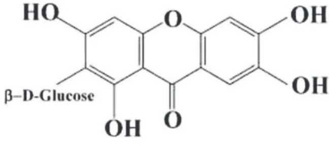
Mangiferin (MGF), a naturally occurring gluco-sylxanthone (Fig. 1) commonly encountered in
sev-eral traditional medicinal plants [16] has been shown to produce multiple pharmacological effects that in-clude antioxidant and anti-inflammatory [2, 4, 32], immunomodulatory [20, 26], and neuroprotective [9, 33] activities. There have been reports that MGF de-creases glutamate-induced neurotoxicity and memory loss induced by scopolamine [23, 27, 39]. We previ-ously demonstrated that MGF affords gastroprotec-tion against absolute ethanol or indomethacin-induced gastric ulceration through an antioxidant mechanism [10]. Impaired antioxidant defenses are suggested to participate in the pathophysiology of schizophrenia and other neurodegenerative conditions. Altered su-peroxide dismutase (SOD) and increased lipid peroxi-dation, measured by the thiobarbituric acid reactive substances (TBARS/MDA), are increased in schizo-phrenic patients [17]. Besides, MGF has the iron com-plexing ability and can protect against Fe2+-citrate in-duced lipid peroxidation [3]. The neurotoxin 6-OHDA is well-known to cause neuronal death by a free radical-mediated mechanism as well as by inhibiting the mitochondrial complex I and IV [18]. As MGF has a wide spectrum of pharmacological activity in-cluding antioxidant and anti-inflammatory activity [26, 32] and accumulating evidence suggests that MGF is neuroprotective, the current study is aimed to examine the cytoprotective role of MGF against 6-OHDA-induced neurotoxicity in cultured rat pri-mary mesencephalic cells and its beneficial effects in the mouse model of ketamine-induced schizhophrenia.
Materials and Methods
Animals
Male Swiss mice (7–8 weeks old, 20–25 g) and adult male/female Wistar rats (4–5 months old) obtained from the Central Animal House of Federal University of Ceará, were used. They were housed in environ-mentally controlled conditions (23 ± 2°C, 12-h light-dark cycle), with free access to standard diet (Purina Chow) and waterad libitum. Rats were mated to ob-tain rats on pregnancy and to collect embryonic mes-encephalic cells, forin vitrostudies. Mice were used forin vivoexperiments aimed to study the effects of drugs in ketamine model of schizophrenia. The experimental protocols were approved by the Animal Mangiferin prevents cytotoxicity and oxidative stress
Vietla S. Rao et al.
(IASP).
Chemicals
Mangiferin (MGF) was extracted and isolated from the bark of Mangifera indica L. (Anacardiaceae) as per procedures reported earlier [10]. The isolated MGF was approximately of 95% purity [4] having the molecular weight (MW) 422.5 and melting point (m.p.) 271°C. Minimum essential medium (MEM), trypsin, MTT [3-(4,5-dimethylthiazol-2-yl)-2,5-di-phenyltetrazolium bromide], cytochalasin-B, propid-ium iodide (PI), ethidpropid-ium bromide (EtBr) and fetal bovine serum (FBS) were purchased from Sigma Chemical Co. (St. Louis, MO, USA). Acridine orange (AO) was purchased from BDH Chemicals Ltd., Poole, England and ketamine from Sigma-Aldrich (MO, USA). MGF was dissolved in 0.02% DMSO and diluted with MEM immediately before use. Other drugs were dissolved in physiological saline or dis-tilled water. Drug concentrations were adjusted forin vivo treatments to give a volume of 10 ml/kg. The dose/concentration selection for MGF and 6-OHDA or ketamine was based on our pilot studies or litera-ture based [28, 37, 39].
Cell culture
Mesencephalic cell culture containing neuronal and non-neuronal cells were obtained from mesencephali-cum of Wistar rat embryos at 17–20 days of preg-nancy [12]. Cells were cultured in MEM supple-mented with FBS (10%), streptomycin (100 mg/ml), penicillin (1,000 IU/ml), actinomycin C (2.5 mg/ml), glucose (11 mM), glutamine (2 mM), and sodium bi-carbonate (24 mM). Cultures were maintained at 37°C in humidified 5% CO2air atmosphere. Seven days after plating, cultures were reutilized for experi-mentation. Media were replaced every 2–3 days and cells were subcultured when 80–90% confluent. Cells were plated at a density of 5 × 10⁴cells per well using 96-well culture plates pre-coated with poly-lysine.
Mesencephalic cell culture plates having a cell den-sity of 5 × 10⁴cells per well were pre-incubated with MGF in concentrations of 10, 30, and 100 µM for 15 min, prior to addition of 6-OHDA (40 µM). Each concentration of MGF was tested in six replicates and repeated three times in separate experiments. Cell viability after exposure to 6-OHDA or MGF (10, 30, and 100 µM) alone, or in combination was deter-mined in culture plates by MTT as described by Mos-mann [36]. This method measures mitochondrial activity based on the reductive cleavage of yellow tetrazolium salt to a purple formazan compound by the dehydrogenase activity of intact mitochondria. Briefly, cells were washed once with PBS before the addition of 0.1 ml of serum free medium containing MTT (1 mg/ml) to each well. After incubation for 3 h, the supernatant was removed and the formazan prod-uct obtained was dissolved in 1 ml dimethyl sulfoxide (DMSO) with stirring for 15 min on a microtiter plate shaker and the absorbance was read at 550 nm. The percentage of viable cells in each treatment group was determined by comparing their respective absorbance with that of control group.
In the analysis of MGF effect on apoptosis/necrosis induced by 6-OHDA, cell culture plates were divided into following groups: Group I, MGF 30 µM; Group II, 6-OHDA 40 µM; Group III, MGF + 6-OHDA – cell cultures of this group were treated with MGF 30 µM for 2 h followed by 6-OHDA 40 µM for 3 h. The cells were then allowed to grow for 24 h. After this time, the cells were pelletted and each sample was mixed with 1 µl of aqueous acridine/ethidium bromide (AO/EtBr) solution (100 µg/ml of each in PBS) just prior to fluorescence microscopy analysis and quantification (Olympus, Tokyo, Japan). Micro-scopic analysis was carried out to provide the cyto-morphological evidence for normal, apoptotic and ne-crotic cells, differentiated by AO/EtBr staining, which were expressed in % of total cells [34, 40]. At least three hundred cells per sample were analyzed.
sulfanila-mide in 1% H3PO4/0.1% N-(1-naphthyl)-ethylenedi-amine dihydrochloride/1% H3PO4/distilled water, 1:1:1:1] or to 100 µl of NaNO2at concentrations rang-ing from 0.75 to 100 µM (standard curve). For the blanks, 100 µl of the Griess reagent was added to 100 µl of the cell culture medium. The absorbance was measured with a reader plate at 560 nm. The standard curve was used for the determination of nitrite con-centrations in samples and expressed in µM.
Ketamine-induced schizophrenia in mice
Schizophrenia was induced in Swiss male mice as de-scribed by Liu et al. [28]. Mice were divided into four groups (n = 8–10 animals/group), Group I served as control and received DMSO (2% in distilled water) as vehicle. Group II animals were treated with ketamine (50 mg/kg, ip, twice a day for 7 consecutive days). Group III animals were subjected to pretreatment with MGF (50 mg/kg/day for 7 days,po, dissolved in vehi-cle 2% DMSO) followed by ketamine injections (as above). Group IV animals were treated with MGF alone (as above). Thirty minutes following the respec-tive treatments on day-7, the animals were first sub-jected to open-field test to note the locomotor activity and behavioral changes. Thereafter, the animals were killed by cervical dislocation. Tissues from the whole brain were immediately excised and homogenized in Tris-HCl buffer 0.01 M (pH 7.4) to get 10% homo-genates. These were then stored in a freezer at –70°C until use for the analysis of IL-6 cytokine and to measure the levels of lipid peroxidation.
Analysis of behavioral alterations in open-field test
Thirty minutes following the last treatment on day-7, the animals from respective groups were subjected to open-field test. The open-field apparatus (30 × 30 × 20 cm) was made of acrylic with transparent walls and black flooring divided into nine squares of equal area. After one minute of acclimatization, each animal from respective treatment groups was evaluated over a 5 min period for the following parameters: number of squares crossed with all the four paws, number of grooming and rearing, and the stereotyped behaviors scored (0–6) according to Setler et al. [41] by a person unaware of the animal treatments.
Analysis of cytokine IL-6 levels and lipid peroxidation in brain tissue
IL-6 cytokine levels in whole brain tissue homogen-ates (10%) were determined with the aid of enzyme immunoassay kit (eBioscience – Mouse IL-6 Ready-SET-Go! Elisa Kit with pre-coated plates). The assay was performed according to manufacturer’s instruc-tions and the cytokine levels were expressed as pg/ml. Lipid peroxidation was measured in terms of malon-dialdehyde (MDA) content following the thiobarbi-turic acid method of Ohkawa et al. [38]. Thiobarbi-turic acid reacts with MDA to form a pink chromo-gen, which can be detected spectrophotometrically at 532 nm. In brief, aliquots (250 µl) of the homogenate obtained previously were put into test tubes and mixed with an equal volume of thiobarbituric acid (1.2%). This mixture was brought to the water bath for 30 min at a temperature ranging from 95 to 100°C and then cooled to room temperature. The tubes were then centrifuged at 3,000 × g for 10 min to precipitate the protein. The absorbance of supernatants was measured at 532 nm. The standard curve was obtained by serial dilutions (1, 0.5, 0.25, 0.12, 0.06, 0.03 mM) using a solution of MDA. The concentration of MDA was expressed as nmol/g tissue.
Statistical analysis
Values were expressed as the means ± SEM. The sig-nificance of the differences between treatments and re-spective controls was analyzed by one-way analysis of variance (ANOVA), followed by the Newman- Keuls multiple comparison test. A p value of 0.05 was con-sidered statistically significant.
Results
Effect of MGF on cytotoxicity induced by 6-OHDA in cell culture
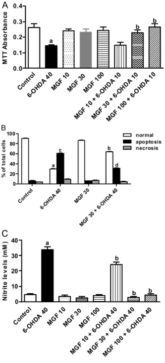
The various concentrations of MGF (10, 30 and 100 µM) did not have a significant impact on cell viability when mesencephalic cells were treated with it for 24 h (Fig. 2A). When cells were treated with 6-OHDA (40 µM), cell viability was markedly reduced (p < 0.01), compared to vehicle-treated con-trol. Incubation of cells with increasing concentra-Mangiferin prevents cytotoxicity and oxidative stress
compared to control group of cells exposed to 6-OHDA alone. Maximum elevation in cell survival was appar-ent at 100 µM MGF concappar-entration.
Fluorescence microscopy analysis of apoptotic cells
To assess the degree of protection by MGF against 6-OHDA-induced apoptosis and necrosis, micro-scopic AO/EtBr dual staining was used. Cells were differentiated into normal (live), apoptotic or necrotic from the uptake of AO/EtBr stain. Normal (live) cells were seen as bright green colored nuclei with intact and uniform cell membrane. Apoptotic cells had or-ange to red nuclei with condensed or fragmented chromatin, whereas necrotic cells showed uniform or-ange to red nuclei. As expected, 6-OHDA largely in-duced apoptosis than necrosis (Fig. 2B). A significant (p < 0.01) increase in apoptosis was observed in cells treated with 6-OHDA (40 µM) alone for 24 h (Fig. 2B). MGF (30 µM) alone treated cells showed no significant differences in the counts of live, apop-totic and necrotic cells when compared with vehicle-treated control. On the other hand, incubation of cells with MGF (30 µM) prior to 6-OHDA (40 µM) for 24 h showed a marked decrease (p < 0.01) in percent apoptosis, when compared with 6-OHDA control.
Nitrite formation
To assess the cytoprotective effect of MGF against 6-OHDA-induced toxicity, the extent of nitrite forma-tion was measured in rat mesencephalic cells super-natant. Treatment of cells with 10–100 µM of MGF alone did not alter the nitrite formation in these cells when compared with normal control group, while treatment of cells with 6-OHDA (40 µM) showed a significant (p< 0.001) and marked increase in ni-trite levels (Fig. 2C). This increase in nini-trite levels by 6-OHDA was significantly (p < 0.05) reduced by MGF (30 and 100 µM) pretreatment.
Effect of MGF treatment on ketamine induced increase in IL-6 and MDA levels
Treatment of animals with ketamine (50 mg/kg, twice a day, po, for 7 days) caused a significant increase Fig. 2.Protective effect of mangiferin (MGF) on 6-OHDA-induced
cytotoxicity in rat mesencephalic cells. (A) Cells were treated with 40 µM 6-OHDA for 24 h in the absence or presence of MGF (10, 30 and 100 µM) and cytotoxicity was analyzed by MTT assay. The results are expressed as the mean ± SEM of three independent experiments.ap < 0.01vs.normal control;bp < 0.01vs.6-OHDA40 (µM). (B) MGF (30 µM) reduces apoptosis and necrosis (assessed by acridine orange/ethidium bromide staining) in rat mesencephalic cells promoted by 6-OHDA 40 (µM).ap < 0.01vs.normal control;
bp < 0.01vs.6-OHDA 40 (µM) normal;cp < 0.01vs.control
apopto-sis;dp < 0.01vs.6-OHDA 40 (µM) apoptosis. (C) MGF (10, 30 and 100 µM) decreases nitrite formation (analyzed by Griess reaction) in rat mesencephalic cells exposed to 40 µM 6-OHDA for 24 h.
ap < 0.001vs.normal control;bp < 0.05vs.6-OHDA 40 (µM)
(p < 0.01) in the levels of IL-6 and MDA in brain tis-sues of schizophrenic group of animals when com-pared to normal control group. Pretreatment with MGF (50 mg/kg/day,po, for 7 days) significantly (p < 0.05) reduced the ketamine associated elevation of IL-6 and MDA levels (Fig. 3A and B).
Effects of MGF treatment on changes in locomotor activity and behaviors
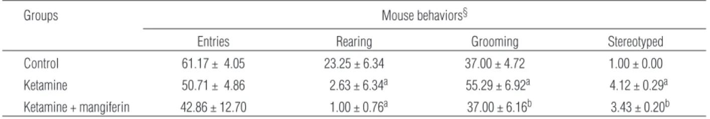
The effects of treatments on behavioral changes in-duced by ketamine are shown in Table 1. Neither MGF nor ketamine treated groups of mice showed a significant change in the number of invasions (en-tries) in open-field area. Treatment with ketamine caused a significant reduction (p < 0.01) in rearing be-havior of animals and MGF was unable to reverse the effect of ketamine. The group treated with ketamine showed significant increase (p < 0.05) in the number of grooming behavior, and the group pretreated with MGF reversed significantly (p < 0.05) this effect. The scores for stereotyped behavior of animals were sig-nificantly higher in all treated groups compared to control (p < 0.01). The group pretreated with MGF re-ceived a significantly lowered levels of scores as compared to ketamine alone (p < 0.05), but unable to attain the levels seen in normal control.
Discussion
As expected, 6-OHDA inhibited cell viability in rat primary mesencephalic cell cultures. 6-OHDA is as-sumed to cause neuronal death by a free radical-mediated mechanism as well as by inhibiting the mi-tochondrial complex I and IV [18]. Our results show that 6-OHDA induced largely the apoptosis rather than necrosis, consistent with an earlier report [12]. MGF alone did not decrease the cell viability even when these cells were exposed to 100 µM for 24 h, in-dicating its non-toxic nature. Interestingly, when cells were pre-treated with MGF before the application of Mangiferin prevents cytotoxicity and oxidative stress
Vietla S. Rao et al.
Fig. 3.Mangiferin (MGF) effects on brain tissue levels of cytokine IL-6 and MDA in normal and schizophrenic mice. Mice in groups (n = 8–10) were treated with vehicle (normal control; 2% DMSO in distilled water, 10 ml/kg), MGF (50 mg/kg/day,pofor 7 days) alone, ketamine (50 mg/kg,ip, twice a day for 7 days) alone or the combination of MGF + ketamine. After the end of 7-day treatment, all animals were sacrificed and whole brain tissues were carefully dissected from each animal and were homogenized to get 10% homogenates. These homogenates were subjected to analysis of cytokine IL-6 (A) and malonaldialdehyde (MDA) (B) levels. The data are expressed as the mean ± SEM of 8–10 animals.ap < 0.01 compared with vehicle-treated control;bp < 0.01 compared with ketamine (one-way ANOVA followed by Student Newman Keul’s test)
Tab. 1.Effect of mangiferin treatment on locomotor behavioral changes in ketamine-induced schizophrenia model mice¥
Groups Mouse behaviors§
Entries Rearing Grooming Stereotyped
Control 61.17 ± 4.05 23.25 ± 6.34 37.00 ± 4.72 1.00 ± 0.00
Ketamine 50.71 ± 4.86 2.63 ± 6.34a 55.29 ± 6.92a 4.12 ± 0.29a
Ketamine + mangiferin 42.86 ± 12.70 1.00 ± 0.76a 37.00 ± 6.16b 3.43 ± 0.20b
¥Mice were treated with mangiferin (50 mg/kg/day,po, in 2% DMSO, for 7 days) followed by ketamine injections (50 mg/kg,ip, twice a day for
almost total and equaled the normal control. MGF in-hibited the apoptosis but showed no significant effect on necrosis. These findings corroborate the earlier studies that demonstrated the protective potential of MGF and antioxidants in general, against 6-OHDA-induced cytotoxicity in various cell lines and primary cultures [37, 42, 46]. A role for NO in the mitochon-dria-mediated apoptosis of dopaminergic neurons after 6-OHDA has recently been described [40]. We ob-served a significantly (p < 0.05) attenuated apoptosis and a decreased NO increase caused by 6-OHDA. When mesencephalic cells rich in neuronal and non-neuronal cells were exposed to 6-OHDA, we observed a significant increase in NO associated with apoptosis, suggesting that NO promotes cell death. However, in mesencephalic cells preconditioned to MGF, NO pro-duction as well as cell death was markedly reduced, which indicates that MGF is cytoprotective.
The brain tissue is extremely sensitive to oxidative damage and oxidative stress can damage neurons and glial cells in a manner similar to other tissues via
products of lipid peroxidation that are neurotoxic. Studies indicate that ketamine activates NADPH-oxidase in brain and show that neuronal production of interleukin-6 (IL-6) is necessary for ketamine-me-diated activation of NADPH-oxidase in brain [7]. Re-moval of IL-6 in neuronal cultures by anti-IL-6 block-ing antibodies, or in vivo by use of IL-6-deficient mice, prevented the increase in superoxide by keta-mine and rescued the interneurons [7]. Our results de-monstrate that oral administration of MGF (50 mg/kg) for 7 days could modulate ketamine induced impaired behaviors in particular, the grooming and stereotyped behaviors as well as the oxidative damage as indi-cated by increase in IL-6 level and lipid peroxidation in brain tissues of ketamine-induced schizhophrenia model mice. However, MGF was unable to reverse the rearing behavioral effect of ketamine that is possi-bly associated with strong sedation. MGF is a xan-thone with various therapeutic activities including an antioxidant. It has been proposed that the catechol moiety with a 6,7-dihydroxylated structure, together with its aromatic bonds, is responsible for its antioxi-dant property [44]. MGF possibly acts as an effective anti-oxidant, mainly on account of its catechol moiety with a 6,7-dihydroxylated structure, which provides it the ability to neutralize reactive oxygen species
gen-and the levels of lipid peroxidation product – malon-dialdehyde, indicates that MGF enhances antioxidant status, consistent with earlier reports that used other experimental models [13, 31, 45].
Studies indicate that MGF being a non-polar poly-phenol can penetrate the blood brain barrier and has the potential to ameliorate the oxidative stress ob-served in neurodegenerative disorders [29] and MGF (20 mg/kg,po) has been proven to improve long-term cholinergic memory deficits by acetylcholinesterase (AChE) inhibition or cholinergic receptor stimulation and inhibition of NF-kB activationin vivo[27].
me-chanisms. Nevertheless, mangiferin treatment over-comes some of the behavioral signs such as grooming and stereotyped activities in ketamine-induced schizophrenia mice.
The experimental models used in this study have some limitations such as the presence of neuronal and non-neuronal cells in rat primary mesencephalic cell cultures and the non-selectivity of ketamine to NMDA receptors affecting respiratory chain function in multi-ple brain regions including striatum, hippocampus and prefrontal cortex, which makes difficult to elucidate the precise protective mechanism of mangiferin. Neverthe-less, mangiferin could counteract the oxidative stress promoted by 6-OHDA in mesencephalic cells in vitro
and in ketamine-induced schizophreniain vivo. Mangi-ferin might serve as a lead compound in developing drugs affective against schizophrenia and other neuro-degenerative diseases.
To conclude, the present work indicated that man-giferin prevents cell death induced by 6-OHDA in rat primary mesencephalic cells, attenuates ketamine as-sociated increase in cytokine IL-6 cytokine and ma-londialdehyde formation in brain tissues, and amelio-rates stereotyped movements, a characterstic of clinical schizophrenia. These findings suggest that mangiferin has a cytoprotective role, at least in part, related to its anti-inflammatory, anti-apoptotic and anti-lipid per-oxidative potential plausibly because of its free radi-cal scavenging and iron chelating ability, which could effectively reduce the oxidative stress.
Conflict of interest statement:
The authors declare that there are no conflicts of interest.
Acknowledgments:
We acknowledge the financial support from National Council of Technological and Scientific Development (CNPq) and Ceará Foundation for the Support of Scientific and Technological Development of the Ceará State (FUNCAP).
References:
1.Adibhatla RM, Hatcher JF: Lipid oxidation and
peroxi-dation in CNS health and disease: from molecular mechanisms to therapeutic opportunities. Antioxid Redox Signal, 2010, 12, 125–169.
2.Amazzal L, Lapôtre A, Quignon F, Bagrel D: Mangiferin protects against 1-methyl-4-phenylpyridinium toxicity mediated by oxidative stress in N2A cells. Neurosci Lett, 2007, 418, 159–164.
3.Andreu GL, Delgado R, Velho J, Curti C, Vercesi AE:
Iron complexing activity of mangiferin, a naturally oc-curring glucosylxanthone, inhibits mitochondrial lipid peroxidation induced by Fe2+-citrate. Eur J Pharmacol, 2005, 513, 47–55.
4.Barreto JC, Trevisan MT, Hull WE, Erben G, de Brito
ES, Pfundstein B, Würtele G et al.: Characterization and quantitation of polyphenolic compounds in bark, kernel, leaves, and peel of mango (Mangifera indicaL.). J Agric Food Chem, 2008, 56, 5599–5610.
5.Becker A, Peters B, Schroeder H, Mann T, Huether G, Grecksch G: Ketamine-induced changes in rat behavior: A possible animal model of schizophrenia. Prog Neuro-psychopharmacol Biol Psychiatry, 2003, 27, 687–700.
6.Beraki S, Kuzmin A, Tai F, Ogren SO: Repeated low dose
of phencyclidine administration impairs spatial learning in mice: blockade by clozapine but not by haloperidol. Eur Neuropsychopharmacol, 2008, 18, 4869–4877.
7.Behrens MM, Ali SS, Dugan LL: Interleukin-6 mediates
the increase in NADPH-oxidase in the ketamine model of schizophrenia. J Neurosci, 2008, 28, 13957–13966.
8.Bubeníková-Valesová V, Horácek J, Vrajová M,
Höschl C: Models of schizophrenia in humans and ani-mals based on inhibition of NMDA receptors. Neurosci Biobehav Rev, 2008, 32, 1014–1023.
9.Campos-Esparza MR, Sánchez-Gómez MV, Matute C:
Molecular mechanisms of neuroprotection by two natu-ral antioxidant polyphenols. Cell Calcium, 2009, 45, 358–368.
10.Carvalho AC, Guedes MM, de Souza AL, Trevisan MT,
Lima AF, Santos FA, Rao VS: Gastroprotective effect of mangiferin, a xanthonoid fromMangifera indica, against gastric injury induced by ethanol and indomethacin in rodents. Planta Med, 2007, 73, 1372–1376.
11. Choi DW, Maulucci-Gedde MA, Kreigstein AR. Gluta-mate neurotoxicity in cortical cell culture. J Neurosci, 1987, 7, 357–368.
12.Choi WS, Yoon SY, Oh TH, Choi EJ, O´Malley KL,
Oh YJ: Two distinct mechanisms are involved in 6-hydroxydopamine-and MPP+-induced dopaminergic
neuronal cell death: role of caspasses, ROS, and JNK. J Neurosci Res, 1999, 57, 86–94.
13.Dar A, Faizi S, Naqvi S, Roome T, Zikr-ur-Rehman S,
Ali M, Firdous S, Moin ST: Analgesic and antioxidant ac-tivity of mangiferin and its derivatives: the structure activ-ity relationship. Biol Pharm Bull, 2005, 28, 596–600.
14.de Oliveira L, Fraga DB, De Luca RD, Canever L, Ghedim
FV, Matos MP, Streck EL et al.: Behavioral changes and mitochondrial dysfunction in a rat model of schizophrenia induced by ketamine. Metab Brain Dis, 2011, 26, 69–77.
15.de Oliveira L, Spiazzi CM, Bortolin T, Canever L,
Petronilho F, Mina FG, Dal-Pizzol F et al.: Different sub-anesthetic doses of ketamine increase oxidative stress in the brain of rats. Prog Neuropsychopharmacol Biol Psychiatry, 2009, 33, 1003–1008.
16.El-Seedi HR, El-Barbary MA, El-Ghorab DM, Bohlin L, Borg-Karlson AK, Göransson U, Verpoorte R: Recent in-sights into the biosynthesis and biological activities of natural xanthones. Curr Med Chem, 2010, 17, 854–901.
17.Gama CS, Salvador M, Andreazza AC, Lobato MI, Berk
M, Kapczinski F, Belmonte-de-Abreu PS: Elevated
se-Mangiferin prevents cytotoxicity and oxidative stress
6-hydroxydopamine neurotoxicity. J Neural Transm Suppl, 1997, 50, 55–56.
19.Green LC, Tanzenbaum SR, Goldman P: Nitrate
synthe-sis in the germ free and conventional rat. Science, 1981, 212, 56–58.
20.Guha S, Ghosal S, Chattopadhyay U: Antitumor,
immu-nomodulatory and anti-HIV effect of mangiferin, a natu-rally occurring glucosylxanthone. Chemotherapy, 1996, 42, 443–451.
21.Gunduz-Bruce H: The acute effects of NMDA antago-nism: from the rodent to the human brain. Brain Res Rev, 2009, 60, 2798–2806.
22.Hetem LA, Danion JM, Diemunsch P, Brandt C: Effect
of a subanesthetic dose of ketamine on memory and con-scious awareness in healthy volunteers. Psychopharma-cology (Berl), 2000, 152, 283–288.
23.Jung K, Lee B, Han SJ, Ryu JH, Kim DH: Mangiferin
ameliorates scopolamine-induced learning deficits in mice. Biol Pharm Bull, 2009, 32, 242–246.
24.Kapur S, Seeman P: NMDA receptor antagonists
keta-mine and PCP have direct effects on the dopaketa-mine D2
and serotonin 5-HT2receptors – implications for models
of schizophrenia. Mol Psychiatry, 2002, 7, 837–844.
25.Lakhan SE, Vieira K: Schizophrenia pathophysiology: are we any closer to a complete model? Ann Gen Psy-chiatry, 2009, 8, 12.
26.Leiro JM, Alvarez E, Arranz JA, Siso IG, Orallo F:In vi-troeffects of mangiferin on superoxide concentrations and expression of the inducible nitric oxide synthase, tu-mour necrosis factor-aand transforming growth factor-b genes. Biochem Pharmacol, 2003, 65, 1361–1371.
27.Lemus-Molina Y, Sánchez-Gómez MV,
Delgado-Hernández R, Matute C:Mangifera indicaL. extract at-tenuates glutamate-induced neurotoxicity on rat cortical neurons. Neurotoxicology, 2009, 30, 1053–1058.
28.Liu WL, Bian S-Z, Gu Z-L, Jiang X-G, Guo C-Y, Zhao
Y-B: Behavior study of ketamine-induced symptoms similar to schizophrenia in mice. J Forensic Med, 2009, 25, 172–175.
29.Mahmood D, Khanam R, Pillai KK, Akhtar M: Reversal
of oxidative stress by histamine H3 receptor-ligands in experimental models of schizophrenia. Arzneimittelfor-schung, 2012, 62, 222–229.
30.Mahmood D, Khanam R, Pillai KK, Akhtar M:
Protec-tive effects of histamine H3-receptor ligands in
schizo-phrenic behaviors in experimental models. Pharmacol Rep, 2012, 64, 191–204.
31.Mandryk M, Fidecka S, Poleszak E, Malec D:
Participa-tion of adenosine system in the ketamine-induced motor activity in mice. Pharmacol Rep, 2005, 57, 55–60.
32.Márquez L, García-Bueno B, Madrigal JL, Leza JC:
Mangiferin decreases inflammation and oxidative dam-age in rat brain after stress. Eur J Nutr, 2012, 51, 729–739.
33.Martínez Sánchez G, Candelario-Jalil E, Giuliani A,
León OS, Sam S, Delgado R, Núñez Sellés AJ:
Mangif-Shi Y, Mogil RJ, Nishioka WK, Green DR: The end of the (cell) line: methods for the study of apoptosis in vi-tro. Methods Cell Biol, 1995, 46, 153–185.
35.Moghaddam B. Recent basic findings in support of
excita-tory amino acid hypotheses of schizophrenia. Prog Neu-ropsychopharmacol Biol Psychiatry, 1994, 18, 859–870.
36.Mosmann T: Rapid colorimetric assay for cellular
growth and survival: application to proliferation and cy-totoxicity assays. J. Immunol Methods, 1983, 65, 5–63.
37.Nobre Júnior HV, Cunha GM, Maia FD, Oliveira RA,
Moraes MO, Rao VS: Catechin attenuates 6-hydroxy-dopamine (6-OHDA)-induced cell death in primary cul-tures of mesencephalic cells. Comp Biochem Physiol C Toxicol Pharmacol, 2003, 136, 175–180.
38.Ohkawa H, Ohishi N, Yagi K: Assay for lipid peroxides in animal tissues by thiobarbituric acid reaction. Anal Biochem, 1979, 95, 351–358.
39.Pardo Andreu GL, Maurmann N, Reolon GK, de Farias
CB, Schwartsmann G, Delgado R, Roesler R: Mangif-erin, a naturally occurring glucoxilxanthone improves long-term object recognition memory in rats. Eur J Phar-macol, 2010, 635, 124–128.
40.Renvoize C, Biola A, Pallardy M, Breard J: Apoptosis:
identification of dying cells. Cell Biol Toxicol, 1998, 14, 111–120.
41.Setler P, Sarau H, McKinzie G: Differential attenuation
of some effects of haloperidol in rats given scopalamine. Eur J Pharmacol, 1976, 39, 117–126.
42.Singh S, Kumar S, Dikshit M: Involvement of the mito-chondrial apoptotic pathway and nitric oxide synthase in dopaminergic neuronal death induced by 6-hydroxy-dopamine and lipopolysaccharide. Redox Rep, 2010, 15, 115–122.
43.Tsai G, van Kammen DP, Chen S, Kelley ME, Grier A, Coyle JT: Glutamatergic neurotransmission involves structural and clinical deficits of schizophrenia. Biol Psychiatry, 1998, 44, 667–674.
44. Wauthoz N, Balde A, Balde ES, Van Damme M, Duez
P: Ethnopharmacology ofMangifera indicaL. Bark and pharmacological studies of its main C-glucosylxanthone, mangiferin, Int J Biomed Pharm Sci, 2007, 1, 112–119.
45.Yoshikawa M, Ninomiya K, Shimoda H, Nishida N, Matsuda H: Hepatoprotective and antioxidative proper-ties ofSalacia reticulata: preventive effects of phenolic constituents on CCl4-induced liver injury in mice. Biol
Pharm Bull, 2002, 25, 72–76.
46.Zhang ZJ, Cheang LC, Wang MW, Lee SM: Quercetin exerts a neuroprotective effect through inhibition of the iNOS/NO system and pro-inflammation gene expression in PC12 cells and in zebrafish. Int J Mol Med, 2011, 27, 195–203.