Adiponectin Efect on The Viability of Human Endometrial
Stromal Cells and mRNA Expression of Adiponectin
Receptors
Somayeh Bohlouli D.V.M, Ph.D1*, Mozafar Khazaei, Ph.D.2, Masoud Teshfam, Ph.D.1, Hosein Hassanpour, Ph.D.3,
1. Department of Veterinary Physiology, Science and Research Branch, Islamic Azad University, Tehran, Iran 2. Fertility and Infertility Research Center, Kermanshah University of Medical Sciences, Kermanshah, Iran 3. Department of Basic Sciences (Veterinary Physiology Division), Faculty of Veterinary Medicine, Shahrekord
University, Shahrekord, Iran
Abstract
Background: Adiponectin is one of the most important adipokines secreted from fatty tissue that has a direct inhibitory effect on the development of cancer cells. Adiponectin plays an important role in human reproduction system and fertility of women. Adi-ponectin concentration decreases in women with endometriosis and endometrial can-cer. The aim of the present study was to investigate the effect of adiponectin on human endometrial stromal cell (HESC) viability as well as mRNA expression of Adipo R1 and Adipo R2 receptors.
Materials and Methods: In this experimental study, eight endometrial biopsies were
taken and stromal cells were separated by enzymatic digestion and cell iltrations. Stro -mal cells of each biopsy were divided into four groups: control, 10, 100, and 200 ng/ml adiponectin concentrations. The effect of adiponectin on viability of the normal HESCs was studied by trypan blue staining and the relative expression levels of Adipo R1 and R2 were analyzed by semi-quantitative reverse transcription polymerase chain reaction (RT-PCR). Data were analyzed by one way ANOVA and unpaired student’s t test and
p<0.05 was considered signiicant.
Results: Adiponectin decreased viability of normal human endometrial stromal cells in a dose and time dependent manner. Expression of Adipo R1 and Adipo R2 receptors did not change in the presence of adiponectin.
Conclusion: Adiponectin can directly inluence the viability of HESCs and decrease
their viability, but it didn’t change expression of adiponectin receptors.
Keywords: Adiponectin, Stromal Cells, Adipo R1, Adipo R2, Endometrium
Citation: Bohlouli S, Khazaei M, Teshfam M, Hassanpour H. Adiponectin effect on the viability of human endometrial stromal cells and mRNA expression of adiponectin receptors. Int J Fertil Steril. 2013; 7(1): 43-48.
Received: 16 May 2012, Accepted: 23 Oct 2012
* Corresponding Address: P.O.Box: 14515-775, Department of Vet-erinary Physiology, Science and Research Branch, Islamic Azad University, Tehran, Iran
Email: [email protected] Royan Institute
International Journal of Fertility and Sterility
Introduction
Adiponectin is one of the most important members of adipokine family which is widely synthesized and secreted by fatty tissue. Various roles have been identified for adiponectin such as regulation of glucose level and lipids homeo-stasis. Furthermore, adiponectin plays a pivotal role in reproductive system (1, 2). Adiponectin is abundantly present in the blood stream and
its concentration in human plasma is 5-30 μg/
ml which comprises 0.01% of all proteins in the plasma (3).
(8). Adiponectin binds to receptors, known as Adipo R1 and Adipo R2 (9). These receptors contain seven transmembrane domains but dif-fer from G-protein coupled receptors structural-ly and functionalstructural-ly. The tendency of adiponec-tin receptors to bind to adiponecadiponec-tin isoforms as well as tissue distribution of these receptors are different (10).
In mice, Adipo R1 exists in different organs such as skeletal muscle, lung, and spleen; whereas Adipo R2 is mainly expressed in liver (11). In human, Adipo R1 and Adipo R2 are ex-pressed in islets of Langerhans, macrophages, adipocytes, and vascular smooth muscles (12- 14).
Various data have indicated that adiponectin is influential in female fertility and plays an important role in female reproductive system. Study has indicated that serum adiponectin lev-el decreases in women with endometriosis (15) and endometrial cancer (16). Also, adiponectin level in peritoneal fluid of endometriosis pa-tients decreased dramatically in advanced en-dometriosis (17).
In histopathological studies of endometriosis tissues, stromal cells and glands are abundant-ly present, but changes of endometrial stromal cells are much more than those of endometrio-sis identifying glands and there is the possibil-ity of the presence of gland-free stromal cells in endometriosis tissue (18). HESCs play pivotal role in female reproductive biology and there is no report on the effect of adiponectin on these cells. The aim of the present study was to ex-amine the effect of adiponectin on human endo-metrial stromal cells and in vitro mRNA expres-sion of adiponectin receptors.
Materials and Methods
Samples
In this experimental study, endometrial tis-sues were taken from women aged 25-35 who had no record of hormonal treatment for three months before surgery and had undergone hys-terectomy surgery or biopsy diagnosis for in-fertility management and reasons other than en-dometrial malignancies such as myoma. Eight samples of normal endometrium in the
secre-tory phase were taken. The Ethics Committee of Kermanshah University of Medical Sciences and Tehran Science and Research Branch of Islamic Azad University accepted the work on human endometrial tissue in this study and all patients signed informed consents.
Separation and culture of human endometrial stromal cells
Stromal cells were separated from endome-trial tissue according to previous work (19, 20). Each endometrial sample was prepared in ster-ile condition and was washed with PBS solu-tion containing 1% antibiotic/antimycotic, and then was chopped mechanically. The sample was incubated with collagenase type I solution (2 mg/ml in DMEM/F12) (Sigma, Germany) for 60-90 minutes. The cell suspension was passed
through 70 and 40 μm cell strainers (BD falcon,
USA) respectively, centrifuged for 15 minutes (2500 rpm) and DMEM/F12 (Gibco, Germany) was added to the cell pellet. Then the suspen-sion was layered on ficoll (Amersham, Swe-den) and was centrifuged for 30 minutes (1500 rpm). The stromal cells were collected and were washed with PBS and were cultured in DMEM/ F12 containing 10% fetal bovine serum (FBS) (Gibco, Belgium), 0.1 mg/ml streptomycin, and 100UI/ml penicillin. The cultures were incubat-ed in a humidifiincubat-ed atmosphere of 95% air and 5% CO2 at 37˚C. After seven days, cell density reached confluency and 1×10⁵ cells were trans-ferred to each well of 24-well culture plate.
Cell treatment
To add adiponectin (high molecular weight, R&D System Minneapolis, MN USA) to stro-mal cells, the media was removed and cells were washed with PBS and incubated with serum-free media overnight and then were treated with adi-ponectin at 0, 10, 100, and 200 ng/ml in 24, 48, and 72 hours for each dose (21).
Evaluation of cells viability
divid-ing the number of non-stained cells by total num-ber of cells multiplied by 100 (22).
Reverse transcription polymerase chain reaction (RT-PCR)
Total RNA was extracted from stromal cells in control group and adiponectin group (100
ng/ml for 48 hours) using RNA puriication kit
(Jena Bioscience, GmbH, Germany). Total RNA
(≥1μg) was used to synthesize complementary DNA (cDNA) in a 20 μl reaction by AccuPow -er® RocketScriptTM RT PreMix kit (BIONEER, Korea) and oligo(dT). The PCR was performed using PCR PreMix kit (BIONEER, Korea) ac-cording to the manufacturer’s instructions. Cycle conditions were as follows: initial
dena-turation at 94˚C for 10minutes; followed by 35 cycles of denaturation at 94˚C for 60 seconds, annealing at 58˚C (GAPDH) and 62˚C) Adipo
R1 and Adipo R2 (for 60 seconds and extension
at 72˚C for 60 seconds, with a inal extension at 72˚C for 10 minutes (Table 1). Since less than 35
cycles produced PCR products at low intensity, the PCR reactions were thought to be still in the exponential phase. Experiments were performed
in triplicate to ensure reproducibility.
Semi-quantitative reverse transcription–poly-merase chain reaction analysis
The expression of target genes was quantiied
against the internal reference gene (GAPDH). Prod-ucts were electrophoresed on a 1.5% agarose gel. Gels were stained with ethidium bromide (10 µg/ mL) and photographed on an ultraviolet transillumi-nator (UVIdoc; Uvitec, Cambridge, UK). Gel imag-es were analyzed using the UN-SCAN-IT program. Semi-quantitative RT-PCR values were presented as a ratio of the density of Adipo R1 and Adipo R2 bands divided by density of GAPDH bands. RT-PCR was performed as three individual replicates.
Statistical analysis
Data are reported as means ± SEM and statistical analysis was done by SPSS (version 16) using one way analysis of variance (ANOVA) followed by
tukey test. The signiicance of differences in ex -pression of mRNA between two groups was deter-mined using the unpaired Student’s t test. P<0.05 was considered signiicant.
Table 1: Characteristics of the primers used for target genes and internal control
RT-PCR product size (bp) Annealing
temperature (˚C) Primer sequences (5′-3′)
Gene
224 58
Forward CCAGGTGGTCTCCTCTGACTTCAAC Reverse AGGGTCTCTCTCTTCCTCTTGTGTGCTC
GAPDH
288 62
Forward AAACTGGCAACATCTGGACC Reverse GCTGTGGGGAGCAGTAGAAG
Adipo R1
300 62
Forward ACAGGCAACATTTGGACACA Reverse CCAAGGAACAAAACTTCCCA
Adipo R2
Results
Effect of Adiponectin on the viability of human endometrial stromal cells
Human endometrial stromal cells were treated with adiponectin (10. 100, and 200 ng/ml) for 24, 48, and 72 hours. Treatment with adiponectin de-creased the viability of stromal cells depending on dose and time (Fig 1). 100 and 200 ng/ml doses in
all of the experiment times indicated a signiicant
difference compared to control group, so cell vi-ability in adiponectin (200 ng/ml) was 76.3% after 72 hours (p<0.001) (Fig 2). Adiponectin (10 ng/
ml) showed signiicant difference only in 48 and 72
hours in comparison with control group (p<0.001).
100 ng/ml (C), and 200 ng/ml (D) adiponectin. As it is indicated, treatment with higher
concentra-tions of adiponectin has resulted in a signiicant
decrease in cells viability in comparison with the control group (A).
Equal numbers of cells were exposed to adi-ponectin (10, 100, and 200 ng/ml) for 24, 48, and 72 hours and cells viability was examined by trypan blue staining method. Adiponectin de-creased cell viability depending on dose and time and caused cell death. Columns marked with
aster-isk indicate the signiicant difference compared to
control group (p<0.001).
Fig 1: Morphology of normal human endometrial stro-mal cells in the presence of adiponectin: Control group: (A ×100), 10 ng/ml group: (B ×100), 100 ng/ml group: (C ×100), 200 ng/ml group: (D ×100).
24 h 48h 72h
V
iability (%)
100 90 80 70 60 50 40 30 20 10 0
Groups
Control 10 ng/ml 100 ng/ml 200 ng/ml
* * * * * * * * * * * *
* * * *
Fig 2: Effect of Adiponectin on viability of normal human endometrial stromal cell. Equal numbers of cells were ex-posed to adiponectin (10, 100, and 200 ng/ml) for 24, 48, and 72 hours and cells viability was examined by trypan blue staining method. Adiponectin decreased cell viability depending on dose and time and caused cell death. Columns
marked with asterisk indicate the signiicant difference com -pared to control group (p<0.001).
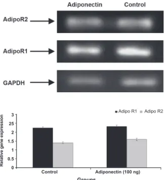
Expression of mRNA AdipoR1 and AdipoR2 in normal human endometrial stromal cells
In this study, expression of Adipo R1 and Adipo R2 in normal human endometrial stromal cells in the secretory phase in the presence and absence of Adiponectin was demonstrated by semi-quantita-tive RT-PCR analysis (Fig 3). The results revealed that expression of Adipo R1 and Adipo R2 mRNA in control group (without Adiponectin) and treat-ment group (100 ng/ml adiponectin) for 48 hours
did not indicate signiicant difference (p>0.05).
3 25 2 1.5 1 0.5 0
Control Adiponectin (100 ng)
Groups
Relative gene expression
Adipo R1 Adipo R2
Fig 3: Expression of Adipo R1 and Adipo R2 in normal hu-man endometrial stromal cells with and without Adiponectin in the secretory phase was demonstrated by semi-quantita-tive RT-PCR analysis. Expression of Adipo R1 and Adipo R2 mRNA in control group and treatment group (100 ng/ml
adiponectin) did not indicate signiicant difference (p>0.05).
Discussion
In our study, the in vitro effect of adiponectin on viability of normal HESCs and expression of Adipo R1 and Adipo R2 receptors was examined.
The indings indicated that adiponectin depending
on dose and time decreased the viability of HESCs
signiicantly. This inding, conirms the indings
reported by Cong et al. which regard to the inhibi-tory effect of adiponectin on the endometrial car-cinoma cell lines (HEC-1-A and RL95-2) in the culture (23) as well as anti-proliferative effects on trophoblast cells and trophoblast cell lines (JEG-3
A B
and BeWO) and decreasing their numbers in the culture (21).
The effects of adiponectin on cell death and decreasing stromal cells count, in this study, was observed with concentrations much lower than normal level which is normally circulating in hu-man blood serum (24, 25). Furthermore, the ob-tained results in this study are compatible with
the indings of previous research regarding the
decreasing impact of adiponectin on the viability of various cancer cells such as breast cancer cell line (MCF7), prostate, endothelial cancer and bone cells (26-29).
Lower level of adiponectin is an independ-ent risk factor in the incidence of infertility and reproduction and different genital cancers in epidemiological studies. The direct and indirect
mechanisms that inluence this phenomenon are
not still well-known (30). However, it seems that adiponectin exerts its biological effects through two receptors named Adipo R1 and Adipo R2. Takemura et al. in 2006 showed the expression of two receptors of adiponectin in the epithelial and endometrial stromal cells of endometrial tis-sue (31). The presence of these two receptors in various normal tissues and cancer cells has been
conirmed (32-34).
In the present study, the expression of Adipo R1 and Adipo R2 mRNA in the absence of adiponec-tin as well as presence of 100 ng/ml adiponecadiponec-tin in normal stromal cells was analyzed. Expression of these two receptors in these cells was observed
which conirms the indings of previous studies in
which the presence of these receptors in endome-trial stromal cells in both secretory and prolifera-tive phases was demonstrated (31). However, the results of our study revealed that adiponectin did
not have signiicant effect on the expression of
Adipo R1 and Adipo R2 receptors.
In another study, the expression of Adipo R1 and Adipo R2 receptors in human normal endometrial and endometrial cancerous tissue in the presence of adiponectin (in vitro) was investigated. The
indings showed that adiponectin decreased cell
proliferation in human endometrial cancerous tis-sue via adiponectin receptors and the level of Ad-ipo R1 expression was higher than that of AdAd-ipo R2 but the level of expression of receptors in
can-cerous tissue did not indicate signiicant difference
compared to normal non-cancerous tissue (30). The recent research has indicated that expression of Adipo R1 in breast cancer cells (32) and human endometrial cancerous tissue (23) is higher than that of Adipo R2.
These findings are compatible with the results of our study. The reason for this could be be-cause of adiponectin binding to Adipo R1 and Adipo R2 receptors and the ability of these recep-tors in activating ligand-dependent AMP-activated protein kinase (AMPK). Activation of AMPK re-sults in decreasing cell proliferation and increasing the number of inhibited cells in G1/G0 phase and consequently inducing cell death (35).
Conclusion
Adiponectin inhibit endometrial stromal cell proliferation in dose and time dependant manner, and cause cell death. It can suggest as anti-endo-metriosis agent.
For further studies on the effect of adiponectin in inhibition of progressive development and prolif-eration of endometriotic cells, endometrial stromal cells of endometriosis patients should be used and the function and expression of its receptors in the development of the disease must be investigated.
Acknowledgement
The authors would like to thank the Fertility and Infertility Research Center, Kermanshah sity of Medical Science and Islamic Azad Univer-sity, Branch of Sciences and Research of Tehran
for funding this work. There is no conlict of inter -est in this study.
References
1. Garaulet M, Hernández-Morante JJ, de Heredia FP, Tébar FJ. Adiponectin, the controversial hormone. Public Health Nutr. 2007; 10(10A): 1145-1150.
2. Zavalza-Gómez AB, Anaya-Prado R, Rincón-Sánchez AR, Mora-Martínez JM. Adipokines and insulin resistance during pregnancy. Diabetes Res Clin Pract. 2008; 80(1): 8-15.
3. Wang Y, Lam KS, Yau MH, Xu A. Post-translational
modi-ications of adiponectin: mechanisms and functional impli -cations. Biochem J. 2008; 409(3): 623-633.
4. Engeli S, Feldpausch M, Gorzelniak K, Hartwig F, Heintze U, Janke J, et al. Association between adiponectin and
mediators of inlammation in obese women. Diabetes.
2003; 52(4): 942-947.
109-114.
6. Otake S, Takeda H, Suzuki Y, Fukui T, Watanabe S,
Ishi-hama K, et al. Association of visceral fat accumulation
and plasma adiponectin with colorectal adenoma:
evi-dence for participation of insulin resistance. Clin Cancer
Res. 2005; 11(10): 3642-3646.
7. Freedland SJ, Sokoll LJ, Platz EA, Mangold LA, Bruzek DJ, Mohr P, et al. Association between serum adiponec-tin, and pathological stage and grade in men undergoing radical prostatectomy. J Urol. 2005; 174(4 pt1): 1266-1270.
8. Ishikawa M, Kitayama J, Kazama S, Hiramatsu T, Hatano K, Nagawa H. Plasma adiponectin and gastric cancer. Clin Cancer Res. 2005; 11(2 Pt 1): 466-472.
9. Yamauchi T, Nio Y, Maki T, Kobayashi M, Takazawa
T, Iwabu M, et al. Targeted disruption of Adipo R1 and Adipo R2 causes abrogation of adiponectin binding and
metabolic actions. Nat Med. 2007; 13(3): 332–339. 10. Kadowaki T, Yamauchi T. Adiponectin and adiponectin
receptors. Endocr Rev. 2005; 26(3): 439-451.
11. Yamauchi T, Kamon J, Ito Y, Tsuchida A, Yokomizo T, Kita
S, et al. Cloning of adiponectin receptors that mediate antidiabetic metabolic effects. Nature. 2003; 423(6941):
762-769.
12. Kharroubi I, Rasschaert J, Eizirik DL, Cnop M.
Expres-sion of adiponectin receptors in pancreatic beta cells. Bi -ochem Biophys Res Commun. 2003; 312(4): 1118-1122. 13. Chinetti G, Zawadski C, Fruchart JC, Staels B. Expression
of adiponectin receptors in human macrophages and reg
-ulation by agonists of the nuclear receptors PPAR alpha,
PPAR gamma, and LXR. Biochem Biophys Res Commun. 2004; 314(1): 151-158.
14. Arita Y, Kihara S, Ouchi N, Takahashi M, Maeda K,
Miya-gawa J, et al. Paradoxical decrease of an adipose-specif -ic protein, adiponectin, in obesity. Biochem Biophys Res Commun. 1999; 257(1): 79-83.
15. Takemura Y, Osuga Y, Harada M, Hirata T, Koga K, Mo-rimoto C, et al. Serum adiponectin concentrations are decreased in women with endometriosis. Hum Reprod. 2005; 20(12): 3510-3513.
16. Soliman PT, Wu D, Tortolero-Luna G, Schmeler KM, Slomovitz BM, Bray MS, et al. Association between adiponectin, insulin resistance, and endometrial cancer. Cancer. 2006; 106(11): 2376-2381.
17. Yi KW, Shin JH, Park HT, Kim T, Kim SH, Hur JY.
Re-sistin concentration is increased in the peritoneal luid of women with endometriosis. Am J Reprod Immunol.
2010; 64(5): 318-323.
18. Takayama K, Zeitoun K, Gunby RT, Sasano H, Carr BR,
Bulun SE. Treatment of severe postmenopausal endo -metriosis with an aromatase inhibitor. Fertil Steril. 1998; 69(4): 709-713.
19. Khazaei M, Chobsaz F, Khazaei S. The effect of differ
-ent doses of clomiphene citrate on morphology and pro
-liferation of human endometrial stromal cells in in-vitro
culture. Babol J Med Sci. 2010; 12(2): 1-12.
20. Esfandiari N, Ai J, Khazaei M, Nazemian Z, Jolly A,
Casper RF. Angiogenesis following three-dimensional culture of isolated human endometrial stromal cells. Int J
Fertil Steril. 2008; 2(1): 19-22.
21. Benaitreau D, Dieudonné MN, Dos Santos E, Leneveu
MC, Mazancourt Pd, Pecquery R. Antiproliferative ef
-fects of adiponectin on human trophoblastic cell lines
JEG-3 and BeWo. Biol Reprod. 2009; 80(6): 1107–1114. 22. Freshney R. Culture of animal cells: A manual of basic
technique. 5Th ed. New York: Wiley-Liss; 2005; 1-8. 23. Cong L, Gasser J, Zhao J, Yang B, Li F, Zhao AZ. Human
adiponectin inhibits cell growth and induces apoptosis in human endometrial carcinoma cells, HEC-1-A and RL95-2. Endocr Relat Cancer. 2007; 14(3): 713-720.
24. Corbetta S, Bulfamante G, Cortelazzi D, Barresi V, Cetin I, Mantovani G, et al. Adiponectin expression in human
fetal tissues during mid- and late gestation. J Clin Endo -crinol Metab. 2005; 90(4): 2397-2402.
25. Kajantie E, Hytinantti T, Hovi P, Andersson S. Cord
plas-ma adiponectin: a 20-fold rise between 24 weeks ges -tation and term. J Clin Endocrinol Metab. 2004; 89(8): 4031-4036.
26. Dieudonne MN, Bussiere M, Dos Santos E, Leneveu MC, Giudicelli Y, Pecquery R. Adiponectin mediates
an-tiproliferative and apoptotic responses in human MCF7
breast cancer cells. Biochem Biophys Res Commun. 2006; 345(1): 271-279.
27. Bråkenhielm E, Veitonmäki N, Cao R, Kihara S, Matsu -zawa Y, Zhivotovsky B, et al. Adiponectin-induced an-tiangiogenesis and antitumor activity involve caspase-mediated endothelial cell apoptosis. Proc Natl Acad Sci USA. 2004; 101(8): 2476-2481.
28. Berner HS, Lyngstadaas SP, Spahr A, Monjo M, Thommesen L, Drevon CA, et al. Adiponectin and its
receptors are expressed in bone-forming cells. Bone.
2004; 35(4): 842-849.
29. Bub JD, Miyazaki T, Iwamoto Y. Adiponectin as a growth inhibitor in prostate cancer cells. Biochem Biophys Res Commun. 2006; 340(4): 1158-1166.
30. Moon HS, Chamberland JP, Aronis K, Tseleni-Balafouta
S, Mantzoros CS. Direct role of adiponectin and adi -ponectin receptors in endometrial cancer: in vitro and ex vivo studies inhumans. Mol Cancer Ther. 2011, 10(12): 2234-2243.
31. Takemura Y, Osuga Y, Yamauchi T, Kobayashi M, Harada
M, Hirata T, et al. Expression of adiponectin receptors
and its possible implication in the human endometrium. Endocrinology. 2006; 147(7): 3203-3210.
32. Körner A, Pazaitou-Panayiotou K, Kelesidis T, Kelesidis I, Williams CJ, Kaprara A, et al. Total and high-molecular-weight adiponectin in breast cancer: in vitro and in vivo studies. J Clin Endocrinol Metab. 2007; 92(3): 1041-1048.
33. Motoshima H, Wu X, Mahadev K, Goldstein BJ.
Adi-ponectin suppresses proliferation and superoxide gen -eration and enhances eNOS activity in endothelial cells treated with oxidized LDL. Biochem Biophys Res Com-mun. 2004; 315(2): 264-271.
34. Luo XH, Guo LJ, Yuan LQ, Xie H, Zhou HD, Wu XP, et al.
Adiponectin stimulates human osteoblasts proliferation and differentiation via the MAPK signaling pathway. Exp
Cell Res. 2005; 309(1): 99-109.
35. Reynolds RK, Hu C, Baker VV. Transforming growth
factor-alpha and insulin-like growth factor-I, but not epi