8-Year Long-Term Outcome Comparison: Two
Ways to Exclude the Internal Iliac Artery
during Endovascular Aorta Repair (EVAR)
Surgery
Han Luo1☯, Bin Huang2☯, Ding Yuan2, Yi Yang2, Fei Xiong2, Guojun Zeng2, Zhoupeng Wu2, Xiyang Chen2, Xiaojiong Du2, Xiaorong Wen3, Chuncheng Liu3, Hongliu Yang1,
Jichun Zhao2*
1West China Medical School of Sichuan University, 37 Guo Xue Alley, Chengdu 610041, Sichuan Province, China,2Department of Vascular Surgery, West China Hospital, 37 Guo Xue Alley, Chengdu 610041, Sichuan Province, China,3Department of Ultrasound, West China Hospital, 37 Guo Xue Alley, Chengdu 610041, Sichuan Province, China
☯These authors contributed equally to this work.
Abstract
Purpose
To evaluate the 8-year long-term outcome after internal iliac artery (IIA) coverage with or without embolization in EVAR.
Patients and Methods
From January 2006 to December 2013, abdominal aortic aneurysm (AAA) subjects that underwent EVAR and IIA exclusion were recruited and analyzed retrospectively. All the subjects were divided into group A or B based on the presence or absence of intraoperative IIA embolization before coverage (group A: without embolization; group B: with emboliza-tion). The 30-day mortality, stent patency, and the incidences of endoleaks and ischemia of the buttocks and lower limbs were compared. The follow-up period was 96 months.
Result
There were 137 subjects (A: 74 vs. B: 63), 124 male (91.1%) and 13 female (9.5%), with a mean age of 71.6 years. There were no significant differences in the early outcomes of intraoperative blood loss (87.23±14.07 ml; A: 86.53±9.57 ml vs. B: 88.06±18.04 ml, p =
.545) and surgery time (87.13±9.25 min; A: 85.99±7.07 min vs. B: 88.48±11.19 min, p =
.130). However, there were significant differences in contrast consumption (65.18±9.85 ml;
A: 61.89±7.95 ml vs. B: 69.05±10.50 ml, p<.001) and intraoperative X-ray time (5.9±0.86
min; A: 5.63±0.49 min vs. B: 6.22±1.07 min, P<.001). The 30-day mortality was
approxi-mately 0.73%. In the follow-up analysis, no significant differences were identified in the
OPEN ACCESS
Citation:Luo H, Huang B, Yuan D, Yang Y, Xiong F, Zeng G, et al. (2015) 8-Year Long-Term Outcome Comparison: Two Ways to Exclude the Internal Iliac Artery during Endovascular Aorta Repair (EVAR) Surgery. PLoS ONE 10(7): e0130586. doi:10.1371/ journal.pone.0130586
Editor:Alberto Aliseda, University of Washington, UNITED STATES
Received:November 11, 2014
Accepted:May 22, 2015
Published:July 20, 2015
Copyright:© 2015 Luo et al. This is an open access article distributed under the terms of theCreative Commons Attribution License, which permits unrestricted use, distribution, and reproduction in any medium, provided the original author and source are credited.
Data Availability Statement:Due to ethical restrictions, data are available from the West China Hospital Ethics Committee for researchers who meet the criteria for access to confidential data.
Funding:This study was funded by the Planned Science and Technology Project of Sichuan Province, China (2012SZ0023).
incidence of endoleak (22 subjects; type I: A: 2 vs. B: 2, p = 1.000; type II: A: 8 vs. B: 4, p = .666; type III: A: 4 vs. B: 3, p = 1.000), occlusion (5 subjects; 4.35%; A: 1 vs. B: 4, p = .180), or ischemia (9 subjects; 7.83%; A: 3 vs. B: 6, p = .301). In the analysis of group B, although there were no significant differences between subjects with unilateral and bilateral IIA embolization, but longer hospital stays were required (P<.001), and a more severe compli-cation (skin and gluteus necrosis) occurred in 1 subject with bilateral IIA embolization.
Conclusion
IIA could be excluded during EVAR. IIA coverage without embolization had a good surgical and prognostic outcome, and this procedure was not different significantly from coverage with embolization in terms of endoleaks, patency and ischemia.
Introduction
Since Dr. Parodi first developed minimally invasive endovascular technology in 1991[1,2] to treat abdominal aortic aneurysms (AAAs), endovascular aortic repair (EVAR) has been the primary choice for AAA. Currently, nearly half of AAA patients undergo EVAR after IIA exclusion[3]. When the distant landing zone in the common iliac artery (CIA) is too short to anchor, the endograft limb should be extended into the external iliac artery (EIA), thus cover-ing the original internal iliac artery (IIA). Although some researchers and doctors advocate preserving the IIA because it is a crucial collateral artery of the lower extremities and pelvis[4– 7], excluding the IIA during EVAR has been proven safe, and it is an effective way to avoid endoleaks[3].
However, the methods for excluding the IIA have recently become controversial. Some studies suggest that IIA embolization with coils is much more effective than direct coverage with a stent in avoiding type II endoleaks[3,8,9]. However, other results show that excluding the IIA without embolization has a similar long-term outcome to the procedure with emboliza-tion[10–12].
Therefore, a retrospective cohort study was designed at our institution to compare the long-term outcome of two exclusion methods (absence or presence of a coil) and to evaluate the effectiveness of IIA exclusion. Here, we would like to report the results of our study.
Patients and methods
The patients that were included in this study were recruited from the Vascular Department of West China Hospital, Sichuan University, between January 2006 and December 2013.
Inclusion criteria. In total, 702 patients with a diagnosis of AAA were included in this study. Cases of AAA were diagnosed via ultrasound or computed tomography (CT), and iso-lated cases were diagnosed by magnetic resonance angiography (MRA). All the repaired AAAs were more than 5.5 cm in diameter or showed rapid growth (>0.5 cm over 6 months).
Exclusion criteria. The exclusion criteria consisted of four parts: A, open surgery; B, uni-lateral or biuni-lateral IIA(s) was (were) not excluded; C, instructions for use (IFU) were not fol-lowed. Three factors regarding the neck of the AAA were selected among the IFU as exclusion criteria, including the 1) length of aneurysm neck15 mm; 2) angulation of infrarenal neck >60°; and 3) neck atheroma with a thickness and length>5 mm (measured via preoperative
D, Surgery time: emergency EVAR, such as EVAR for ruptured AAA (rAAA). Here, rAAA indicates an emergent unstable hemodynamic condition during surgery, which would nega-tively influence a surgeon’s decision whether to preserve the IIA.[15] Moreover, emergent EVAR is a life-saving surgery; therefore, surgeons would try to shorten the operation time to cover the aneurysm leak as soon as possible.
Based on the inclusion and exclusion criteria, 137 subjects were enrolled into the final comparison analysis. All the enrolled subjects were divided into two groups according to the presence or absence of intraoperative embolization IIA before coverage. Subjects without embolization were included in group A, and those with embolization were placed in group B.
Internal iliac artery (IIA) management
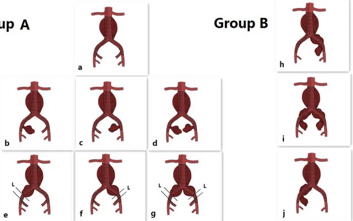
1. Indications for IIA exclusion without embolization (Group A):
1) Standard AAA [not coupled with common iliac artery aneurysm (CIAA)], but the land-ing zone in CIA was too short to anchor.
2) AAA coupled with IIAA.
3) AAA coupled with CIAA (unilateral or bilateral), CIAA did not invade the original IIA,
Fig 1.*Indications for group A (a, b, c, d, e, f, g): a: Standard abdominal aortic aneurysm (AAA; not coupled with common iliac artery aneurysm, CIAA), but the landing zone in the common iliac artery (CIA) was too short to anchor.b, c, d: Standard AAA coupled with internal iliac artery aneurysm (IIAA). e, f, g: AAA coupled with CIAA (unilateral or bilateral), and CIAA did not invade the original IIA, and the distance (L) from the distal end of the CIAA to the original IIA was less than 10–15 mm without stenosis or ectasia.*Indications for group B (h, I, j): AAA coupled with CIAA (unilateral or bilateral), and the
CIAA invaded the original internal iliac artery (IIA).*Stents were supplied by Medtronic Inc (TALENT and ENDURANT series). Coils were supplied by Johnson & Johnson Company (Amplatzer).
andL(shown inFig 1, the distance from the distal end of the CIAA to the original IIA) was less than 10–15 mm without stenosis or ectasia (a, b, c, d, e, f, g).
2. Indications for IIA exclusion with embolization (Group B):
AAA coupled with CIAA that invaded the original IIA, andLwas less than 10–15 mm (h, i, j).
The Indications for IIA embolization are presented inFig 1
Postoperative management after IIA exclusion. After EVAR, antiplatelet, anticoagula-tion and vasodilator therapies are essential for the patients (Aspirin, 100 mg/day; low molecu-lar weight heparin, 600 mg/day; and PGE2, 40μg/day); simultaneously, the skin temperature
of the buttocks and limbs should be carefully checked, especially after IIA exclusion. If the skin temperature is lower than normal or tenderness and skin necrosis appear, reconstruction of the IIA should be considered in a timely manner.
Follow-up: After EVAR, discharged subjects were followed at the 1st, 3rd, 6th, and 12th months, with annual visits thereafter for CT evaluations. The recorded endpoints included 30-day mortality, stent patency, and the occurrences of endoleaks and ischemic complications, such as ischemic colitis, spinal cord infraction, skin necrosis and chronic buttock claudication. The follow-up period ceased in June 2014.
Analysis Method. All the statistical analyses were performed using SPSS version 16 (SPSS Inc, Chicago, IL). The data are presented as the mean±standard deviation for continuous vari-ables and as the frequency (percentage) for categorical varivari-ables, which were compared using the two-sample t-test, Fisher exact test, and Pearson Chi-square test where appropriate. Overall survival and patency curves were generated using the Kaplan-Meier method, and the log-rank test was used to compare the differences. Differences with a P value<.05 were considered to be significant.
Ethics
This study was approved by the Ethics Committee of West China Hospital, Sichuan University. All the study participants provided written informed consent stating that the clinical data could be used in clinical research.
Results
Baseline comparisons
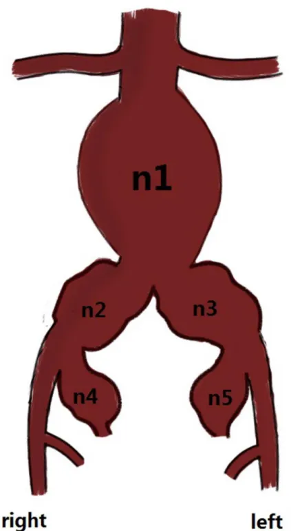
Seventy-four subjects were in group A, and 63 subjects were in group B. Among the 137 conse-cutive subjects, 124 (91.1%) were male, and 13 (9.5%) were female, with mean ages of 71.56 and 71.67 years, respectively. The vascular morphologic characteristics of the 137 subjects are shown inFig 2. General patient information is provided inTable 1.
Fig 2.*n1: abdominal aortic aneurysm (AAA); n2: right common iliac artery aneurysm (CIAA); n3: left CIAA; n4: right internal iliac artery aneurysm (IIAA); and n5: left IIAA.*n1: 47; n1+2: 17; n1+3: 5; n1+4: 6; n1+5: 3; n1+2+3: 25; n1+2+5: 7; n1+2+4: 2; n1+3+4: 1; n1+3+5:1; n1+4+5: 3; n1+2+3+4: 4; n1+2+4+5: 2; n1+2+3+5: 5; and n1+2+3+4+5: 9. n1+2represents an AAA that invaded the right common iliac artery; the others were considered likely.*Fig 2 is just an illustration; we cannot show the exact anatomical details.
Early outcome comparison
All 137 patients underwent a successful EVAR, and there were no conversions to laparotomy. Fifty-four subjects (39.4%) received general anesthesia, 17 subjects (12.4%) had epidural anes-thesia, and 66 subjects (48.2%) received local anesthesia. There was no significant difference in intraoperative blood loss (87.23±14.07 ml; A: 86.53±9.57 ml vs B: 88.06±18.04 ml, p = .545) or surgery time (87.13±9.25 min; A: 85.99±7.07 min vs B: 88.48±11.19 min, p = .130), and there were no blood transfusions during the operations. However, contrast consumption (65.18±9.85 ml; A: 61.89±7.95 ml vs B: 69.05±10.50 ml, p<.001) and intraoperative X-ray time (5.9±0.86 min; A: 5.63±0.49 min vs B: 6.22±1.07 min, P<.001) were significantly differ-ent. The 30-day mortality was approximately 0.73%. The early outcome comparisons are shown inTable 3.
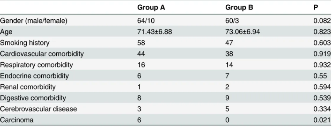
Table 1. General information.
Group A Group B P
Gender (male/female) 64/10 60/3 0.082
Age 71.43±6.88 73.06±6.94 0.823
Smoking history 58 47 0.603
Cardiovascular comorbidity 44 38 0.919
Respiratory comorbidity 16 14 0.932
Endocrine comorbidity 6 7 0.55
Renal comorbidity 1 2 0.594
Digestive comorbidity 8 9 0.539
Cerebrovascular disease 3 5 0.334
Carcinoma 6 0 0.021
Hypertension was defined as a pre-hemorrhage blood pressure documented as>140 mmHg systolic or
>90 mmHg diastolic or the use of an anti-hypertensive agent. CAD and cerebral artery disease were defined by medical history or CTA of the coronary and cerebral artery, respectively. Respiratory failure was defined as an oxygen PaO2 greater than 80 mmHg (11 kPa) and/or a carbon dioxide PaCO2 less than 45 mmHg (6.0 kPa) or the need for intubation. Renal insufficiency was defined as serum creatinine2 mg/dL. COPD, emphysema, bronchitis, and pulmonary bullae were mainly defined by medical history.
p<0.05 was considered statistically significant. doi:10.1371/journal.pone.0130586.t001
Table 2.
Group A Group B P
Diameter of neck 21.5±2.9 21.2±3.8 0.635
Length of neck 25.5±7.5 25.0±8.6 0.737
Diameter of aneurysm 56.5±14.7 51.8±17.8 0.110
Length of aneurysm 78.5±19.6 87.5±29.5 0.259
Diameter of left CIA 24.4±10.6 24.7±10.6 0.880
Diameter of right CIA 26.5±7.9 26.6±9.1 0.956
Diameter of left IIA 26.1±4.2 25.6±11.2 0.831
Diameter of right IIA 23.8±5.3 26.5±14.8 0.411
CIA: Common Iliac Artery; IIA: Internal Iliac Artery mm: millimeter
Outcome of follow-up
The study spanned 8 years, from June 2006 to June 2014, and the mean follow-up period was 61.2 months. No deaths or graft-associated deaths occurred during the follow-up period. Twenty-one patients were lost during follow-up, and 115 patients were followed. The overall incidences of endoleak, occlusion, and ischemic complications were 19.1%, 5.22%, and 4.35%, respectively. The comparisons of the incidences of endoleak, stent occlusion and ischemic complications between groups A and B during follow-up are shown inTable 4. The follow-up comparison between groups A and B is presented inFig 3(a, b, c).
Endoleak occurred in 22 cases (A: 13 vs B: 9), with 4 Type I cases (A: 2 vs B: 2), 12 Type II cases (A: 8 vs B: 4), and 7 Type III cases (A: 4 vs B: 3). Type I and III endoleaks simultaneously occurred in one case. One subject in group A with a Type II endoleak underwent an endovas-cular intervention for an increased aneurysm and newly developed CIAA. In the other 21 cases, the endoleak disappeared or shrunk, and the size of the aneurysm did not increase during follow-up. Furthermore, the survival analysis revealed that there was no significant difference in the long-term incidence of endoleak between group A and B (P = .537;Fig 4A). Therefore, intraoperative embolization of the IIA with coils before coverage did not decrease the long-term incidence of type II endoleak.
Table 3. Early outcome comparisons between groups A and B.
Group A Group B P
Anesthetics
General 25 29 0.163
Epidural 10 7 0.797
Local 34 32 0.610
Intraoperative blood loss 86.53±9.57 88.06±18.04 0.545
Surgery time 85.99±7.07 88.48±11.19 0.130
Contrast consumption 61.89±7.95 69.05±10.50 <.001
Intraoperative X-ray time 50.63±0.49 60.22±1.07 <.001
30-day mortality 1 0 1.000
p<0.05 was considered statistically significant.
Intraoperative blood loss and contrast consumption were measured in milliliters, and surgery time and intraoperative X-ray time were measured in minutes.
doi:10.1371/journal.pone.0130586.t003
Table 4. Incidences of complications in groups A and B.
Total Group A Group B P
Endoleak 22 13 9 0.647
Type I 4 2 2 1.000
Type II 12 8 4 0.666
Type III 7 4 3 1.000
Occlusion 5 1 4 0.180
Surgery 2 0 2 1.000
Conservative 3 1 2 1.000
Ischemic Complication `9 3 6 0.301
p<0.05 was considered statistically significant.
Only 5 subjects (A: 1 vs B: 4) suffered from stent occlusion, and 2 symptomatic subjects underwent surgery (A: 0 vs B: 2, femoral-femoral bypass surgery and embolectomy surgery). Fig 3. a: No significant difference was found in the survival analysis of endoleak between groups A and B (A: group A, B: group B) (P = .537).Internal iliac artery (IIA) coverage with embolization did not reduce the long-term risk of endoleak. b: No significant difference was identified in the survival analysis of patency between groups A and B (A: group A, B: group B) (P = .143). The incidence of occlusion during follow-up was not significantly different between groups A and B. c: No significant difference was found in the survival analysis of ischemic complications between groups A and B (A: group A, B: group B) (P = .260). However, the incidence of ischemic complications was higher in group B than in group A (A: 4.84% vs B: 11.11%), and more severe ischemic complications occurred in group B.
doi:10.1371/journal.pone.0130586.g003
Fig 4. There was no significant difference in the incidence of postoperative ischemia between groups B1 (subjects with unilateral IIA exclusion, n = 4) and B2 (bilateral IIA exclusion, n = 2).However, B1 was obviously different from B2 in terms of hospital stays and the severity of the ischemic complications. The two subjects in B2 had hospital stays of 12 and 17 days; by contrast, the hospital stays of the subjects in B1 were 3, 5, 5, and 6 days (P<
.001). A severe ischemic complication (gluteal skin necrosis) occurred in one subject in group B2 with a claudication distance of less than 100 meters. Gluteal soreness with a claudication distance of approximately 150 meters occurred in another subject in group B2. By contrast, gluteal ischemia and limb ischemia in group B1 were mild.
Ischemic syndrome did not appear in the two patients thereafter. The 3 asymptomatic subjects (A: 1 vs B: 2) received conservative therapy. During conservative therapy, the ischemic symp-toms were aggravated: the dermal temperature of the lower extremities decreased in the 2nd month after EVAR and then recovered at the end of the 6thmonth. Two other cases did not have any uncomfortable feelings during follow-up. The survival analysis revealed no significant differences between groups A and B in terms of stent occlusion (P = .143;Fig 4B), indicating that coverage with or without embolization does not affect long-term stent patency. Overall, the 5-year stent patency rate in our institution was 95.7%.
Ischemic complications, such as pelvic ischemia, spinal cord infraction, limb and gluteal ischemia, were a critical concern during follow-up. During the follow-up period, spinal cord infraction and pelvic ischemia were not observed in any subjects. Gluteal ischemia (A: 1 vs B: 4, P = 1.000) and limb ischemia (A: 2 vs B: 3, P = 1.000) after EVAR were observed in 9 subjects (A: 3 vs B: 6), and the incidence was 7.83%. One subject in group B simultaneously presented with buttocks claudication and limb ischemia and underwent a femoral-femoral bypass and embolectomy surgery 4 months after EVAR. Eight other subjects received conservative ther-apy. After 2–12 months of conservative therapy, ischemic syndrome was relieved in 8 cases. A comparison of the incidence of ischemic complications between groups A and B is shown in
Table 5. Furthermore, no significant differences in ischemic complications between groups A and B were identified in the survival analysis (P = .26). Similarly, there were no significant dif-ferences in gluteal ischemia or limb ischemia based on the survival analysis. However, the inci-dence of ischemic complications was higher in group B than in group A (A: 4.84% vs B: 11.11%). Moreover, as shown inFig 4C, there was a tendency towards chronic ischemia being more likely in group B and acute ischemia being more likely in group A. This trend will be dis-cussed later in the discussion section.
The subgroup survival analysis (Fig 4) of group B suggested that there was no significant dif-ference in the incidence of postoperative ischemia between groups B1 (subjects with unilateral IIA exclusion; n = 4) and B2 (bilateral IIA exclusion; n = 2). However, although there were no numerical differences between these subgroups, B1 obviously differed from B2 in terms of pital stays and the severity of the ischemic complications. Two subjects in B2 stayed in the hos-pital for 12 and 17 days; by contrast, the patients in B1 stayed for 3, 5, 5, and 6 days (P<.001). A severe ischemic complication (gluteal skin necrosis) appeared in one subject in group B2 with a claudication distance of less than 100 meters. Gluteal soreness with a claudication
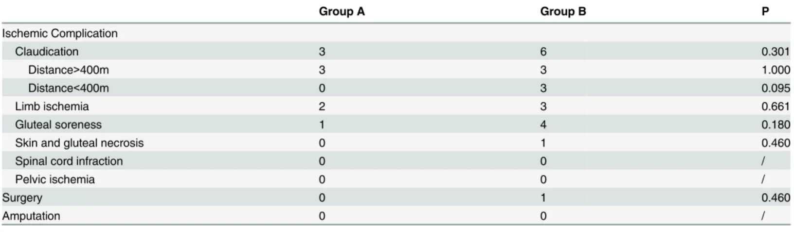
Table 5. Ischemic complications and ischemia-related index comparisons.
Group A Group B P
Ischemic Complication
Claudication 3 6 0.301
Distance>400m 3 3 1.000
Distance<400m 0 3 0.095
Limb ischemia 2 3 0.661
Gluteal soreness 1 4 0.180
Skin and gluteal necrosis 0 1 0.460
Spinal cord infraction 0 0 /
Pelvic ischemia 0 0 /
Surgery 0 1 0.460
Amputation 0 0 /
p<0.05 was considered statistically significant.
distance of approximately 150 meters occurred in another subject in group B2. By contrast, glu-teal ischemia and limb ischemia in group B1 were mild.
Discussion
The incidence of AAA is increasing, and it is particularly obvious in the elderly. With the development of new techniques and increases in standards of living, more AAA patients would like to undergo an EVAR[3]. However, the fate of IIA remains disputable. It is controversial whether excluding the IIA with a coil is more effective at preventing type II endoleak after EVAR. Moreover, long-term results (greater than five years) are seldom reported but are cru-cial for clinical practice and guidance. Accordingly, this type of research is meaningful.
IIA exclusion may lead to gluteal and colonic ischemia and erectile dysfunction (ED)[7,8]. Therefore, a comprehensive preoperative evaluation of blood supply in the lower extremities and buttocks is critical for surgery and prognostics, and IIA reconstruction must be performed when necessary, or postoperative ischemia is inevitable. In Dr. Rana’s research, freedom from buttock claudication was higher after open repair than after endovascular repair (79% vs 59%; P = 0.05), and reconstructions for IIA flow preservation had very good long-term patency. Every effort should be made to preserve IIA flow, as it leads to significantly better outcomes in terms of pelvic ischemic symptoms and buttock claudication[7].
In this study, there was no obvious significant difference in 5-year-long prognosis between group A (coverage without embolization) and group B (coverage with embolization). However, blood loss, surgery time, X-ray time, and contrast consumption were superior in group A com-pared to group B. Thus, patients in group A would likely receive a better long-term prognosis with less blood loss and contrast consumption as well as reduced surgery and X-ray times, which would be greatly beneficial for the patient. This likely conclusion was supported by Dr. Papazoglou’s research[12].
Furthermore, chronic ischemia rarely occurred in group A. A possible reason may be that the permeability of and unsolidified adherence to covered stents contributed to little blood flow into the IIA, leading to slow thrombosis in the IIA. Until a solidified embolism forms, there is enough time to establish collateral circulation, which ensures adequate blood supply to the lower extremities and buttocks. However, IIA coverage with embolization in group B caused fast, even acute, embolism that was not beneficial for the formation of collateral circula-tion, especially when bilateral IIA embolizations were performed. However, this tendency needs to be confirmed in future studies. If confirmed, it will be better to cover bilateral IIAs directly in staged surgery.
severe ischemia (skin necrosis). Overall, ischemia appears more frequently in cases with bilat-eral IIA exclusion, and severe ischemia occurs more readily3.
Unfortunately, the evidence and outcome would be more powerful if this study was a ran-domized controlled trial (RCT) or included more participants. However, the results of our study are consistent with those of previous scientific studies, which encourages us to conduct a larger cohort study to confirm the current findings. In addition, the levels of the IIA exclusion with a coil were not exactly the same. Most IIAs were excluded at the trunk, which creates the lowest risk of ischemia, according to some research studies[18–20]. However, fewer than five subjects had exclusions at the branch level. Although some studies showed significant differ-ences between patients with IIA embolization at different levels, the effect of the embolization level is negligible. If an iliac branch device (IBD) could be introduced into routine clinical prac-tice in our hospital, it would be interesting to compare patients with and without IIA preserva-tion. Interestingly, no significant differences in ischemic complications between patients with IIA embolization and with an IBD were reported in a recent study[21].
Conclusion
In the traditional view, IIA embolization is an effective way to reduce the incidence of endo-leaks, especially type II endoleaks. However, based on the present study, IIA coverage without embolization does not increase the long-term incidence of endoleak. Moreover, although there was no significant difference in ischemic complications with and without coil embolization, the differences in the severity of the ischemic complications and the reduced contrast consumption and X-ray time between the groups suggest that direct coverage is more effective and
beneficial.
Acknowledgments
This study was funded by the Planned Science and Technology Project of Sichuan Province, China (2012SZ0023).
Author Contributions
Conceived and designed the experiments: JCZ HL. Performed the experiments: BH DY YY GJZ FX ZPW XYC XRW CCL. Analyzed the data: HL HLY. Contributed reagents/materials/ analysis tools: XRW CCL. Wrote the paper: HL HLY. Follow-up of patients after EVAR: ZPW XYC XJD.
References
1. Parodi JC, Palmaz JC, Barone HD. Transfemoral intraluminal graft implantation for abdominal aortic aneurysms. Annals of vascular surgery. 1991; 5(6):491–9. Epub 1991/11/01. doi:10.1007/bf02015271
PMID:1837729.
2. Yao JS, Eskandari MK, Parodia J. Transfemoral intraluminal graft implantation for abdominal aortic aneurysms, 1991: two decades later. Annals of vascular surgery. 2012; 26(7):895–905. Epub 2012/09/
05. doi:10.1016/j.avsg.2012.06.001PMID:22944565.
3. Schoder M, Zaunbauer L, Holzenbein T, Fleischmann D, Cejna M, Kretschmer G, et al. Internal iliac artery embolization before endovascular repair of abdominal aortic aneurysms: frequency, efficacy, and clinical results. AJR American journal of roentgenology. 2001; 177(3):599–605. Epub 2001/08/23.
doi:10.2214/ajr.177.3.1770599PMID:11517053.
4. Chun JY, Mailli L, Abbasi MA, Belli AM, Gonsalves M, Munneke G, et al. Embolization of the internal iliac artery before EVAR: is it effective? Is it safe? Which technique should be used? Cardiovascular and interventional radiology. 2014; 37(2):329–36. Epub 2013/06/19. doi:10.1007/s00270-013-0659-2
PMID:23771327.
of the International Society of Endovascular Specialists. 2002; 9(4):488–92. Epub 2002/09/12. doi:10.
1583/1545-1550(2002)009<0488:aheptp>2.0.co;2PMID:12223010.
6. Cochennec F, Marzelle J, Allaire E, Desgranges P, Becquemin JP. Open vs endovascular repair of abdominal aortic aneurysm involving the iliac bifurcation. J Vasc Surg. 2010; 51(6):1360–6. Epub 2010/
03/30. doi:10.1016/j.jvs.2010.01.032PMID:20347547.
7. Rana MA, Kalra M, Oderich GS, de Grandis E, Gloviczki P, Duncan AA, et al. Outcomes of open and endovascular repair for ruptured and nonruptured internal iliac artery aneurysms. Journal of Vascular Surgery. 2014; 59(3):634–44. doi:10.1016/j.jvs.2013.09.060PMID:24571938
8. Criado FJ, Wilson EP, Velazquez OC, Carpenter JP, Barker C, Wellons E, et al. Safety of coil emboliza-tion of the internal iliac artery in endovascular grafting of abdominal aortic aneurysms. J Vasc Surg. 2000; 32(4):684–8. Epub 2000/09/30. doi:10.1067/mva.2000.110052PMID:11013031.
9. Wolpert LM, Dittrich KP, Hallisey MJ, Allmendinger PP, Gallagher JJ, Heydt K, et al. Hypogastric artery embolization in endovascular abdominal aortic aneurysm repair. J Vasc Surg. 2001; 33(6):1193–8.
Epub 2001/06/05. PMID:11389417.
10. Heye S, Nevelsteen A, Maleux G. Internal iliac artery coil embolization in the prevention of potential type 2 endoleak after endovascular repair of abdominal aortoiliac and iliac artery aneurysms: effect of total occlusion versus residual flow. Journal of vascular and interventional radiology: JVIR. 2005; 16(2 Pt 1):235–9. Epub 2005/02/17. doi:10.1097/01.rvi.0000143842.36512.dfPMID:15713924.
11. Wyers MC, Schermerhorn ML, Fillinger MF, Powell RJ, Rzucidlo EM, Walsh DB, et al. Internal iliac occlusion without coil embolization during endovascular abdominal aortic aneurysm repair. J Vasc Surg. 2002; 36(6):1138–45. Epub 2002/12/07. doi:10.1067/mva.2002.129639PMID:12469044. 12. Papazoglou KO, Sfyroeras GS, Zambas N, Konstantinidis K, Kakkos SK, Mitka M. Outcomes of
endo-vascular aneurysm repair with selective internal iliac artery coverage without coil embolization. J Vasc Surg. 2012; 56(2):298–303. Epub 2012/05/11. doi:10.1016/j.jvs.2011.08.063PMID:22572010. 13. Nakai M, Sato M, Sato H, Sakaguchi H, Tanaka F, Ikoma A, et al. Midterm results of endovascular
abdominal aortic aneurysm repair: comparison of instruction-for-use (IFU) cases and non-IFU cases. Japanese journal of radiology. 2013; 31(9):585–92. Epub 2013/06/14. doi:
10.1007/s11604-013-0223-7PMID:23760672.
14. Hoshina K, Hashimoto T, Kato M, Ohkubo N, Shigematsu K, Miyata T. Feasibility of endovascular abdominal aortic aneurysm repair outside of the instructions for use and morphological changes at 3 years after the procedure. Annals of vascular diseases. 2014; 7(1):34–9. Epub 2014/04/11. doi:10.
3400/avd.oa.13-00073PMID:24719660; PubMed Central PMCID: PMCPmc3968413.
15. Mehta M, Taggert J, Darling RC 3rd, Chang BB, Kreienberg PB, Paty PS, et al. Establishing a protocol for endovascular treatment of ruptured abdominal aortic aneurysms: outcomes of a prospective analy-sis. J Vasc Surg. 2006; 44(1):1–8; discussion Epub 2006/07/11. doi:10.1016/j.jvs.2006.02.057PMID:
16828417.
16. Lin PH, Bush RL, Chaikof EL, Chen C, Conklin B, Terramani TT, et al. A prospective evaluation of hypo-gastric artery embolization in endovascular aortoiliac aneurysm repair. J Vasc Surg. 2002; 36(3):500–
6. Epub 2002/09/10. PMID:12218973.
17. Johnston KW. Multicenter prospective study of nonruptured abdominal aortic aneurysm. Part II. Vari-ables predicting morbidity and mortality. J Vasc Surg. 1989; 9(3):437–47. Epub 1989/03/01. PMID:
2646460.
18. Bratby MJ, Munneke GM, Belli AM, Loosemore TM, Loftus I, Thompson MM, et al. How safe is bilateral internal iliac artery embolization prior to EVAR? Cardiovascular and interventional radiology. 2008; 31 (2):246–53. Epub 2007/10/25. doi:10.1007/s00270-007-9203-6PMID:17957407.
19. Engelke C, Elford J, Morgan RA, Belli AM. Internal iliac artery embolization with bilateral occlusion before endovascular aortoiliac aneurysm repair-clinical outcome of simultaneous and sequential inter-vention. Journal of vascular and interventional radiology: JVIR. 2002; 13(7):667–76. Epub 2002/07/18.
PMID:12119323.
20. Kritpracha B, Pigott JP, Price CI, Russell TE, Corbey MJ, Beebe HG. Distal internal iliac artery emboli-zation: a procedure to avoid. J Vasc Surg. 2003; 37(5):943–8. Epub 2003/05/21. doi:10.1067/mva.
2003.251PMID:12756337.