Dysfunction: A Systematized Approach
Anatomia Radiológica da Disfunção Erétil Arteriogénica:
Uma Abordagem Sistematizada
1. Departamento de Radiologia. Hospital Saint Louis. Lisboa. Portugal.
2. Centro de Investigação de Angiomorfologia. Departamento de Anatomia. Faculdade de Ciências Médicas da Universidade Nova de Lisboa. Lisboa. Portugal. Recebido: 15 de Dezembro de 2012 - Aceite: 25 de Fevereiro de 2013 | Copyright © Ordem dos Médicos 2013
José António PEREIRA1, Tiago BILHIM1,2, Hugo RIO TINTO1, Lúcia FERNANDES1, João MARTINS PISCO1, João
GOYRI-O’NEIL2
Acta Med Port 2013 May-Jun;26(3):219-225
RESUMO
Introdução: A disfunção erétil é uma doença com elevada prevalência existindo crescente interesse na sua terapêutica endovascular.
Devido à complexidade do sistema arterial pélvico masculino, o conhecimento anatómico é fundamental. Avaliou-se a aplicabilidade da classificação de Yamaki na avaliação de doentes com disfunção erétil arteriogénica usando a Angiografia Tomográfica Computorizada e a Angiografia Digital de Subtração.
Métodos: Análise retrospetiva dos achados imagiológicos de Angiografia Tomográfica Computorizada e Angiografia Digital de
Subtra-ção em 21 doentes do sexo masculino, com suspeita de disfunSubtra-ção erétil arteriogénica, que foram submetidos a embolizaSubtra-ção pélvica seletiva numa única instituição. A função erétil foi avaliada através do IIEF-5. O padrão de bifurcação da Artéria Ilíaca Interna foi car-acterizado de acordo com a classificação de Yamaki. O diagnóstico da disfunção erétil arteriogénica foi feita baseado na presença de lesões ateroscleróticas da Artéria Ilíaca Interna e da Artéria Pudenda Interna.
Resultados: A idade média foi de 67,2 anos; a média do IIEF foi 10,6 pontos. A Angiografia Tomográfica Computorizada e a
An-giografia Digital de Subtração permitiram a classificação de todos os 42 lados pélvicos de acordo com a classificação de Yamaki. Vinte e quatro lados pélvicos foram classificados como Grupo A (57%), nove como Grupo B (21,5%) e nove como Grupo C (21,5%). A Angiografia Digital de Subtração detectou 19 Artérias Pudendas Internas anormais (lesões ateroscleróticas) (45%). A Angiografia Tomográfica Computorizada detectou 24 Artérias Pudendas Internas anormais (57%).
Conclusão: Os achados por Angiografia Tomográfica Computorizada e Angiografia Digital de Subtração incluem estenoses e
oclu-sões da Artéria Ilíaca Interna e da Artéria Pudenda Interna. A classificação de Yamaki tem reprodutibilidade radiológica e permite o reconhecimento da Artéria Pudenda Interna em doentes com disfunção erétil arteriogénica.
Palavras-chave: Artérias/anatomia; Disfunção Eréctil; Angiografia Digital de Subtracção; Tomografia Computorizada.
AbStRACt
Introduction: Erectile Dysfunction is a highly prevalent disease and there is growing interest in its endovascular treatment. Due to
the complexity of the male pelvic arterial system, thorough anatomical knowledge is paramount. We evaluated the applicability of the Yamaki classification with Computerized Tomography Angiography and Digital Subtraction Angiography in the evaluation of patients with arteriogenic Erectile Dysfunction, illustrating the arterial lesions that can cause Erectile Dysfunction.
Methods: Single-center retrospective analysis of the Computerized Tomography Angiography and Digital Subtraction Angiography
imaging findings in 21 male patients with suspected arteriogenic Erectile Dysfunction that underwent selective pelvic arterial emboliza-tion. Assessment of erectile function was achieved using the IIEF-5. The branching patterns of the Internal Iliac Artery were classified according to the Yamaki classification. The diagnosis of arteriogenic Erectile Dysfunction was based on the presence of atherosclerotic lesions (stenoses and/or occlusions) of the Internal Iliac Artery or the Internal Pudendal Arteries.
Results: The mean patient age was 67.2 years; with a mean IIEF of 10.6 points. Computerized Tomography Angiography and Digital
Subtraction Angiography findings allowed classification of all the 42 pelvic sides according to the Yamaki classification. Twenty-four pelvic sides were classified as Group A (57%), 9 as Group B (21.5%) and 9 as Group C (21.5%). The Digital Subtraction Angiography detected 19 abnormal Internal Pudendal Arteries (with atherosclerotic lesions) (45%). The Computerized Tomography Angiography detected 24 abnormal Internal Pudendal Arteries (57%).
Conclusion: Computerized Tomography Angiography and Digital Subtraction Angiography findings of arteriogenic Erectile Dysfunction
include stenotic and occlusive lesions of the Internal Iliac Artery and Internal Pudendal Artery. The Yamaki classification is radiologically reproducible and allows easy recognition of the Internal Pudendal Artery in patients with arteriogenic Erectile Dysfunction.
Keywords: Arteries/anatomy & histology; Erectile Dysfunction/radiography; Angiography, Digital Subtraction; Tomography, X-Ray
Computed.
ARtIGO ORIGINAL
INtRODUCtION
Erectile dysfunction (ED) is the persistent inability to achieve or maintain an erection sufficient for satisfactory sexual performance.1 ED is highly prevalent, affecting more
than 150 million men worldwide.2 In Portugal, most recent
surveys indicate a prevalence between 5 - 28% depend-ing on the definition.3 As the male population ages and
awareness of the problem increases, it is expected that the prevalence will at least double in forthcoming years.4
desire and satisfaction).5 A simplified version of the
ques-tionnaire has been developed, consisting of only five items (IIEF-5), which has been shown to be a practical tool for ED diagnosis and classification.6
ED can have an organic (vascular, hormonal, neurolog-ic and drug induced), psychogenneurolog-ic or mixed etiology, and although in the past it was thought to be primarily due to psychogenic factors, it is now generally acknowledged that organic causes are present in most patients.7 Vascular ED
can be caused by dysfunction of the penile veno-occlusive mechanism or from penile arterial insufficiency (arteriogenic ED).
With the evidence that arteriogenic ED and cardiovascu-lar disease share common vascucardiovascu-lar abnormalities and that ED may be a predictor of major adverse cardiac events8, 9
there has been growing interest on the screening and treat-ment of this condition that seems to be more prevalent than previously estimated.
Among the various techniques that can assess the pe-nile vascular supply in patients with suspected arteriogenic ED, Digital Subtraction Angiography (DSA) and Computer-ized Tomography Angiography (CTA) have shown a valu-able role in the diagnosis and treatment of arteriogenic
ED.10-12 Color Doppler ultrasound is useful in
characteriza-tion of not only the small penile arteries but also in the as-sessment of the venous sytem.13
Recent studies have shown that internal pudendal artery revascularization is a feasible procedure that can improve arterial inflow and result in improvement of erectile func-tion. However due to the complexity and extreme variability of the male pelvic arterial system (with frequent variations as the accessory pudendal arteries), the internal pudendal artery is not readily identifiable in some cases.14 The
inter-nal pudendal artery is the major provider of penile blood to the corpora cavernosa, arising from the anterior division of the internal iliac artery. The Yamaki classification, which is based on the branching patterns of the main collaterals of the internal iliac artery (internal pudendal, superior and in-ferior gluteal arteries), seems to be the most simple and re-producible classification of this complex vascular system,15
enabling readily recognition of these arteries with imaging studies.16
To our knowledge, we found no reports using this classi-fication as a systematized method for identifying the internal pudendal artery in patients with arteriogenic ED. Therefore, in this study, we evaluated the applicability of the Yamaki classification with CTA and DSA in the evaluation of patients with arteriogenic ED. We also illustrate the variety and dis-tribution of arterial lesions that can cause ED using CTA and DSA imaging findings.
PAtIENtS AND MEtHODS
Single-center retrospective analysis (January 2010 - December 2011) of the CTA and DSA imaging findings in 21 male patients with Benign Prostatic Hyperplasia (BPH) and suspected arteriogenic ED. These patients underwent prostatic artery embolization for symptomatic relief of lower
urinary tract symptoms (LUTS). All patients underwent CTA previously to DSA.
Assessment of erectile function was achieved using the IIEF-5. The questionnaire’s score determined the presence of ED: absent (> 21), present (≤ 21).6
CTA examinations were performed using 16 spiral GE®
scanners in all patients in the supine position. Power set-tings were 100 – 120 kV and 200 – 300 mA, matrix of 512 x 512 pixels, collimation of 16 x 1.25 mm (slice thickness 0.5 mm), and pitch of 1.3. Iodine contrast injection of 120 cc (at a concentration of 350 mg/mL iodine), at an injection rate of 5 mL/s using bolus triggering in the abdominal aorta (above the renal arteries) was performed in every patient. Post-processing using maximum intensity projections (MIP) and volume rendering (VR) with 3D reconstructions were performed.
DSA was performed in all patients by a single femoral approach, usually the right side, using the Roberts uterine catheter (RUC, Cook, Bloomington, IN). DSA was first per-formed in the aorta to visualize both pelvic sides and com-mon iliac arteries (injection volume 30 mL, injection rate of 15 mL/s). Afterwards, contra-lateral (usually the left) internal pudendal artery was selectively catheterized and digital an-giography (injection volume 6 mL, injection rate of 3 mL/s) was performed in the artery origin in neutral position and repeated with left anterior oblique projection (35°) and cau-dal-cranial angulation (10°). Same side internal pudendal artery (usually the right) was selectively catheterized after performing the Waltman loop on the catheter with same side (usually the right) anterior oblique projection (35°) and caudal-cranial angulation (10°). One vascular and interven-tional radiologist evaluated all CTA and DSA examinations (with 3 years of experience).
Internal iliac arteries were classified using the Yamaki classification15 (modified from the Adachi’s classification)
in four groups. In Group A the internal iliac artery divides into two major branches, the superior gluteal artery and the common trunk of the inferior gluteal and internal pudendal arteries (anterior division). In Group B the internal iliac artery divides into a posterior branch with the superior and inferior gluteal arteries, with the internal pudendal artery having an independent origin. In Group C all three main internal iliac branches have independent origins at the same location. In Group D, the superior gluteal and internal pudendal artery have common origin and the inferior gluteal artery arises independently. Besides these three main branches, other important vessels, namely the obturator and accessory pu-dendal arteries were assessed.
The diagnosis of arteriogenic ED was based on the presence of atherosclerotic lesions (stenoses and/or occlu-sions) of the internal iliac or internal pudendal arteries. The arteries were classified as abnormal or normal considering the presence or absence of stenoses and/or occlusions, respectively. When an artery presented with both stenosis and occlusion, we considered the worst prognostic lesion (occlusion) as the most relevant one.
ARtIGO ORIGINAL RESULtS
We evaluated 21 patients with a mean age of 67.2 years old, range 57 - 78 years. The mean ± standard deviation of the IIEF score in the studied group was 10.6 ± 5.1 points (range 5 - 20 points).
When interpreting the images, we found that the CTA 3D VR and the sagittal MIP reformats where the most useful as they allowed good characterization of the internal iliac artery main branches and clear depiction and visualization of the whole trajectory of the internal pudendal arteries. On DSA the ipsilateral anterior oblique projection (35°) with slight caudal-cranial angulation (10°) was found to be the most elucidative incidence with a perfect correlation with the CTA reformats.
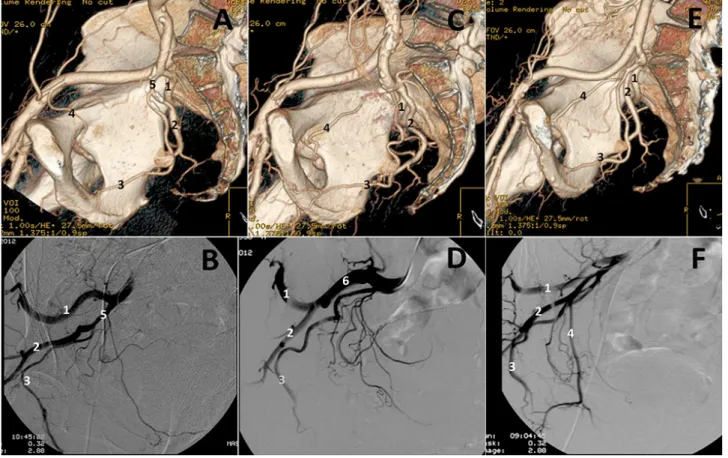
The CTA and DSA findings allowed classification of all 42 pelvic sides according to the Yamaki classification. Twenty-four pelvic sides were classified as Group A (57%), 9 as Group B (21.5%) and 9 as Group C (21.5%). We did not find any Group D pattern (Fig. 1).
The superior gluteal artery usually is the largest branch, with a thick trunk and a very typical trajectory, describing a superiorly concave arch, as it passes under the superior border of the great sacro-sciatic foramen, exiting the pelvis. The inferior gluteal artery is also a large branch, with
variable origins regarding the type of internal iliac bifurca-tion, usually at the level of the upper border of the great sacro-sciatic foramen. It has a trajectory downwards and outwards, exiting the pelvis to the inferior aspect of the glu-teal region.
The internal pudendal artery is the main supplier of blood to the corpora cavernosa. It arises from the anterior division of the internal iliac artery and has a typical concave trajec-tory curving under the sciatic notch allowing its recognition16
(Fig.s 2, 3). The artery enters the perineum via the lesser sciatic foramen and runs along the lateral wall of the ischio-rectal fossa between the split layers of the obturator fascia in Alcock’s canal to the inferior pubic ramus. It gives rise to the muscular, inferior rectal and perineal-scrotal collateral branches. From the origin of the perineal-scrotal artery, most authors call it the penile artery, giving rise to the bulbar and the urethral (spongiosal) arteries. The penile artery has 2 terminal branches, the deep artery of the penis (cavernosal artery) and the dorsal artery of the penis. The dorsal artery of the penis is responsible for the blood supply of the penile skin and glans penis. The cavernosal arteries on the other hand enters the corpora cavernosa at the hilum and gives rise to multiple small helicine arteries that drain directly into the vascular lacunar spaces of the corpora cavernosa.
Figure 1 - Branching patterns of the internal iliac artery.
A. CTA 3D reformat and b. Selective DSA of right internal iliac artery showing a Group A branching pattern. Note that the internal pudendal
and the inferior gluteal arteries have a common origin on the gluteal-pudendal trunk (5). C. CTA 3D reformat and D. selective DSA of right
internal iliac artery showing a Group B branching pattern. Note the posterior division common gluteal trunk (6) that originates the superior
and inferior gluteal arteries. E. CTA 3D reformat and F. selective DSA of right internal iliac artery showing a Group C branching pattern.
ARtIGO ORIGINAL
We found the obturator artery arising from the IIA in 25 pelvic sides (60%), and from the inferior epigastric artery in 17 pelvic sides (40%). There were 11 accessory pudendal arteries (26.2%) identified.
Of the evaluated patients, using DSA as the gold-stan-dard method, we found a total of 21 arteries with atheroscle-rotic lesions. Two lesions were detected both on the CTA and DSA and were localized in the proximal internal iliac artery (1 occlusion and 1 stenosis) (Fig. 4). Of the 42 inter-nal pudendal arteries evaluated, the DSA evaluation found 19 abnormal and 23 normal internal pudendal arteries. Of these abnormal arteries, 11 were occlusions and the other 8 were stenoses. On the CTA we found 24 abnormal and 18 normal internal pudendal arteries. Of the abnormal arteries, 13 were occlusions and 11 were stenosis (Fig. 5). We found a patient with an occlusion of the common gluteal-pudendal trunk that was considered as an occlusion of the internal pudendal artery (Fig. 6).
Considering DSA the gold-standard technique, the CTA’s sensibility and specificity for detecting lesions on the internal pudendal arteries was 95% and 73%, respectively. Also the positive predictive value (PPV) was 75% and the negative predictive value (NPV) was 94%, i.e. CTA has a relatively high false-positive rate but a low false-negative rate when assessing lesions of the internal pudendal arteries.
DISCUSSION
Knowledge of the anatomy and anatomical variations of the arteries of the pelvic region has very important clinical relevance. Associations between anatomical variations of the internal pudendal artery and erectile dysfunction have been described.17 Recognizing the internal pudendal
ar-tery is essential for accurate diagnosis of arteriogenic ED and to perform safe and effective revascularization proce-dures.14,18-20 The presence of diffuse atherosclerotic lesions
of the pelvic vessels frequently found in arteriogenic ED in-creases the difficulty in correctly identifying and distinguish-ing the internal iliac collaterals.
The Yamaki classification was created using cadaveric studies15 and has been applied with various angiographic
modalities, namely magnetic resonance angiography (MRA), CTA and DSA.16 Based on our experience we find
it the most simple and reproducible classification of this complex vascular system. This systematic approach allows easy recognition of the main collaterals of the internal iliac artery, even in the presence of specific settings, like the dif-fuse atherosclerotic changes associated with arteriogenic ED.
In our study we found 24 pelvic sides classified as Group A (57%), 9 as Group B (21.5%) and 9 as Group C (21.5%). Like in the Yamaki study,15 our most frequent branching
pattern was Group A, however with different percentages (57% versus the 80% of the Yamaki study). There were also Figure 2 - Accessory pudendal arteries.
A. Selective DSA of left internal iliac artery. b. Selective DSA of
the anterior division of the left internal iliac in the same patient. Note that an accessory pudendal artery, originating in the obturator
artery, becomes apparent after the contrast injection. C, D.
Selec-tive DSA of an acessory pudendal artery. Note its typical convex trajectory behind the pubic bone. See the retrograde opacification of the proper internal pudendal artery. 1.Superior gluteal artery 2. Inferior gluteal artery 3.Internal pudendal artery. 4.Obturator artery 5.Accessory pudendal artery.
Figure 3 - Anatomy of the internal pudendal artery.
A. Sagittal MIP and b. 3D VR of the left side depicting the normal
anatomy of the internal pudendal artery. Note the typical concave
trajectory curving under the sciatic notch (thick arrow). C.
Angiog-raphy of left internal iliac artery and D. Selective DSA of the left
ARtIGO ORIGINAL
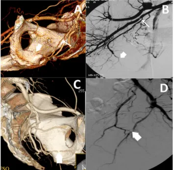
Figure 4 - Examples of lesions found on the internal iliac arteries.
A. CTA 3D VR reformat and b. CTA MIP reformat showing short
stenosis of the right internal iliac artery. Note also severe athero-sclerosis of the inferior segment of the abdominal aorta and
com-mon iliac arteries. C. Pigtail pelvic DSA showing the correlation with
CTA findings. D. CTA 3D VR reformat of another patient showing
focal occlusion of the left hypogastric artery (thick arrow) and mul-tiple stenosis of the internal pudendal artery (arrows).
Figure 5 - Examples of lesions found on the internal pudendal ar-teries.
A.CTA 3D reformat and b. selective DSA of the right internal iliac
artery showing a stenosis (arrow) and an occlusion (thick arrow) of the origin and of the perineal segment of the right internal pudendal
artery, respectively C. CTA 3D reformat and D. selective DSA of left
hypogastric arteryshowing occlusion of the perineal segment of the
internal pudendal artery (thick arrow).
differences in Groups B and C, as we found equal preva-lence in our study. According to Yamaki and other studies,16
Group B should be the second most frequent, followed by Group C. As expected, we did not find any Group D, as Ya-maki only found 1 in 645 pelvic sides. In our opinion these differences may be due to the significantly different size of the samples, the fact that our study is not cadaveric and we only included male patients with arteriogenic ED. The higher prevalence of type B and C bifurcations in this study may imply that these anatomical variations may be asso-ciated with ED. Further studies are needed to assess this hypothesis.
As mentioned earlier the Yamaki classification is com-posed of 4 groups (A, B, C and D) based on the branching patterns of the internal pudendal, the superior and inferior gluteal arteries. Another prominent artery that is frequently encountered, deserving mention is the obturator artery. Its recognition is usually straightforward as it follows a distinct trajectory, running forwards, into the obturator foramen exit-ing the pelvis and givexit-ing rise to two terminal branches that form approximately a 90° angle (Fig. 1). Due to its variabil-ity, like in most studies, we did not to include the obturator artery in the branching pattern classification of the internal iliac artery.
A common and relevant anatomical variation in patients with arteriogenic ED is the presence of accessory pudendal arteries (APAs). These are arteries superior to the pelvic diaphragm, that pass posterior to the pubic bone to finally enter the penile hilum and provide unilaterally or bilaterally arterial blood to the corpora cavernosa.21 They may arise
from the hypogastric, the internal or external iliac or from the obturator arteries. They can be recognized because un-like the internal pudendal artery, they have a typical convex trajectory behind the pubic bone (Fig. 2). In our study we found 11 APAs (26%). The true prevalence of APAs is un-clear and varies between 4% and 75%, depending on the anatomical definitionand the used methodology.12,22 Also its
clinical relevance remains to be determined;23,24 therefore
we are not able to discuss with confidence the significance of our results.
The pathophysiology behind arteriogenic ED is that ath-erosclerotic disease of the internal pudendal or hypogastric arteries may limit the increase of blood flow required to fill the corpora cavernosa in order to achieve erection. DSA remains the gold standard method for diagnosis of arterio-genic ED as it is the technique that allows the best detail of the terminal branches of the internal pudendal artery. How-ever, it has some disadvantages like being a semi-invasive and relatively expensive procedure. Furthermore it requires a longer examination time and a larger amount of contrast medium when compared to CTA.
ARtIGO ORIGINAL
Dynamic penile color Doppler ultrasound is the least in-vasive technique for the diagnosis of vascular ED.25,26
How-ever its popularity among clinicians is low due to its cost and lack of standardization. Also, like all ultrasound evalua-tions it is operator–dependent and incorrect diagnosis due to increased sympathetic stimulation and high anatomical variability has been reported. The Doppler analysis of the cavernously arteries in the flaccid penis is a relatively cheap alternative however its validity remains to be proven.27,28
This study has several limitations. It is retrospective and we did not apply a standardized pre-procedure evaluation of ED causes. We looked for atherosclerotic lesions (ste-noses and/or occlusions) of the internal pudendal arteries and found similar results to previous studies10,11; however
we did not characterize similar lesions in the smaller penile vessels, like the cavernosal and dorsal arteries of the penis, as we found it to be beyond our main objective.
In this study we characterized the radiological anatomy of the male pelvic arteries in a specific subset of patients with suspected arteriogenic ED. We found that even with the presence of the atherosclerotic lesions typically associated to this condition, the Yamaki classification is a reproducible and systematic approach that allows easy identification of the main collaterals of the internal iliac artery, namely the internal pudendal artery, a requisite that is paramount for
the diagnosis and endovascular treatment of this condition.
CONCLUSION
Arteriogenic ED is a condition characterized by ath-erosclerotic disease of the pelvic arteries. CTA and DSA findings associated with this condition include stenotic and occlusive lesions of the internal iliac and internal pudendal arteries. Knowledge of male pelvic anatomy and its varia-tions is paramount in the diagnosis of arteriogenic ED. The Yamaki classification is a simple and reproducible classifi-cation that allows easy recognition of the internal pudendal artery that may be useful for the diagnosis and treatment of patients with arteriogenic ED.
ACKNOWLEDGMENt
The authors would like to acknowledge Lam C for the valuable linguistic and translational contribution to this study, particularly in the English translation of an article pub-lished in Chinese.
CONFLICt OF INtEREStS None stated.
FUNDING SOURCES None stated Figure 6 - Occlusion of the anterior division of the right internal iliac artery (type A bifurcation)
A.CTA 3D reformat and b. selective DSA of the right internal iliac artery showing an occlusion of the anterior division (thick arrow). Note
the distal repermeabilization of the inferior gluteal and internal pudendal arteries. 1. Internal iliac artery 2.Superior gluteal artery 3. Anterior division (gluteal-pudendal trunk) 4.Inferior gluteal artery 5.Internal pudendal artery 6.Obturator artery 7.Prostatic artery.
REFERENCES
1. NIH Consensus Conference. Impotence. NIH Consensus Development Panel on Impotence. JAMA. 1993;270:83-90.
2. McKinlay JB. The worldwide prevalence and epidemiology of erectile dysfunction. Int J Impot Res. 2000;12:6-11.
3. Vendeira P, Pereira NP, Tomada N, Carvalho LF. Estudo EPISEX-PT/ Masculino: prevalência das disfunções sexuais masculinas em Portu-gal. Cad Sexol. 2011;4:15-22.
4. Ayta IA, McKinlay JB, Krane RJ. The likely worldwide increase in erectile
dysfunction between 1995 and 2025 and some possible policy conse-quences. BJU Int. 1999;84:50-6.
5. Rosen RC, Riley A, Wagner G, Osterloh IH, Kirkpatrick J, Mishra A. The international index of erectile function (IIEF): a multidimensional scale for assessment of erectile dysfunction. Urology. 1997;49:822-30. 6. Rosen RC, Cappelleri JC, Smith MD, Lipsky J, Pena BM. Development
ARtIGO ORIGINAL Int J Impot Res. 1999:11:319-26.
7. Lue TF. Erectile dysfunction. N Engl J Med. 2000:342:1802-13. 8. Gazzaruso C, Solerte SB, Pujia A, Coppola A, Vezzoli M, Salvucci F, et
al. Erectile dysfunction as a predictor of cardiovascular events and death in diabetic patients with angiographically proven asymptomatic coronary artery disease: a potential protective role for statins and 5-phosphodies-terase inhibitors. J Am Coll Cardiol. 2008;51:2040-4.
9. Bonetti PO, Lerman LO, Lerman A. Endothelial dysfunction: a marker of atherosclerotic risk. Arterioscler Thromb Vasc Biol.2003;23:168-75. 10. Lan YS, Jiang J, Jiang R.[Multi-slice spiral computed tomography
an-giography of internal pudendal arteries: imaging and value]. Zhonghua Nan Ke Xue. 2012;18:296-301.
11. Kawanishi Y, Muguruma H, Sugiyama H, Kagawa J, Tanimoto S, Ya-manaka M, et al., Feasibility of multi-slice computed tomography in the diagnosis of arteriogenic erectile dysfunction. BJU Int. 2001;88:390-5. 12. Gray RR, Keresteci AG, St Louis EL, Grosman H, Jewett MA, Rankin
JT, et al. Investigation of impotence by internal pudendal angiography: experience with 73 cases. Radiology. 1982;144:773-80.
13. Fitzgerald SW, Erickson SJ, Foley WD, Lipchik EO, Lawson TL. Color Doppler sonography in the evaluation of erectile dysfunction. Radio-graphics. 1992;12:3-17.
14. Rogers JH, Goldstein I, Kandzari DE, Kohler TS, Stinis CT, Wagner PJ, et al. Zotarolimus-eluting peripheral stents for the treatment of erectile dysfunction in subjects with suboptimal response to phosphodiester-ase-5 inhibitors. J Am Coll Cardiol. 2012;60:2618-27.
15. Yamaki K, Saga T, Doi Y, Aida K, Yoshizuka M. A statistical study of the branching of the human internal iliac artery. Kurume Med J. 1998;45:333-40.
16. Bilhim T, Casal D, Furtado A, Pais D, O’Neill JE, Pisco JM. Branching patterns of the male internal iliac artery: imaging findings. Surg Radiol Anat. 2011;33:151-9.
17. Kawanishi Y, Muguruma H, Sugiyama H, Kagawa J, Tanimoto S, Yama-naka M, et al. Variations of the internal pudendal artery as a congenital contributing factor to age at onset of erectile dysfunction in Japanese. BJU Int. 2008;101: 581-7.
18. Valji K, Bookstein JJ. Transluminal angioplasty in the treatment of
arte-riogenic impotence.Cardiovasc Intervent Radiol. 1988;11:245-52. 19. Zumbe J, Drawz G, Wiedemann A, Grozinger K, Engelmann U.
Indi-cations for penile revascularization and long-term results. Andrologia. 1999;31:83-7.
20. Babaev A, Jhaveri RR. Angiography and endovascular revasculariza-tion of pudendal artery atherosclerotic disease in patients with medically refractory erectile dysfunction. J Invasive Cardiol. 2012;24:236-40. 21. Secin FP, Karanikolas N, Touijer AK, Salamanca JI, Vickers AJ,
Guillon-neau B. Anatomy of accessory pudendal arteries in laparoscopic radical prostatectomy. J Urol. 2005;174:523-6.
22. Mulhall JP, Secin FP, Guillonneau B. Artery sparing radical prostatecto-my-myth or reality? J Urol. 2008;179:827-31.
23. Park BJ, Sung DJ, Kim MJ, Cho SB, Kim YH, Chung KB, et al. The incidence and anatomy of accessory pudendal arteries as depicted on multidetector-row CT angiography: clinical implications of preoperative evaluation for laparoscopic and robot-assisted radical prostatectomy. Korean J Radiol. 2009;10:587-95.
24. Box GN, Kaplan AG, Rodriguez E Jr, Skarecky DW, Osann KE, Finley DS, et al. Sacrifice of accessory pudendal arteries in normally potent men during robot-assisted radical prostatectomy does not impact po-tency. J Sex Med. 2010;7:298-303.
25. Corona G, Fagioli G, Mannucci E, Romeo A, Rossi M, Lotti F, et al. Pe-nile doppler ultrasound in patients with erectile dysfunction (ED): role of peak systolic velocity measured in the flaccid state in predicting arterio-genic ED and silent coronary artery disease. J Sex Med. 2008;5:2623-34.
26. Coelho MF, Santos PB. Erectile dysfunction of vascular cause: statistical evaluation on the plurimetabolic syndrome’s risk factors and their cor-relation with penile eco-doppler rates. Acta Med Port. 2011;24:379-82. 27. Mancini M, Bartolini M, Maggi M, Innocenti P, Forti G. The presence
of arterial anatomical variations can affect the results of duplex so-nographic evaluation of penile vessels in impotent patients. J Urol. 1996;155:1919-23.