Iliac vein compression syndrome: literature review
Síndrome de compressão da veia ilíaca: revisão de literatura
Leonardo Pessoa Cavalcante1,2, José Emerson dos Santos Souza2, Raquel Magalhães Pereira3,
Marcos Velludo Bernardes1, Alan Maurice da Silva Amanajás2, Marcos Henrique Parisati1,
Ricardo Dias da Rocha1, Antônio Oliveira de Araújo1,2
Abstract
Iliac vein compression syndrome is a clinical condition in which the right common iliac artery extrinsically compresses the left common iliac vein. he syndrome predominantly afects young women between their 2nd and 4th decades of life. In view of the syndrome’s potential complications, it should be recognized/diagnosed and treated in symptomatic patients before it causes irreversible damage to patients’ venous systems. Noninvasive methods, such as venous color Doppler US are reasonable screening methods, but angiotomography and magnetic resonance angiography are more reliable diagnostic tools and the method of choice for conirmation of diagnosis remains multi-plane phlebography with measurement of pressure gradients. Endovascular treatment (angioplasty with placement of self-expanding stents) is safe and efective and can replace open surgical reconstruction and/or anticoagulation alone.
Keywords: May-hurner Syndrome; iliac artery; iliac vein; venous insuiciency; phlebography; angioplasty.
Resumo
A Síndrome de Compressão da Veia Ilíaca (SCVI) é uma situação clínica na qual a artéria ilíaca comum direita comprime extrinsecamente a veia ilíaca comum esquerda. Há uma predominância em mulheres jovens, entre a segunda e a quarta décadas de vida. Levando-se em consideração as complicações potenciais da síndrome, esta deve ser reconhecida/ diagnosticada e tratada, em pacientes sintomáticos, antes que cause alterações irreversíveis no sistema venoso do paciente. Métodos não invasivos, como o US-Doppler colorido, quando realizados por examinadores experientes, são métodos de triagem razoáveis; porém, a angiotomograia e a angiorressonância são mais idedignas. O método de escolha para a conirmação diagnóstica consiste na lebograia, em múltiplas incidências, com aferição de gradientes pressóricos. O tratamento endovascular (angioplastia com colocação de stent autoexpansível) é seguro e efetivo, podendo substituir a reconstrução cirúrgica aberta e/ou a anticoagulação isolada.
Palavras-chave: Síndrome de May-hurner; artéria ilíaca; veia ilíaca; insuiciência venosa; lebograia; angioplastia.
1Universidade Federal do Amazonas – UFAM, Hospital Universitário Francisca Mendes – HUFM, Manaus, AM, Brazil.
2Universidade Federal do Amazonas – UFAM, Hospital Universitário Getúlio Vargas – HUGV, Hospital Universitário Francisca Mendes – HUFM, Manaus, AM, Brazil. 3Universidade Federal do Amazonas – UFAM, Manaus, AM, Brazil.
Financial support: None.
Conlicts of interest: No conlicts of interest declared concerning the publication of this article. Submitted: March 21, 2014. Accepted: September 08, 2014.
INTRODUCTION
Iliac vein compression syndrome (IVCS) is a clinical condition in which the right common iliac artery causes extrinsic compression of the left common iliac vein and which presents clinically as pain and edema in the left lower limb or even as left iliofemoral venous thrombosis.1
Cockett & Thomas2,3 point out that this disease is
one example of how a minor anatomic anomaly can
be the underlying cause of an extremely signiicant
pathological injury. Extrinsic compression of the left common iliac vein at this level has been given a variety of names, including May-Thurner syndrome, iliac venous compression syndrome, iliocaval compression syndrome and Cockett’s syndrome.4,5
This extrinsic compression is not infrequently the cause of venous abnormalities in the left lower limb, which are very often considered as ‘primary’ conditions because of the failure to diagnose the compressive disease.6 The true prevalence of this
condition is unknown because, as phlebography has been substituted by noninvasive methods (primarily Color Doppler US) for diagnosis of venous diseases of the lower limbs, many cases of chronic
venous insuficiency and/or left iliofemoral venous
thrombosis that are linked with IVCS have not been diagnosed etiologically.7
When the right common iliac artery crosses over the left common iliac vein it appears to induce partial obstruction of the vein in two different ways: 1) simple mechanical obstruction, caused by compression of the vein between the artery and the vertebral body ; and 2) extensive intimal hypertrophy of the vein can result from repeated compressions caused by the overlying arterial pulse, causing a certain degree of shear stress between the anterior and posterior walls of the vein.6,8-10
Since IVCS is an under-diagnosed entity and one that causes venous abnormalities of the left lower limb with greater frequency than is generally thought, the objective of this article is to review the most important points related to the condition, from epidemiology to the treatment options currently available.
A bibliographic review of Brazilian and International studies was conducted after running searches on the MEDLINE (PUBMED), SCIELO and BIREME databases. The search strategy employed for MEDLINE was as follows: thurner syndrome”[MeSH Terms] OR (“may-thurner”[All Fields] AND “syndrome”[All Fields]) OR “may-thurner syndrome”[All Fields] OR
(“thurner”[All Fields] AND “syndrome”[All Fields]) OR “may thurner syndrome”[All Fields]) OR (“may-thurner syndrome”[MeSH Terms] OR (“may-thurner”[All Fields] AND “syndrome”[All Fields]) OR “may-thurner syndrome”[All Fields] OR (“iliocaval”[All Fields] AND “compression”[All Fields] AND “syndrome”[All Fields]) OR “iliocaval compression syndrome”[All Fields]) OR thurner syndrome”[MeSH Terms] OR (“may-thurner”[All Fields] AND “syndrome”[All Fields]) OR “may-thurner syndrome”[All Fields] OR (“cockett”[All Fields] AND “syndrome”[All Fields]) OR “cockett syndrome”[All Fields]). Studies were selected for review on the basis of reading their abstracts, characterizing this study as a narrative review.
EPIDEMIOLOGY
Although the true prevalence of IVCS is unknown, it is estimated that this condition is present in 2 to 5% of patients who have venous disease of the lower limbs.7,10 The reason why there is an imbalance in the
normal anatomic relationship between artery and
vein, interfering with venous low, is also unknown.4
When the incidence of IVCS in patients with deep vein thrombosis of the left leg has been investigated, the rates observed have varied from 18 to 49%.11
Many studies have reported evidence that left lower limb deep vein thrombosis prevalence rates are 3 to 8 times greater than the prevalence rates for the right limb,2,12 and IVCS is one possible explanation for this
higher prevalence.
In 1943, Ehrich & Krumbhaar13 conducted
anatomic dissection of 412 cadavers, inding lesions
obstructing the left common iliac vein in 23.8% of them. From a histological perspective, these lesions were not recanalized thrombosis, rather they were made up of elastin and collagen, without
cellular inlammatory iniltrate or ibrosis. They also
observed that 33.8% of these lesions occurred after 10 years of age, concluding that they were acquired (and not congenital).
This syndrome predominately affects females and, in 1965, Cockett & Thomas2 described it as
predominantly affecting young women in their second to fourth decades of life. In Brazil, Marques et al.8
studied 20 patients with the syndrome, inding that
80% of them were female and reporting a mean age of 34.4 years. Surgery, pregnancy and prolonged rest have been described as acute trigger factors.14
Based on autopsy findings, May & Thurner1
“spurs” at the level at which the right common iliac artery crosses the left common iliac vein anteriorly. Since iliac vein thrombosis is more common on the left side, it is believed that the compression and the “spurs” described by May & Thurner are responsible for many of these cases.
DIAGNOSIS
Clinical status
Iliac vein compression syndrome generally manifests as progressive edema of the left lower limb in individuals who do not apparently have risk factors for deep vein thrombosis and, in some cases, even when patients are on anticoagulation.14
Symptoms can vary from significant venous claudication to vague and mild complaints that are often attributed to primary chronic venous insufficiency. Physical examination may find edema, although generally moderate, and, more rarely, cutaneous abnormalities compatible with
chronic venous insufficiency (varicose veins/
lipodermatosclerosis/venous ulcers).9,10,15,16
A literature review conducted by Moudgill et al.17
in 2009 conirmed that the disease predominantly
affects female patients. By the time they are diagnosed, the majority of patients present with acute deep vein thrombosis in the left lower limb14
and a small proportion will present with edema or
non-speciic pain in the left lower limb, with no
documented venous thrombosis.
Sandri16 and Boyd18 also reported that patients
with signs/symptoms of left iliofemoral venous
thrombosis caused by extrinsic venous compression tend to be young women, in their second to fourth decades of life, after prolonged immobilization or pregnancy. Clinical features includes persistent rhizomelic edema of the left lower limb, with or without other signs of venous hypertension.
Therefore, IVCS should be suspected when patient history and physical examination, supplemented by venous Color Doppler US, are incompatible with deep vein thrombosis in a young woman with edema of the left lower limb.9
Imaging
Patients with unilateral left-side edema are generally sent for venous Color Doppler US of the left lower limb, to rule out deep vein thrombosis, and abdominal computed tomography, to rule out a pelvic mass as cause of compression.19
Ultrasonography is subject to significant
limitations for identiication of IVCS, since it is
technically dificult to view the pelvic veins because
of their deep location “hidden” behind the bladder, the intestines and adipose tissues.9 It has been
observed that even experienced ultrasound specialists
ind it dificult to see the iliac veins in up to 20% of
scans.20
In response to the dificulty of directly viewing
the compression, Barros & Coelho21 have described
some indirect signs that can be seen on Doppler ultrasonography and can help to diagnose this clinical
condition: 1) low volume in the right common iliac vein 40% greater than the low volume in the left
common iliac vein; 2) ratio of peak velocity in the left common femoral vein to peak velocity in the right less than 0.9.
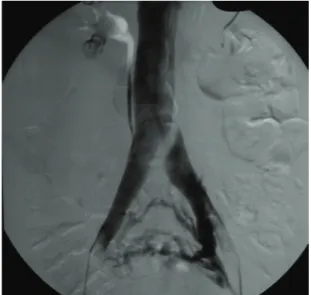
Under normal circumstances, there should be essentially no difference between pressures in the inferior vena cava and in the iliac veins, unless
there is a stenosis with hemodynamic signiicance.
A pressure gradient of 2 mmHg or more (measured using an intraluminal catheter during phlebographic examination) is a significant finding and, in combination with the presence of pelvic collateral circulation (Figure 1), has been used to conirm IVCS diagnoses.11,20
Wolpert et al.19 analyzed a small group of
24 patients with magnetic resonance angiography and phlebography, achieving a good diagnostic correlation between the two, and emphasized the
reduced invasivity of the irst imaging exam and its
ability to enable assessment of other pelvic structures.
The diagnostic method of choice for conirmation
of IVCS has traditionally been phlebography;9,11,14,22
but Raju & Neglen23 compared phlebography
with intravascular ultrasonography (IVUS) and reported that phlebography was less sensitive than
IVUS, inding that the two examinations offered,
respectively, 66% and 90% capacity to detect this clinical condition.
Additionally, both extension and severity of the obstructive venous injury appear worse when seen on IVUS than according to phlebography
indings, and Neglén and Raju24 have shown that
even severe obstructions can be underdiagnosed using phlebography. These authors investigated 304 limbs using phlebography with venous pressure measurement and IVUS and concluded that IVUS appears to be superior to single-plane phlebography for morphological diagnosis of stenotic lesions of the iliac veins. They also claim that further studies should be conducted to investigate the hemodynamic
signiicance of lesions detected by IVUS in order to
determine whether the results can aid in the decision of whether or not to intervene. Despite its apparent superiority, IVUS is still an expensive technique and many authors do not have this technology available or only employ it in selected cases.16
In view of the potential complications of IVCS (deep vein thrombosis and chronic venous
insuficiency), this syndrome must be recognized/
diagnosed in patients who exhibit symptoms related to it, to provide the opportunity for treatment before irreversible damage to the venous system occurs.11,25
TREATMENT
There is no consensus in the literature with respect to treatment of patients with IVCS who have not yet developed deep vein thrombosis. However, since stasis is one of the three pillars of Virchow’s triad, these patients probably have a greater chance of developing venous thromboembolism.18 On the
other hand, there is a certain degree of consensus in the literature10,11,26 that symptomatic IVCS should
always be treated.
Treatment options to improve symptoms and prevent thromboembolic phenomena consist of techniques to relieve the compression with either open surgery or with endovascular techniques, with the objective of alleviating the venous stasis in the left lower limb. If an invasive procedure is not chosen, a small number of authors advocate anticoagulation alone.
Since IVCS is a progressive disease, capable of causing incapacitating complications over the long term, an aggressive approach to achieving decompression should be strongly considered.4,11
Historically, several different surgical techniques have been employed to relieve symptoms and correct the resulting venous obstruction, the majority involving bypasses constructed with autologous veins,16,27 retropositioning of the iliac artery28 and
excision of the venous intraluminal spurs with subsequent venous patch angioplasty,7,11 among other
techniques.
More recently, endovascular techniques have been employed both to treat iliofemoral venous thrombosis and to repair the anatomic abnormality underlying IVCS.10,11 The irst report of exclusively endovascular
treatment of IVCS was published by Michel et al.29 in
1994. They treated the syndrome with percutaneous transluminal angioplasty and stent placement.
Cunha Jr et al.30 described a case of an
18-year-old female patient who was successfully treated endovascularly by placement of a self-expanding elgiloy stent in the left common iliac vein to counteract the extrinsic venous compression caused by pulsation of the overlying right common iliac artery. An intraluminal stent was also implanted successfully by Foit et al.14 (in a case with concomitant
thrombolysis) in a male patient who was diagnosed with IVCS 4 days after surgery for a tibial plateau fracture.
Sandri31 studied a series of 54 patients with IVCS
treated with endovascular techniques, observing total or near total resolution of symptomology in 52 of them (92.5%), conferring a considerable improvement in quality of life.
Moudgill et al.17 analyzed data from 113 patients
(72% women), the majority treated with catheter-guided thrombolysis followed by intravascular stent placement, observing a 95% mean technical success rate and a 96% mean 1-year patency rate. O’Sullivan et al.20 also reported that endovascular
treatment of IVCS is safe and effective and
can substitute surgical reconstruction and/or
anticoagulation alone. However, in view of the fact that these patients are mostly females and are generally of reproductive age, more studies are needed to assess the long-term patency of these stents.
More recent treatments, such as percutaneous transluminal angioplasty with self-expanding stent placement, have been linked with improved treatment success13 because, in addition to treating
enable alleviation of the extrinsic mechanical compression.9,11,32
Reports such as those described above and also retrospective and prospective studies of case series have shown that endovascular treatment is now the
irst-line approach.9,11 The majority of authors11,17
recommend a minimum of 6 month anticoagulation after stent placement in the venous system, unless
there is a speciic contraindication to doing so.
Migration of stents placed in the iliofemoral venous axis is a rare, but devastating, complication that has been described in the literature.33 If this type
of embolization occurs, removal can be accomplished either percutaneously or by open surgery, depending on the site.
Conservative treatment simply involves pain control, with or without drug prophylaxis for deep vein thrombosis. Since there are no reports of spontaneous resolution of IVCS to date, the decision to limit intervention to conservative treatment can be
a dificult one to take for a patient (generally young
and active), who suffers from discomfort in the left lower limb at rest or during physical exercise.18
Anticoagulation alone has not proven effective to control symptoms in symptomatic patients.14
Due to the rarity of this diagnosis, there are no randomized studies or large follow-up studies
comparing open/endovascular/clinical treatments
and, therefore, even though the endovascular approach is becoming more and more widely accepted, there is no consensus in the literature with regard to the most appropriate treatment for this disease.18 Raju34 reviewed the contemporary
literature, analyzing studies that described treatment of a total of approximately 1,500 patients and concluded that endovascular treatment for IVCS is a safe and effective alternative, compared with traditional open surgery treatment.
FINAL COMMENTS
Before patients are labeled as suffering from
primary venous valve insuficiency, it is important
to remember that persistent edema of the left lower limb or even just ‘sensation of heaviness’ in the left lower limb (particularly in young patients) may be the result of IVCS.9,10,11
The true prevalence of this disease is very likely to be higher than that described in the literature, because in many patients with left iliofemoral venous thrombosis this syndrome may be a hidden etiologic agent that is often neglected during work-up.31
Noninvasive methods (Color Doppler-US) are reasonable options for screening, if used with the objective of detecting this disease and performed by experienced examiners. However, angiotomography and magnetic resonance angiography are the most reliable imaging tests and remain the methods of
choice for diagnostic conirmation, particularly here
in Brazil where IVUS is not widely available yet, while the gold standard imaging method remains multi-plane phlebography with measurement of pressure gradients.
The endovascular approach is currently in the
process of becoming the irst line treatment, but
treatment must still be tailored to patients, choosing the best option for their individual characteristics and clinical status.
REFERENCES
1. May R, Thurner J. The cause of the predominantly sinistral occurrence of thrombosis of the pelvic veins. Angiology. 1957;8(5):419-27. http://dx.doi.org/10.1177/000331975700800505. PMid:13478912
2. Cockett FB, Thomas ML. The iliac compression syndrome. Br J Surg. 1965;52(10):816-21. http://dx.doi.org/10.1002/ bjs.1800521028. PMid:5828716
3. Cockett FB, Thomas ML, Negus D. Iliac vein compression.—Its relation to iliofemoral thrombosis and the post-thrombotic syndrome. Br Med J. 1967;2(5543):14-9. http://dx.doi.org/10.1136/ bmj.2.5543.14. PMid:6020994
4. Cil BE, Akpinar E, Karcaaltincaba M, Akinci D. Case 76: May-Thurner syndrome. Radiology. 2004;233(2):361-5. http://dx.doi. org/10.1148/radiol.2332030152. PMid:15516613
5. Gil Martín AR, Carreras Aja M, Arrieta Ardieta I, Labayen Azparren I. Cockett’s syndrome, May-Thurner syndrome, or iliac vein compression syndrome. Radiologia. 2014;56(5):e5-8. PMid:22621823
6. Heniford BT, Senler SO, Olsofka JM, Carrillo EH, Bergamini TM. May-Thurner syndrome: management by endovascular surgical techniques. Ann Vasc Surg. 1998;12(5):482-6. http://dx.doi. org/10.1007/s100169900189. PMid:9732429
7. Taheri SA, Williams J, Powell S, et al. Iliocaval compression syndrome. Am J Surg. 1987;154(2):169-72. http://dx.doi. org/10.1016/0002-9610(87)90172-3. PMid:3631389
8. Marques MA, Silveira PRM, von Ristow A, et al. Prevalência de marcadores de trombofilia em pacientes portadores da síndrome de May-Thurner e trombose de veia ilíaca comum esquerda. J Vasc Bras. 2010;9(4):229-32. http://dx.doi.org/10.1590/ S1677-54492010000400004.
9. Melo CCS, Barros MVL, Yepez JAR. Tratamento endovascular na Síndrome de May-Thurner: relato de caso e revisão da literatura. Rev Bras Ecocardiogr Imagem Cardiovasc. 2012;25(2):122-5.
10. Kalu S, Shah P, Natarajan A, Nwankwo N, Mustafa U, Hussain N. May-thurner syndrome: a case report and review of the literature. Case Rep Vasc Med. 2013;2013:740182. http://dx.doi. org/10.1155/2013/740182. PMid:23509664
12. Patel NH, Stookey KR, Ketcham DB, Cragg AH. Endovascular management of acute extensive iliofemoral deep venous thrombosis caused by May-Thurner syndrome. J Vasc Interv Radiol. 2000;11(10):1297-302. http://dx.doi.org/10.1016/S1051-0443(07)61304-9. PMid:11099239
13. Ehrich WE, Krumbhaar EB. A frequent obstructive anomaly of the mouth of the common iliac vein. Am Heart J. 1943;26(6):737-50. http://dx.doi.org/10.1016/S0002-8703(43)90285-6.
14. Foit NA, Chen QM, Cook B, Hammerberg EM. Iliofemoral deep vein thrombosis after tibial plateau fracture fixation related to undiagnosed May-Thurner syndrome: a case report. Patient Saf Surg. 2013;7(1):12. http://dx.doi.org/10.1186/1754-9493-7-12. PMid:23628366
15. Akers DL Jr, Creado B, Hewitt RL. Iliac vein compression syndrome: case report and review of the literature. J Vasc Surg. 1996;24(3):477-81. http://dx.doi.org/10.1016/S0741-5214(96)70205-7. PMid:8808971
16. Sandri GA. Tratamento endovascular das obstruções venosas crônicas do segmento iliocaval. J Vasc Bras. 2011;10(2):137-44. http://dx.doi.org/10.1590/S1677-54492011000200008.
17. Moudgill N, Hager E, Gonsalves C, Larson R, Lombardi J, DiMuzio P. May-Thurner syndrome: case report and review of the literature involving modern endovascular therapy. Vascular. 2009;17(6):330-5. http://dx.doi.org/10.2310/6670.2009.00027. PMid:19909680
18. Boyd DA. Unilateral lower extremity edema in May-Thurner syndrome. Mil Med. 2004;169(12):968-71. PMid:15646188.
19. Wolpert LM, Rahmani O, Stein B, Gallagher JJ, Drezner AD. Magnetic resonance venography in the diagnosis and management of May-Thurner syndrome. Vasc Endovascular Surg. 2002;36(1):51-7. http://dx.doi.org/10.1177/153857440203600109. PMid:12704525
20. O’Sullivan GJ, Semba CP, Bittner CA, et al. Endovascular management of iliac vein compression (May-Thurner) syndrome. J Vasc Interv Radiol. 2000;11(7):823-36. http://dx.doi.org/10.1016/ S1051-0443(07)61796-5. PMid:10928517
21. Barros FS, Coelho NA. Síndrome compressiva da veia ilíaca comum esquerda pela artéria ilíaca comum direita. In: Engelhorn CA, Morais D Fo, Barros FS, Coelho NA. Guia prático de ultrassonografia vascular. Rio de Janeiro: Di Livros; 2011. p. 219-23.
22. Timi JRR, Kenegusuku J, Souza PC, et al. Achados radiológicos na syndrome de compressão da veia ilíaca comum esquerda. Radiol Bras. 1993;26:53-5.
23. Raju S, Neglen P. High prevalence of nonthrombotic iliac vein lesions in chronic venous disease: a permissive role in pathogenicity. J Vasc Surg. 2006;44(1):136-43, discussion 144. http://dx.doi.org/10.1016/j.jvs.2006.02.065. PMid:16828437
24. Neglén P, Raju S. Intravascular ultrasound scan evaluation of the obstructed vein. J Vasc Surg. 2002;35(4):694-700. http://dx.doi. org/10.1067/mva.2002.121127. PMid:11932665
25. Oğuzkurt L, Ozkan U, Tercan F, Koç Z. Ultrasonographic diagnosis of iliac vein compression (May-Thurner) syndrome. Diagn Interv Radiol. 2007;13(3):152-5. PMid:17846991.
26. Neglén P, Raju S. Balloon dilation and stenting of chronic iliac vein obstruction: technical aspects and early clinical outcome. J Endovasc Ther. 2000;7(2):79-91. http://dx.doi.org/10.1583/1545-1550(2000)007<0079:BDASOC>2.3.CO;2. PMid:10821093
27. Palma EC, Esperon R. Vein transplants and grafts in the surgical treatment of the postphlebitic syndrome. J Cardiovasc Surg (Torino). 1960;1:94-107. PMid:14429961.
28. Calnan JS, Kountz S, Pentecost BL, Shillingford JP, Steiner RE. Venous obstructions in the aetiology of lymphoedema praecox.
Br Med J. 1964;2(5403):221-6. http://dx.doi.org/10.1136/ bmj.2.5403.214-b. PMid:14150911
29. Michel C, Laffy PY, Leblanc G, Bonnet D. [Treatment of Cockett syndrome by percutaneous insertion of a vascular endoprosthesis (Gianturco)]. J Radiol. 1994;75(5):327-30. PMid:8051686.
30. Cunha JR Jr, Neves DQ, Fontes FA, et al. Tratamento endovascular da síndrome de compressão da veia ilíaca (May-Thurner) – relato de caso. J Vasc Bras. 2011;10:72-6.
31. Sandri JL. Síndrome de May-Thurner – tratamento endovascular. In: Brito CJ. Cirurgia vascular: cirurgia endovascular, angiologia. Rio de Janeiro: Revinter; 2014. p. 1998-2009.
32. Loukas M, Shah R, Esmaeili E, Bangeholm A, Tubbs RS, Jordan R. A case of May-Thurner syndrome. Folia Morphol (Warsz). 2008;67(3):214-7. PMid:18828105.
33. Mullens W, De Keyser J, Van Dorpe A, et al. Migration of two venous stents into the right ventricle in a patient with May-Thurner syndrome. Int J Cardiol. 2006;110(1):114-5. http://dx.doi. org/10.1016/j.ijcard.2005.05.070. PMid:16005088
34. Raju S. Best management options for chronic iliac vein stenosis and occlusion. J Vasc Surg. 2013;57(4):1163-9. http://dx.doi. org/10.1016/j.jvs.2012.11.084. PMid:23433816
Correspondence Leonardo Pessoa Cavalcante Hospital Universitário Francisca Mendes - Serviço de Cirurgia Vascular/Endovascular Av. Camapuã, 108 – Cidade Nova II CEP 69097-720 – Manaus (AM), Brazil E-mail: [email protected]
Author information LPC - Head, Endovascular Surgery Service, Hospital Universitário Francisca Mendes (HUFM), Universidade Federal do Amazonas (UFAM). JESS and AMSA - Vascular surgeons from the Medical Residence Program, Vascular and Endovascular Surgery Service, Hospital Universitário Francisca Mendes (HUFM), Hospital Universitário Getúlio Vargas (HUGV), Universidade Federal do Amazonas (UFAM). RMP - Medical student, Universidade Federal do Amazonas (UFAM). MVB - Head, Vascular Surgery Service, Hospital Universitário Francisca Mendes (HUFM), Universidade Federal do Amazonas (UFAM). MHP and RDR - Vascular surgeons, Vascular and Endovascular Surgery Service, Hospital Universitário Francisca Mendes (HUFM), Universidade Federal do Amazonas (UFAM). AOA - Resident physician from the Medical Residence Program, Vascular and Endovascular Surgery Service, Hospital Universitário Francisca Mendes (HUFM), Hospital Universitário Getúlio Vargas (HUGV), Universidade Federal do Amazonas (UFAM).
Author contributions Conception and design: LPC Analysis and interpretation: LPC, MVB, RDR Data collection: AMSA, MHP Writing the article: LPC, RMP, JESS Critical revision of the article: MVB, AMSA, RDR, AOA Final approval of the article*: LPC, JESS, RMP, MVB, AMSA, MHP, RDR, AOA Statistical analysis: N/A. Overall responsibility: LPC