www.reumatologia.com.br
REVISTA BRASILEIRA DE
REUMATOLOGIA
R E V B R A S R E U M A T O L . 2 0 1 3 ;5 4 ( 1 ): 6 5 – 6 7
Case report
“Milk of calcium”: a rare presentation of calcinosis
☆Tania Caroline Monteiro de Castro
a, Roberto Guarniero
b,
Maria Fernanda de Azevedo Giacomin
c, Marília Bergstron Lenzi Meneghin
c,
Guilherme Bottino Martins
c, Simone de Andrade Lotufo
ca Hospital Infantil Municipal Menino Jesus, São Paulo, SP, Brazil
b Faculdade de Medicina da Universidade de São Paulo, São Paulo, SP, Brazil c Hospital Infantil Menino Jesus, São Paulo, SP, Brazil
a r t i c l e i n f o
Article history:
Received on 7 October 2011 Accepted on 14 May 2013
Keywords: Calcinosis Dermatomyositis Resonance imaging
Systemic lupus erythematosus Adolescent
a b s t r a c t
Rheumatic diseases such as juvenile dermatomyositis (JDM), juvenile sistemic lupus ery-thematosus (JSLE) and sistemic sclerosis may have calcium deposits in the subcutaneous and muscle tissues known as calcinosis. Extensive calcium-laden l uid collections referred as “milk of calcium” are rare forms of calcinosis in JDM. We describe a 15-year old patient with overlap syndrome (sclerodermatomyositis and JSLE), whose magnetic resonance im-aging (MRI) showed perimusculares l uid collections in the lower limbs. During surgery, we observed the presence of whitish l uid collection suggestive of “milk of calcium”.
© 2014 Elsevier Editora Ltda. All rights reserved.
☆
Center: Hospital Infantil Municipal Menino Jesus - Instituto de Responsabilidade Social Hospital Sírio-Libanês. * Corresponding author.
E-mail: [email protected] (T.C.M.d. Castro).
0482-5004/$ - see front matter. © 2014 Elsevier Editora Ltda. All rights reserved. http://dx.doi.org/10.1016/j.rbre.2014.02.010
“Milk of calcium”: uma apresentação rara de calcinose
Palavras-chave: Calcinose Dermatomiosite
Imagem por ressonância magnética Lúpus eritematoso sistêmico Adolescente
r e s u m o
Algumas doenças reumáticas, como dermatomiosite juvenil (DMJ), lúpus eritematoso sis-têmico juvenil (LESJ) e esclerose sistêmica (ES), podem apresentar depósitos de cálcio nos tecidos subcutâneo e muscular, lesões conhecidas como calcinoses. Extensas coleções lí-quidas de cálcio referidas como milk of calcium são formas raras de calcinoses presentes na DMJ. Descrevemos um paciente de 15 anos de idade, com diagnóstico de síndrome de sobreposição ou overlap (esclerodermatomiosite e LESJ), cuja ressonância magnética (RM) evidenciou coleções líquidas perimusculares em membros inferiores e que, durante proce-dimento cirúrgico, foi observada a presença de coleção líquida esbranquiçada sugestiva de milk of calcium.
66
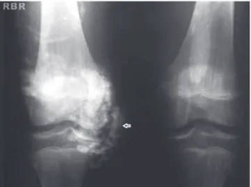
R E V B R A S R E U M A T O L . 2 0 1 3 ;5 4 ( 1 ): 6 5 – 6 7Fig. 1 – Calcinosis lesion on the right knee.
Introduction
The superposition or overlap syndrome is a clinical entity in which two or more symptoms of autoimmune diseases are identii ed in the same patient.1 These patients may have
clinical or laboratory features of two or more of the following conditions: Juvenile dermatomyositis (JDM), juvenile systemic lupus erythematosus (JSLE), systemic sclerosis (SSc) and ju-venile idiopathic arthritis (JIA). The coexistence of JSLE and esclerodermatomiositis is less frequent. These patients have heterogeneous clinical course and usually do not develop pulmonary i brosis.2 Calcinosis is a complication of some
rheumatic diseases. In SSc, calcii cations occur mainly in lo-calized scleroderma.3 In JDM, these calcinosis are associated
with some risk factors such as treatment delay, vasculopa-thy and severe disease.3,4 Patients with JSLE may also
pres-ent dystrophic calcii cations similar to those found in SSc and JDM.3 Little is known about its pathophysiology and there is
no known effective treatment.4 A liquid collection known as
"milk of calcium" is a very rare form of calcinosis. This term was originally used to characterize l uid-i lled collections of calcium found in the gall bladder and kidneys.5 Is speculated
that these l uid collections occur as a result of shearing of calcii ed tissues with resultant formation of pseudobursae containing liquid.5,6
This paper reports the case of a patient with superposition or overlap syndrome of esclerodermatomiositis and JSLE, that presented during its evolution perimuscular l uid collections in the right and left thigh, later with surgical drainage.
Case report
EJAS, male, 15 years old, brown color, born and living in Sao Paulo.
Four years ago, the patient was affected by arthritis in the hands, elbows and ankles lasting for four months. The prob-lem had migratory character, worsening during movement and improving with rest, being associated with loss of muscle strength in the upper limbs, sclerodactyly, Gottron's papules, ulcerated lesions on hands and feet and a convulsive episode. The patient began his monitoring at the Rheumatology Clinic of the Hospital Infantil Municipal Menino Jesus, where the following laboratory tests were performed: coarse speckled ANA 1/320, anti-SM and anti-RNP positive, CBC unchanged, CPK = 189 U/L (normal 26-190 U/L), LDH = 302 U/L (normal <480 U/L) and urine type 1 normal, ESR = 55 mm/h (normal 0-25 mm/h), PCR = 58.4 mg/L (normal <8.0 mg/L) and aldolase slightly increased to 11.9 U/L (normal up to 8.8 U/L).
Still in diagnostic investigation with a probable diagnosis of overlap (esclerodermatomiositis and JSLE), the patient was initiated on naproxen, chloroquine and corticosteroid, but re-mained unattended for two years. Upon return, the patient had a history of Raynaud's phenomenon, malar rash and sei-zures. Then, we chose to introduce methotrexate and carba-mazepine, and to increase the dose of corticosteroids. Then a painless bilateral progressive increase of the thighs began, more at the left side and extending from the proximal thigh to the knee, without signs of inl ammation; in this occasion,
nodules in the right arm and left arm were noted. The nail-fold capillaroscopy revealed SD pattern, with great number of ectasias and deletion III grade, compatible with JDM, mixed connective tissue disease (MCTD) or systemic sclerosis (SSc).
X-rays were obtained from elbows and lower limbs, show-ing radio-opaque material in the soft tissues at the elbows and along the quadriceps muscles (Fig. 1). Ultrasonography (US) was performed on the thighs, showing a bilateral ante-rior liquid lamina, dividing the subcutaneous muscle planes, more pronounced on the left, where were noticed debris in suspension. A needle puncture was performed and the cy-tology revealed granular eosinophilic material, lots of red blood cells and degenerative inl ammatory cells, presence of numerous crystals, DHL = 3,463 U/L and negativity for malig-nancy.
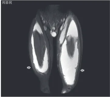
Magnetic resonance imaging (MRI) of the thighs was re-quested. This study showed massive l uid collections in peri-muscular spaces in the anterior and posterior regions of the right thigh and extensive l uid collection in perimuscular spaces in the anterior region of the left thigh plus adjacent intramuscular edema and edema of fatty planes of the medial region the of left thigh (Fig. 2). The patient was referred for surgical drainage, performed in the left thigh 3 months ago. During the procedure, there was leakage of large amounts of a milky liquid material. The microscopy revealed acellular granular eosinophilic material. The diagnosis of "milk of cal-cium" was established. Alendronate 70 mg /week was intro-duced, being in use for 2 months.
Discussion
Calcinosis or calcii cations of non-articular tissues are mani-festations of some rheumatic diseases, such as dermatomyo-sitis, systemic lupus erythematosus, systemic sclerosis and MCTD; these conditions can be painful and very debilitating, sometimes resulting in functional disability.6 In a multicenter
67
R E V B R A S R E U M A T O L . 2 0 1 3 ;5 4 ( 1 ): 6 5 – 6 7
Fig. 2 – “Milk of calcium” - hyperintense on STIR image.
rare form of calcinosis found in patients with JDM. Its recogni-tion and its differential diagnosis, especially with soft tissue infections and deep abscesses, are needed.6 These collections
may persist for months and spontaneously drain away liquid contents through the skin.5 The exact mechanism of the
for-mation of collections of "milk of calcium" in JDM is unknown. Hesla et al., who described the US i ndings in these l uid collec-tions, speculated whether they are the result of the formation of pseudobursae with liquid content among the various planes of calcii ed tissues.5 These authors described two patients with
severe JDM whose US results revealed extensive liquid collec-tions i lled with calcium in the subcutaneous and intermuscu-lar tissues of thighs and calves. The authors suggest that these l uid collections are more frequent in patients with more se-vere involvement of the disease.5
Brown et al. described the i ndings of computed tomog-raphy (CT) and MRI of a l uid collection in the left calf region in an adolescent diagnosed with JDM. A CT scan showed ex-tensive calcii cation of the subcutaneous tissues and inter-muscular septum bilaterally and the presence of subcutane-ous and intermuscular l uid collections in the left calf. MRI revealed subcutaneous and intermuscular l uid collections in the left calf with hypointense on T1 and hyperintense on T2.6
Sanson et al. described a teenager with JDM who presented l uid collection in her wrist, in MRI scan.8
The radiography is an important tool in the visualization of calcii cations and the US scan characterizes the l uid col-lections in relation to their nature and their extent, but MRI can demonstrate both as well as their global relationship to the surrounding soft tissues in any plane.8 The dystrophic
calcii cations are present as hypointense on T1 and T2. The liquid collections of "milk of calcium" are demonstrated as hyperintense in T2 sequences.8
The differential diagnosis of l uid collections of "milk of calcium" must be done with pyomyositis, that may involve the subcutaneous tissue, deep muscle tissue and intermus-cular spaces. Fever and leukocytosis are not always present at the onset of clinical manifestations of pyomyositis. In the US scan, these collections can be ecolucent, hypoechoic or with the presence of debris.5
Many drugs have been used to treat calcinosis, such as hydroxychloroquine, intravenous immunoglobulin, cyclospo-rine, inl iximab and alendronate. The results of these thera-pies have been contradictory and consistent benei ts have not been observed.3,9 Despite the difi culty of diagnosis of
this clinical case, the presence of clinical manifestations and laboratory changes of more than a rheumatic disease in the same patient suggests the diagnosis of overlap syndrome. To our knowledge, in the literature there are only three cases re-ported of "milk of calcium" in JDM patients and no case report in patients with overlap syndrome. Liquid collection of "milk of calcium" is a rare complication of JDM and even more rare of overlap syndrome, and its differentiation with infectious processes such as abscesses and pyomyositis should always be remembered.
Conl icts of interest
The authors declare no conl icts of interest.
R E F E R E N C E S
1. Jury EC, D’ Cruz D, Morrow WJW. Autoantibodies and overlap syndromes in autoimmune rheumatic disease. J Clin Pathol 2001; 54(5): 340-7.
2. Rodríguez-Reyna TS, Alarcón-Segovia D. Overlap syndromes in the context of shared autoimmunity. Autoimmunity 2005; 38(3): 219-23.
3. Boulman N, Slobodin G, Rozenbaum M, Rosner I. Calcinosis in rheumatic diseases. Semin Arthritis Rheum 2005; 34(6): 805-12.
4. Mukamel M, Horev G, Mimouni M. New insight into calcinosis of juvenile dermatomyositis: A study of composition and treatment. J Pediatr 2001; 138(5):763.
5. Hesla RB, Karlson LK, McCauley RGK. Milk of calcium l uid collection in dermatomyositis: Ultrasound i ndings. Pediatric Radiology 1990; 20(5):344-6.
6. Brown AL, Murray JG, Robinson SP, Rooney MM. Case report: Milk of calcium complicating juvenile dermatomyositis - Imaging features. Clin Radiol 1996; 51(2): 147-9.
7. Sato JO, Sallum AM, Ferriani VP, Marini P, Sacchetti SB, Okuda EM e cols. A Brazilian registry of juvenile dermatomyositis: onset features and classii cation of 189 cases. Clin Exp Rheumatol 2009; 27(6): 1031-8.
8. Samson C, Soulen RL, Gursel E. Milk of calcium l uid collections in juvenile dermatomyositis: MR characteristics. Pediatr Radiol 2000; 30(1): 28-9.