SUMMARY
Objective: he purpose of this study was to investigate the diferences between P wave disper-sion, aortic elastic properties and transthoracic echocardiographic indings in the young and old football players compared to control groups in order to asses the inluence of regular sportive activity on aortic distensibility and its potential efect on atrial electrophysiology. Methods: We recruited 42 young football players with a training history of many years. he control group was formed by 27 healthy sedentary men. Twenty-three healthy retired football players of a professional football club aged over 50 years were included in the study as old group and 18 subjects over 50 year old who did not perform regular exercise when they were young were included in the control group of old subjects. Results: he heart rate and ejection fraction were decreased in the young football players. here were no signiicant diferences in the aortic elastic parameters and P wave dispersion between young football players and control group. But in old subjects with sustained participation in regular sportive activity, the signiicant diference of let ventricular dimension, wall thickness and systolic functions detected in the young group disappeared while increase in the let atrial diameter became signiicant. Conclusion: Potential efect of aortic elastic properties which changes with age, on atrial electrophysiology through increasing P wave dispersion was shown.
Keywords: Sports medicine; atrial ibrillation; cardiac electrophysiology.
RESUMO
Propriedades da aorta e electroisiologia atrial em futebolistas proissionais ativos e aposentados
Objetivo: Com este estudo pretendeu-se levar a cabo um ensaio clínico que permitisse inves-tigar as diferenças entre a dispersão da onda P (DOP), as propriedades elásticas da aorta e os resultados da ecocardiograia transtorácica em futebolistas proissionais ativos e reformados, face a grupos de controle, de modo a avaliar a inluência da atividade desportiva regular na dis-tensibilidade aórtica e o seu potencial efeito na electroisiologia atrial. Métodos: Para este estudo foram recrutados 42 jovens futebolistas proissionais com um histórico de treino de vários anos. O grupo-controle foi constituído por 27 homens saudáveis e sedentários. No grupo composto por indivíduos mais velhos, foram incluídos 23 futebolistas proissionais já aposentados, sau-dáveis e com mais de 50 anos de idade e, como grupo-controle, 18 indivíduos com mais de 50 anos de idade que nunca izeram qualquer tipo de exercício físico regular quando eram mais novos. Resultados: A frequência cardíaca e fração de ejeção eram menores nos jovens futebo-listas proissionais. Não se veriicaram diferenças signiicativas nos parâmetros de elasticidade da aorta e a dispersão da onda P entre os jovens futebolistas proissionais e o grupo-controle. Mas, já no caso do grupo dos indivíduos mais velhos com uma atividade desportiva regular, a diferença signiicativa na dimensão ventricular esquerda, espessura das paredes e funções sis-tólicas detectada no grupo jovem e ativo desapareceu, enquanto o aumento no diâmetro atrial esquerdo tornou-se expressivo. Conclusão: Demonstrou-se com este estudo o efeito potencial das propriedades elásticas da aorta, que se alteram com a idade, na electroisiologia atrial por meio do aumento da dispersão da onda P.
Unitermos: Medicina esportiva; ibrilação atrial; eletroisiologia cardíaca. Study conducted at Dr Siyami
Ersek Cardiovascular and Thoracic Surgery Training and Research Hospital (Department of Arrhytmia), Istanbul, Turkey
Submitted on: 01/20/2011
Approved on: 03/02/2011
Correspondence to: Hakan Hasdemir Department of Cardiology, Dr. Siyami Ersek Cardiovascular and Thoracic Surgery Training and Research Hospital,
Istanbul, Turkey Phone: +90 (216) 542 44 44
Fax: +90 (216) 337 97 19 [email protected]
Conlict of interest: None.
Aortic properties and atrial electrophysiology in the young and old
football players
HAKAN HASDEMIR1, MUSTAFA YILDIZ2, GOKHAN METIN3, HASAN KASAP4, BANUŞAHIN YILDIZ5, BARIŞ YAYLAK1, AYŞEGÜL ÖZYURT6
1 MD, Cardiologist, Department of Cardiology, Dr. Siyami Ersek Cardiovascular and Thoracic Surgery Training and Research Hospital, Istanbul, Turkey 2 MD, PhD, Cardiologist, Department of Cardiology, Kartal Koşuyolu Yüksek Ihtisas Educational and Research Hospital, Istanbul, Turkey
3 MD, PhD, Physiologist, Department of Physiology, Istanbul University Cerrahpaa Medical Faculty, Istanbul, Turkey 4 MD, Cardiologist, Department of Cardiology, Umut Hospital, Istanbul, Turkey
INTRODUCTION
Football is one of the most popular and widely viewed sports in the world. Executing such movements during performance, both aerobic and anaerobic metabolic sys-tems appear to be involved throughout a game1. Widely used in sports medicine, the transthoracic echocardiog-raphy allows quantitative assessment of cardiac structure and function for the athletes. he elite athletes oten ex-hibit some changes in the heart, called athlete’s heart2,3. Athlete’s heart is an enlarged heart related to repeated strenuous exercise. As a result of exercise, the heart will expand physiologically by enlarging chambers, increas-ing muscle mass and increasincreas-ing the volume of blood pumped per stroke. he athletes heart is associated with some types of electrocardiogram (ECG) abnormalities, such as sinus bradycardia, sinus arrhythmia, early re-polarization, irst-degree atrioventricular block and let atrial enlargement4.
Atrial ibrillation is the most frequent cause of pro-longed palpitations in young competitive athletes, even including those performing elite sport activity5. Endur-ance sport practice increases the probability of sufer-ing of atrial ibrillation, ater adjustsufer-ing for other risk factors6. he possible mechanisms explaining the asso-ciation remain speculative. Atrial ectopic beats, inlam-matory changes, and atrial size have been suggested6. P wave dispersion, detected from the surface ECG, have been though to relect let atrial enlargement and altered conduction7,8. P wave dispersion and P wave maximal duration, that relect the activation of atrial muscle and may depend primarily upon the mass of tissue excited, have been used in the assessment of the risk for atrial ibrillation which is characterized by nonhomogeneous and discontinuous atrial conduction7,8. P wave dispersion was deined as the diference between the longest and the shortest P wave duration recorded from multiple difer-ent surface ECG leads7. he clinical signiicance of P wave duration has been demonstrated in many clinical condi-tions, especially in paroxysmal atrial ibrillation8. P wave dispersion has been shown to be inluenced by the auto-nomicnervous systemactivation, which induces changes in let atrial size and the velocity of impulse propagation9.
Arterial compliance plays a role in determining both arterialsystolic and diastolic pressure and therefore, in a clinicalcontext, inluences let ventricular size and func-tion and coronaryblood low10-12. Aerobic exercise has well-documentedeicacy for cardiovascular risk reduc-tion, and it appears thatat least part of its beneit derives from modiication of arterialproperties13. Non-invasive ultrasound techniques such as echocardiography and aortic pulse wave velocity are used to evaluate vascular system and cardiovascular condition11,12.
No studies cited in the literature suggest the young and old football players are at increased risk for atrial
i-brillation and arterial stifness. he purpose of this study was to investigate and compare P wave duration, P wave dispersion, aortic elastic properties (aortic strain, aortic distensibility) and transthoracic echocardiographic ind-ings in the young and old football players.
METHODS
SUBJECT’SPOPULATION
We recruited 42 young football players with a training history of many years. hey were the members of a local football team and they were regularly maintaining sport-ive activities and training programs. Twenty-seven healthy sedentary men formed the control group. Healthy and over 50 years old retired 23 football players of a profes-sional football club were included in the study as old group and 18 subjects over 50 years old who did not perform regular exercise when they were young were included in the control group of old subjects. A detailed history was taken and each participant underwent a systemic physical examination to exclude cardiovascular or other relevant disease before attending to the study. Any subjects who had a history of cardiovascular or any other systemic dis-orders such as hyperlipidemia, hyperglycemia, anemia and those on medications known to alter cardiac conduction were excluded from the study. All subjects were non smok-ers and non alcoholics. All subjects gave their consent for inclusion in the study. he investigation conforms to the principles outlined in the Declaration of Helsinki.
BODYMASSINDEXMEASUREMENT
Body mass index (kg/m²) was calculated dividing body weight in kilograms by square of body height in meters.
BLOODPRESSUREMEASUREMENT
he arterialblood pressure of the subjects was measured by the same clinician. he subjects were in the supine posi-tion and had rested at least 20 minutes before the measure-ment. he blood pressure was measured, using a mercury sphygmomanometer with a cuf appropriate to the arm circumference (Korotkof phase I for systolic blood pres-sure and V for diastolic blood prespres-sure). Blood prespres-sure measurements were performed twice for each subject and their mean was used for statistical analysis.
Pulse pressure = systolic blood pressure - diastolic blood pressure
Mean blood pressure = [systolic blood pres-sure + 2 x diastolic blood pressure] / 3
P WAVEDISPERSIONMEASUREMENT
ob-tained manually by two of the investigators using calipers and magnifying lens for accurate deinition of the ECG delection as deined in previous study7,8. he onset of the P wave was deined as the point of the irst visible upward departure of the trace from the bottom of the baseline. he return to the baseline of the bottom of the trace in wave was considered to be the end of the P wave. P maximum in any of the 12 lead surface ECGs was measured and used as a marker of prolonged atrial conduction time. he dif-ference between P wave maximum and P wave minimum durations was deined as P wave dispersion.
TRANSTHORACICECHOCARDIOGRAPHY
A Vivid 3 cardiovascular ultrasound system [3S sector probe (1.5 - 3.6 MHz), GE] was used for transthoracic echocardiographic evaluation14. Echocardiography was performed with the subject in the lateral decubitus posi-tion. Interventricular septal thickness (IVS), let ventricle posterior wall thickness (LVPW), let ventricle end-dia-stolic (LVED) and end-syend-dia-stolic diameters (LVES), let atri-al diameter (LAD), aortic root diameter and aortic vatri-alve openness were measured in the parasternal long-axis view. he measurements were obtained from two-dimensional guided M-mode recordings. he pulsed Doppler sample volume was positioned at the mitral lealet tips. Early dia-stolic peak low velocity (E) (m/s) and late diadia-stolic peak low velocity (A) (m/s) were measured by transmitral Dop-pler imaging. Ater the routine conventional echocardio-graphic examination was performed, subjects were placed in a mild recumbent position, and the ascending aorta was recorded in the two-dimensional guided M-mode trac-ings. Aortic diameters were recorded 3 cm above the aortic valve by M-mode echocardiography. Aortic systolic diam-eter was ddiam-etermined at the time of the full opening of the aortic valve, and aortic diastolic diameter was determined at peak QRS. he same blinded investigator performed the echocardiography and the echocardiograms were analyzed by two blinded cardiologists.
Aortic strain was calculated as follows: Aortic strain = (AoS - AoD) / AoD
(AoS, systolic aortic diameter, AoD, diastolic aortic diameter)
Aortic distensibility was calculated as follows: Aortic distensibility = 2 x (AoS - AoD) / (AoD x PP) (PP, pulse pressure)15,16
STATISTICALANALYSIS
he statistical analysis was done using the SPSS (version 16.0) ready-to-use programme. All values were expressed as mean ± standard deviation. Mann-Whitney U non-parametric tests were done. Signiicance limit was accept-ed as p ≤ 0.05.
RESULTS
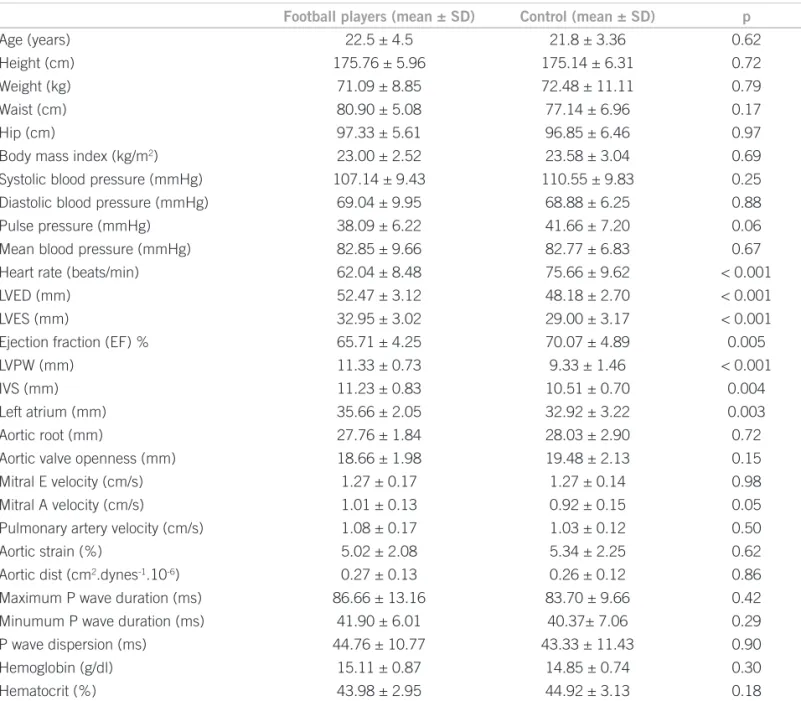
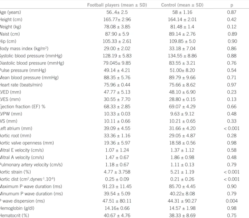
In the young football players group, LVED, LVES, LVPW, IVS and LAD were increased in compared to healthy sed-entary subjects (p <0.001; p < 0.001; p < 0.001; p = 0.004; p = 0.003; respectively). On the contrary; the heart rate and ejection fraction were decreased in the young foot-ball players group (p < 0.001; p = 0.005; respectively). here were no signiicant diferences in the aortic elastic parameters and P wave dispersion between two groups (p > 0.05) (Table 1). In the old football players group while LVED, LVES, LVPW, IVS were not signiicantly dif-ferent from their control group, LAD was signiicantly increased (p < 0.001). Furthermore, the diference in heart rate and ejection fraction disappeared along with aging. Although a signiicant diference in aortic elastic properties and P wave dispersion was not revealed in young groups, P wave dispersion was signiicantly higher in old football players in compared to old control group (p = 0.004). Whereas, aortic strain was signiicantly lower in old football players group (p < 0.001), aortic distensi-bility was signiicantly higher in compared to old control group (p < 0.001) (Table 2).
DISCUSSION
those without atrial ibrillation. It was also shown that elite junior athletes have a signiicantly greater let atrial diam-eter as in our study4,25. herefore, it would be expected that P wave dispersion and P wave duration could be increased in the young football players. In contrast, we found no signiicant diferences in P wave duration and P wave dis-persion between young football players and control group. However, some studies showed that maximal let atrial diameter is not a signiicant predictor of atrial ibrillation and there is no correlation between iltered P wave dura-tion and let atrial enlargement8,26. But the signiicant asso-ciation which was shown in between P wave dispersion and LAD in old group in this study proved the inluence of aging on atrial electrophysiology, independent of increase in LAD. he aorta is a complex organ with multiple functions. It acts as both a conduit and an elastic bufering chamber. By virtue of its elastic properties, this vessel inluences let
ventricular functions and coronary blood low10,11. It has been reported that aortic distensibility is decreased in pa-tients with coronary heart disease10,11. On the other hand, some studies have shown an increase or decrease in these properties in trained athletes27,28. Dynamic exercise may play a critical role in aortic elastic properties and its caliper of the arterial lumen27. Altered arterial elastical responses to cardiac cycle may be due to changes of hemodynamic pa-rameters and arterial wall properties27. he changed vascu-lar response (increased arterial distensibility) might contin-ue at rest period because of adaptation to regular exercise. In contrast, Bertovic et al.28 showed that whole body arterial compliance is lower in strength-trainedmen than in age-matched sedentary controls. he lower arterial compliance of the athleticgroup was the higher pulse pressure (systolic blood pressure - diastolic blood pressure), measured both peripherallyat the brachial artery and centrally at the
com-Football players (mean ± SD) Control (mean ± SD) p
Age (years) 22.5 ± 4.5 21.8 ± 3.36 0.62
Height (cm) 175.76 ± 5.96 175.14 ± 6.31 0.72
Weight (kg) 71.09 ± 8.85 72.48 ± 11.11 0.79
Waist (cm) 80.90 ± 5.08 77.14 ± 6.96 0.17
Hip (cm) 97.33 ± 5.61 96.85 ± 6.46 0.97
Body mass index (kg/m2) 23.00 ± 2.52 23.58 ± 3.04 0.69
Systolic blood pressure (mmHg) 107.14 ± 9.43 110.55 ± 9.83 0.25
Diastolic blood pressure (mmHg) 69.04 ± 9.95 68.88 ± 6.25 0.88
Pulse pressure (mmHg) 38.09 ± 6.22 41.66 ± 7.20 0.06
Mean blood pressure (mmHg) 82.85 ± 9.66 82.77 ± 6.83 0.67
Heart rate (beats/min) 62.04 ± 8.48 75.66 ± 9.62 < 0.001
LVED (mm) 52.47 ± 3.12 48.18 ± 2.70 < 0.001
LVES (mm) 32.95 ± 3.02 29.00 ± 3.17 < 0.001
Ejection fraction (EF) % 65.71 ± 4.25 70.07 ± 4.89 0.005
LVPW (mm) 11.33 ± 0.73 9.33 ± 1.46 < 0.001
IVS (mm) 11.23 ± 0.83 10.51 ± 0.70 0.004
Left atrium (mm) 35.66 ± 2.05 32.92 ± 3.22 0.003
Aortic root (mm) 27.76 ± 1.84 28.03 ± 2.90 0.72
Aortic valve openness (mm) 18.66 ± 1.98 19.48 ± 2.13 0.15
Mitral E velocity (cm/s) 1.27 ± 0.17 1.27 ± 0.14 0.98
Mitral A velocity (cm/s) 1.01 ± 0.13 0.92 ± 0.15 0.05
Pulmonary artery velocity (cm/s) 1.08 ± 0.17 1.03 ± 0.12 0.50
Aortic strain (%) 5.02 ± 2.08 5.34 ± 2.25 0.62
Aortic dist (cm2.dynes-1.10-6) 0.27 ± 0.13 0.26 ± 0.12 0.86
Maximum P wave duration (ms) 86.66 ± 13.16 83.70 ± 9.66 0.42
Minumum P wave duration (ms) 41.90 ± 6.01 40.37± 7.06 0.29
P wave dispersion (ms) 44.76 ± 10.77 43.33 ± 11.43 0.90
Hemoglobin (g/dl) 15.11 ± 0.87 14.85 ± 0.74 0.30
Hematocrit (%) 43.98 ± 2.95 44.92 ± 3.13 0.18
SD, standard deviation; LVED, left ventricle diastolic diameter; LVES, left ventricle systolic diameter; LVPW, left ventricle posterior wall thickness in diastole; IVS, interventricular septum thickness in diastole; Dist, distensibility
mon carotid artery.he diference in pulse pressure was at-tributable to both greatersystolic and lower diastolic arterial blood pressure in the athletes.he higher systolic pressure of the athletic group was maintainedat maximal exercise, indicating a greater aterload during aerobicexercise in the trained group. However, the muscular strength-trainedathletes have similar (as in our study) or lower pres-sures than the sedentary population29,30.
Endurance exercise-trained middle-aged/older adults demonstrate lower large elastic artery stifness and greater endothelium-dependent dilatation (EDD) than their sedentary peers. With daily brisk walking, previously sedentary middle-aged/older adults show reduced stif-ness and improved EDD. he mechanisms underlying the efects of regular aerobic exercise on large elastic artery stifness with aging are mostly unknown, but are likely to include changes of the composition of the arterial wall.
Enhanced EDD in older adults who exercise is mediated by increased nitric oxide bioavailability associated with reduced oxidative stress. Aerobic exercise also may pro-tect arteries in advanced ageing by increasing resistance to the efects of other cardiovascular diseases risk factors like LDL-cholesterol.
In conclusion, while the beneicial efects of exercise to aortic compliance were not evident in young footballers yet, they were evidently maintained in old football players group.
STUDY LIMITATIONS
here might be some limitations in this study. Firstly, we did not measure sympathetic and parasympathetic system activa-tion. Secondly, P wave dispersion measurement errors done with manual evaluation may be potential bias for observed results. However, manual measurement of P wave dispersion has been well accepted and used in several studies7,8.
Football players (mean ± SD) Control (mean ± SD) p
Age (years) 56..4± 2.5 58 ± 1.16 0.87
Height (cm) 165.77± 2.96 164.14 ± 2.01 0.42
Weight (kg) 78.08 ± 3.85 81.48 ± 1.4 0.12
Waist (cm) 87.90 ± 5.9 89.14 ± 2.76 0.89
Hip (cm) 105.33 ± 2.61 109.85 ± 5.0 0.90
Body mass index (kg/m2) 29.00 ± 2.02 33.18 ± 7.04 0.86
Systolic blood pressure (mmHg) 128.19 ± 5.83 134.55 ± 8.86 0.88
Diastolic blood pressure (mmHg) 79.045± 9.85 83.55 ± 3.21 0.76
Pulse pressure (mmHg) 49.14 ± 4.21 51.00± 8.20 0.54
Mean blood pressure (mmHg) 88.35 ± 5.76 89.79 ± 9.66 0.71
Heart rate (beats/min) 75.96 ± 0.44 75.66 ± 8.62 0.97
LVED (mm) 47.77 ± 5.13 48.10 ± 6.90 0.23
LVES (mm) 30.55 ± 7.70 28.80 ± 0.15 0.13
Ejection fraction (EF) % 68.33 ± 2.85 69.07 ± 4.29 0.66
LVPW (mm) 10.33 ± 0.03 9.63 ± 9.12 0.48
IVS (mm) 10.11 ± 0.66 10.21 ± 0.65 0.33
Left atrium (mm) 39.09 ± 4.55 31.66 ± 4.20 < 0.001
Aortic root (mm) 33.36 ± 1.16 29.05 ± 4.87 0.28
Aortic valve openness (mm) 19.36 ± 5.97 18.58 ± 0.56 0.98
Mitral E velocity (cm/s) 1.07 ± 1.24 1.37 ± 1.12 0.58
Mitral A velocity (cm/s) 1.47 ± 0.67 1.86 ± 0.98 0.48
Pulmonary artery velocity (cm/s) 1.18 ± 0.67 1.11 ± 0.13 0.79
Aortic strain (%) 4.77 ± 3.758 5.21 ± 1.19 < 0.001
Aortic dist (cm2.dynes-1.10-6) 0.25 ± 0.09 0.21 ± 0.26 < 0.001
Maximum P wave duration (ms) 91.23 ± 11.45 85.70 ± 4.45 0.90
Minumum P wave duration (ms) 39.54 ± 5.09 40.22± 8.08 0.79
P wave dispersion (ms) 47.51 ± 80.11 44.31 ± 90.27 0.004
Hemoglobin (g/dl) 14.16± 0.66 14.57 ± 1.98 0.98
Hematocrit (%) 40.67 ± 4.76 38.33 ± 8.69 0.75
Table 2 – Anthropometric, hemodynamic, laboratory, echocardiographic and electrocardiographic values in the old groups
REFERENCES
1. Ciuti C, Marcello C, Macis A, Onnis E, Solinas R, Lai C et al. Im- Im-proved aerobic power by detraining in basketball players mainly trained for strength. Sports Med Training Rehab.1996;6:325-35. 2. Longhurst JC, Kelly AR, Gonyea WJ, Mitchell JH. Echo
cardio-graphic let ventricular masses in distance runners and weight liters. J Appl Physiol. 1980;48:154-62.
3. Colan SD, Sanders SP, MacPherson D, Borow KM. Let ventricular diastolic function in elite athletes with physiologic cardiac hypertro-phy. J Am Coll Cardiol. 1985;6:545-9.
4. Crouse SF, Meade T, Hansen BE, Green JS, Martin SE. Electrocardio-grams of collegiate football athletes. Clin Cardiol. 2009;32:37-42. 5. Furlanello F, Pedrinazzi C, Inama G, De Ambroggi L, Cappato R.
he intriguing problem of atrial ibrillation in competitive athletes. Minerva Cardioangiol. 2008;56:659-66.
6. Mont L, Elosua R, Brugada J. Endurance sport practice as a risk fac-tor for atrial ibrillation and atrial lutter. Europace 2009;11:11-7. 7. Dilaveris PE, Gialafos EJ, Sideris SK, heopistou AM,
Andrikopou-los GK, Kyriakidis M et al. Simple electrocardiographic markers for the prediction of paroxysmal idiopathic atrial ibrillation. Am Heart J. 1998;135:733-8.
8. Dilaveris PE, Gialafos EJ, Andrikopoulos GK, Richter DJ, Papaniko-laou V, Poralis K et al. Clinical and electrocardiographic predictors of
recurrent atrial ibrillation. Pacing Clin Electrophysiol. 2000;23:352-8. 9. Cheema AN, Ahmed MW, Kadish AH, Goldberger JJ. Efects of au-tonomic stimulation and blockade on signal-averaged P-wave dura-tion. J Am Coll Cardiol. 1995;26:497-502.
10. Stefanadis C, Wooley CF, Bush CA, Kolibash AJ, Boudoulas H. Aor-tic distensibility abnormalities in coronary artery disease. Am J Car-diol. 1987;59:1300-4.
11. Hirai T, Sasayama S, Kawasaki T, Yagi S. Stifness of systemic arter-ies in patients with myocardial infarction. A non-invasive method to predict coronary atherosclerosis. Circulation 1989;80:78-86. 12. Asmar R, Benetos A, Topouchian J, Laurent P, Pannier B, Brisac AM
et al. Assessment of arterial distensibility by automatic pulse wave
velocity measurement. Validation and clinical application studies. Hypertension 1995;26:485-90.
13. Vaitkevicius PV, Fleg JL, Engel JH, OConnor FC, Wright JG, Lakatta LE et al. Efects of age and aerobic capacity on arterial stifness in healthy adults. Circulation 1993;88(pt 1):1456-62.
14. Pearlman AS, Gardin JM, Martin RP, Parisi AF, Popp RL, Quinones MA et al. Guidelines for optimal physician training in echocardiog-raphy. Recommendations of the American Society of Echocardiog-raphy Committee for Physician Training in EchocardiogEchocardiog-raphy. Am J Cardiol. 1987;60:158-63.
15. Stefanadis C, Stratos C, Boudoulas H, Kourouklis C, Toutouzas P. Distensibility of the ascending aorta: comparison of invasive and noninvasive techniques in healthy men and in men with coronary artery disease. Eur Heart J. 1990;11:990-6.
16. Stefanadis C, Stratos C, Vlachopoulos C, Marakas S, Boudoulas H, Kallikazaros I et al. Pressure-diameter relation of the human aorta: a new method of determination by the application of a special ultra-sonic dimension catheter. Circulation 1995;92:2210-9.
17. Fagard RH. Athlete’s heart: a meta-analysis of the echocardiographic experience. Int J Sports Med. 1996;17(Suppl 3):S140-4.
18. Fagard R, Van Den Broeke C, Amery A. Let ventricular dynam-ics during exercise in elite marathon runners. J Am Coll Cardiol. 1989;14:112-8.
19. Smith ML, Hudson DL, Graitzer HM, Raven PB. Exercise training bradycardia: the role of autonomic balance. Med Sci Sports Exerc. 1989;21:40-4.
20. Zehender M, Meinertz T, Keul J, Just H. ECG variants and cardiac ar-rhythmias in athletes: clinical relevance and prognostic importance. Am Heart J. 1990;119:1378-91.
21. Coumel P. Autonomic inluences in atrial tachyarrhythmias. J Car-diovasc Electrophysiol. 1996;7:999-1007.
22. Buchheit M, Gindre C. Cardiac parasympathetic regulation: respec-tive associations with cardiorespiratory itness and training load. Am J Physiol Heart Circ Physiol. 2006;291:H451-H8.
23. Buchheit M, Simon C, Piquard F, Ehrhart J, Brandenberger G. Ef-fects of increased training load on vagal-related indexes of heart rate variability: a novel sleep approach. Am J Physiol Heart Circ Physiol. 2004;287:H2813-H8.
24. Chen YJ, Tai CT, Chiou CW, Wen ZC, Chan P, Lee SH et al. Induc-ibility of atrial ibrillation during atrioventricular pacing with vary-ing intervals: role of atrial electrophysiology and the autonomic ner-vous system. J Cardiovasc Electrophysiol. 1999;10:1578-85. 25. Sharma S, Maron BJ, Whyte G, Firoozi S, Elliott PM, McKenna WJ.
Physiologic limits of let ventricular hypertrophy in elite junior ath-letes: relevance to diferential diagnosis of athletes heart and hyper-trophic cardiomyopathy. J Am Coll Cardiol. 2002;40:1431-6. 26. Ishimoto N, Ito M, Kinoshita M. Signal-averaged P-wave
abnormali-ties and atrial size in patients with and without idiopathic paroxys-mal atrial ibrillation. Am Heart J. 2000;139(4):684-9.
27. Kasikcioglu E, Kayserilioglu A, Olaz H, Akhan H. Aortic distensibil-ity and let ventricular diastolic functions in endurance athletes. Int J Sports Med. 2005;26:165-70.
28. Bertovic DA, Waddell TK, Gatzka CD, Cameron JD, Dart AM, King-well BA. Muscular strength training is associated with low arterial compliance and high pulse pressure. Hypertension 1999;33:1385-91. 29. Fleck SJ, Dean LS. Resistance-training experience and the pressor
response during resistance exercise. J Appl Physiol. 1987;63:116-20. 30. Longhurst JC, Kelly AR, Gonyea WJ, Mitchell JH. Cardiovascular