Official Journal of the Brazilian Psychiatric Association Volume 34 • Supplement 2 • October/2012
Psychiatry
Revista Brasileira de Psiquiatria
Abstract
The pathophysiology of neurodegenerative diseases (ND) such as Alzheimer’s disease (AD) and Parkinson’s disease (PD) has not yet been completely elucidated. However, in the past few years, there have been great knowledge advances about intra-and extracellular proteins that may display
impaired function or expression in AD, PD and other ND, such as amyloid beta (Aβ), α-synuclein,
tau protein and neuroinlammatory markers. Recent developments in the imaging techniques of
positron emission tomography (PET) and single photon emission computed tomography (SPECT) now allow the non-invasive tracking of such molecular targets of known relevance to ND in vivo.
This article summarizes recent indings of PET and SPECT studies using these novel methods, and discusses their potential role in the ield of drug development for ND as well as future clinical
applications in regard to differential diagnosis of ND and monitoring of disease progression.
New molecular targets for PET and SPECT imaging in
neurodegenerative diseases
Marcel Benadiba,
1,2,3Gert Luurtsema,
1Lauro Wichert-Ana,
3,4Carlos Alberto Buchpigel,
3,5Geraldo Busatto Filho
2,31 Department of Nuclear Medicine and Molecular Imaging, University Medical Center Groningen,
University of Groningen, Groningen, Netherlands
2 Laboratory of Psychiatric Neuroimaging (LIM 21), Department of Psychiatry,
Faculdade de Medicina, Universidade de São Paulo, Brazil
3 Núcleo de Apoio à Pesquisa em Neurociência Aplicada (Center for Interdisciplinary
Research on Applied Neurosciences: NAPNA), Brazil
4 Department of Internal Medicine, Faculdade de Medicina de Ribeirão Preto,
Universidade de São Paulo, Ribeirão Preto, SP, Brazil
5 Center of Nuclear Medicine, Department of Radiology, Faculdade de Medicina da
Universidade de São Paulo, São Paulo, SP, Brazil
Received on October 7, 2011; accepted on February 29, 2012.
DESCRIPTORS Positron emission tomography;
Single photon emission tomography;
Neurodegenerative diseases;
Neuroinlammation;
Brain lipid metabolism. ARTICLE
Corresponding author: Prof. Dr. Geraldo Busatto Filho. Rua Dr. Ovídio Pires de Campos, S/Nº - 2º andar; CMN – Centro de Medicina Nuclear; CEP 05403-010 São Paulo, SP, Brasil. Phone: (+55 11) 2661-8132; Fax: (+55 11) 3082-1015. E-mail: [email protected]. 1516-4446 - ©2012 Elsevier Editora Ltda. All rights reserved.
Introduction
Positron emission tomography (PET) and single photon emission computed tomography (SPECT) are in vivo imag-ing techniques that allow the non-invasive trackimag-ing of brain pathophysiological processes underlying various neurological and psychiatric disorders. These techniques have also been successfully used in various aspects of drug development, including the understanding of the mechanism of action of pharmacological agents in the central nervous system (SNC), their dosage regimens and thresholds for clinical response and emergence of side-effects.1
Several studies over the past decades have shown that PET and SPECT methods can reliably map neurochemical processes of interest in the brain, including the density and
afinity of postsynaptic receptors for neurotransmitters such
as dopamine, serotonin and others, as well as presynaptic transporters for these transmitters, precursors such as L-DOPA and transmitter degrading enzymes. Such approach has provided invaluable information about neurochemi-cal abnormalities involved in psychiatric and neurologineurochemi-cal disorders, as well as helping to elucidate the mechanism of action of the pharmacological agents commonly used to treat these conditions.
More recently, technological advances have enabled the use of PET and SPECT to probe a number of other intra- and extra-cellular proteins that may display impaired function or expression in brain diseases. Such advances have moved the neurological and psychiatric uses of PET and SPECT from a
strict neurochemical imaging role to a much more lexible and comprehensive proile of applications, providing knowledge
about molecular brain mechanisms that may be much closer to the pathophysiological essence of neurological and
psy-chiatric disorders than supericial neurotransmitter changes.
One of the most promising applications of such new PET and SPECT methods regards to the investigation of patho-physiological aspects of neurodegenerative disorders (NDs). This is of great relevance given the large prevalence of NDs such as Alzheimer’s disease (AD) and Parkinson’s disease (PD) in elderly life, as well as the fact that a greater knowledge about the pathophysiology of these disorders may help in the development of novel pharmacological treatments capable of interfering with their molecular pathological substrate. Taken those issues into account, this review will focus on perspec-tives for new PET and SPECT tracers developed to allow the mapping of intracellular and extracellular mechanisms of particular relevance to AD, PD and other NDs.
Molecular brain imaging with PET and SPECT:
basic principles
In order to allow the eficient visualization, characteriza -tion and quantitative measurements of relevant biological processes in the brain, PET and SPECT techniques demand the development of suitable probes that can be labeled with a positron emitting isotope (in the case of PET) or photon emitting isotope (in the case of SPECT). Importantly, because of their limited spatial resolution, the use of computed tomography (CT) or magnetic resonance imaging (MRI) is often required. Functional and structural techniques can be easily fused using special software, by creating parametric images. However, the development of hybrid systems where
functional techniques are fully integrated with structural cross-sectional methods also helped to attenuate the lack of anatomical resolution of PET and SPECT. These parametric images give both anatomical and functional information,
allowing the identiication of regions which exhibit differ -ences in the uptake of labeled compounds. Anyway, the most employed radioisotopes for labeling PET probes are carbon-11 (11C) and luorine-18 (18F), differing basically in
their half-lives and maximum energy. The irst (11C) must
be produced by an on-site cyclotron located near to the PET imaging facility due to the very short physical half-life (20 minutes). However, the longer half-life of 18F (110
min-utes) allows the delivery of 18F-labeled ligands to a broader
list of PET facilities located in the same town, or even in neighborhood cities. For SPECT imaging, probes can be labeled with iodine-123 (123I) or technetium-99m (99mTc);2
these isotopes have much longer half-lives than those used in PET imaging, avoiding the need for a nearby cyclotron.
Having crossed the blood-brain barrier (BBB) after
intra-venous injection, the radiolabeled compound accumulates
in certain parts of the brain, depending on the biological process being tracked. Both PET and SPECT are equipped with distinct radiation detectors that are placed in close
proximity to the head after injection of the radioligand,
and the data collected by such detectors are transformed to generate three-dimensional tomographic maps display-ing the regional distribution of radioactivity emitted by the brain. In order to be suitable for in vivo brain imaging with PET or SPECT, a radiopharmaceutical compound (also called radiotracer, due to its sub-pharmacological dose) needs to
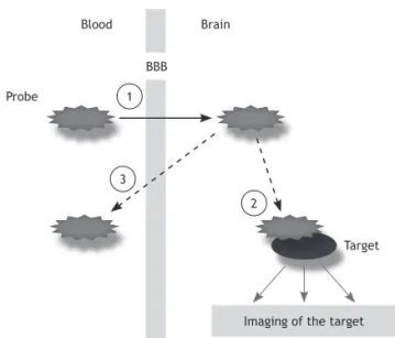
be able to bind speciically to its target (binding potential
of a drug) (Figure 1), otherwise the accuracy of the imaging
information obtained may be impaired. By deinition, bind -ing potential (BP) is a pivotal measure in the use of PET to
Figure 1 The basic requirements for suitable target-imag-ing agents include: (1) prompt crosstarget-imag-ing of the blood-brain barrier; (2) selective binding to the target molecules; and (3) clear and contrasting signals between target and non-target molecules.4
Probe
Target
Imaging of the target BBB
Brain
1
2 3
measure the density of “available” receptors, e.g. to assess the occupancy by drugs or to characterize abnormalities in receptor distribution in association with neuropsychiatric disorders. Thus, BP is a combined measure that depends on
receptor density as well as on drug-receptor afinity.3
Amyloid imaging tracers
Extracellular senile plaques are protein aggregates formed by the misbalance between the production and clearance of proteins or peptides in brain tissue of AD patients. Amyloid
beta (Aβ), released from the cleavage of the amyloid precur -sor protein (APP), is the most important constituent present in these plaques and represents the main hallmark that char-acterizes the neuropathological diagnosis of AD. The cleavage of APP can be performed by several proteases or peptidase proteins. Among these, secretases, especially gamma (which contains presenilins, nicastrin, anterior pharynx defective-1,
and presenilin enhancer-2) and beta (β-site APP cleaving
enzyme 1, BACE1), are the most important enzymes, with their activity being responsible for the excessive release
of the highly amyloidogenic 42 amino acid variant (Aβ42)
peptide. In contrast, APP cleavage promoted by the α-type
secretases disintegrin and metalloproteinase (ADAM) 10 and 17 contributes to the formation of soluble neuroprotective
fragments known as Sα-APP.5
The great advances in the knowledge about the molecular basis of AD, described above, have led to enormous interest in the development of PET and SPECT tracers that could be useful for in vivo imaging of Aβ plaques in the human brain. The irst PET tracer developed to bind speciically to ibrillar Aβ plaques was the 11C-labeled Pittsburgh Compound-B ([11C]
PIB). Up until now, this has been the best characterized and most widely used PET tracer for the study of amyloid deposits in the human brain, both in AD and in other NDs. The several potential roles of in vivo amyloid imaging techniques in AD are outlined in Table 1.
Since a deinite diagnosis of AD can only be conirmed by
post-mortem neuropathological examination, diagnostic tools that can be used to give support to a suspicion of AD in an
individual with memory deicits and other features of cogni -tive decline are highly valuable. Several studies have shown a marked degree of [11C]PIB retention in the association cortex
of mild AD patients compared to healthy controls6–9 (Figure 2).
Figure 2 PET images obtained after intravenous injection of 11C-labeled Pittsburgh Compound B (PiB) in a patient with
These indings established PET imaging with [11C]PIB as a
use-ful imaging tool to aid in the diagnostic conirmation of early
AD.9,10 However, it should be noted that amyloid deposition
is not pathognomonic of AD, being for instance found in a proportion of cognitively healthy elderlies. Nevertheless, a negative [11C]PIB PET result is highly informative to rule out
the diagnosis of AD.
The usefulness of [11C]PIB imaging with PET for assessing
the progression of AD has not been as well established. For instance, an interesting 2-year follow-up study of AD patients
revealed that there was no signiicant increase in [11C]PIB
uptake over time, although individually some patients showed clear increases.8 Such pattern of results indicates Aβ deposi
-tion plateaus when clinical dementia is already established. PET investigations with [11C]PIB are also clinically useful to
aid in the distinction between AD and other dementias. Most notably, patients with frontotemporal dementia (FTD) have generally normal [11C]PIB uptake (although occasional FTD
patients may display increased brain uptake).11,12
Individuals with objective cognitive decline not severe enough to fulill the criteria for established dementia re -ceive the diagnosis of minor cognitive impairment (MCI).13
Subjects diagnosed as suffering from MCI have a high risk
of developing dementia, with an estimated rate of conver-sion to AD of approximately 12% per year.13 A number of PET
studies have shown that a subpopulation of subjects with
MCI shows increased levels of [11C]PIB uptake to the same
degree as seen in patients with AD.7,14,15 In addition, recent
investigations demonstrate that increased [11C]PIB uptake in
the brain of MCI patients is highly predictive of subsequent conversion to AD.16
Also noteworthy, most patients with dementia with Lewy bodies (DLB) demonstrate increased [11C]PIB uptake in the
brain.17 Recent reports have shown that [11C]PIB holds
prom-ise to help in the discrimination of DLB patients from those with PD, PD with dementia (PDD), PD with mild cognitive
impairment (PD-MCI), and healthy control subjects (HCS).17,18
However, [11C]PIB retention did not differ across the
diagno-ses of PDD, PD-MCI, PD, and HCS.18 Importantly, one study
reported that the increased [11C]PIB retention in the brains of
DLB patients is largely attributable to the binding of [11C]PIB
to Aβ plaques and not to α-synuclein, the primary structural component of Lewy body ibrils.19
Finally, two other important areas of potential use for amyloid imaging tracers include drug development and moni-toring of treatment effects (Table 1). For example, one study of AD patients treated with phenserine, an anticholinesterase compound, showed an improvement in cognition that was
not however accompanied by signiicant changes in mean
cortical [11C]PIB retention in the brain.20
Given the longer half-life of 18F-labeled probes in comparison to [11C]labeled compounds (110 minutes vs.
20 minutes), there has been a great degree of interest in the past few years in the development of 18F-labeled
com-pounds for brain amyloid imaging with PET, which could be transported from radiopharmaceutical production facilities to other PET imaging sites. A number of such 18F-labeled amyloid imaging tracers, including [18F]lutemetamol (an 18F-labeled derivative of PIB), [18F]AV-45 (lorbetapir) and
[18F]BAY 94-9172 (lorbetaben), are already in an advanced
stage of clinical trials.21–24 Noteworthy, lorbetapir has been
recently approved by the FDA in the United States for PET imaging of the brain in adults under evaluation for AD and other causes of cognitive decline.25 Clinical studies indicate
that these 18F-labeled amyloid imaging tracers provide good
discrimination between AD patients and healthy subjects.
However, they still present some technical limitations,
in-cluding a relatively low degree of speciic binding in vivo, as well as a high level of white matter binding in healthy human brains which reduces the contrast between cortical
and non-cortical speciic uptake of the tracer.
Furthermore, two technetium (99mTc) and rhenium (Re)
labeled ligands have been recently synthesized for Aβ im -aging with SPECT. They are derived from the compounds (2-(1-(6-(dialkylamino)naphthalen-2-yl)ethylidene)malo-nonitrile (DDNP) and 1-(6-(dialkylamino)naphthalen-2-yl) ethanone (ENE). However, these compounds showed low
afinities for Aβ aggregates and require further reinements
in order to improve their diffusion through the BBB.26 In
ad-dition, it is also important to mention that SPECT has lower sensitivity and spatial resolution while compared to PET, and this might also promote possible differences in accuracy between these two techniques.
Tau imaging tracers
In addition to the extracellular Aβ plaques mentioned above, intracellular neuroibrillary tangles (NFTs), composed of ila -ments of microtubule-binding hyper-phosphorylated protein tau, are also an important hallmark of neurodegenerative disorders including AD, being preferentially located in the hippocampus and associative cortical regions.27,28 Previous
neuropathological research suggests that the deposition of NFTs occurs before the manifestation of clinical symptoms in
AD and is suficient to provide a neuropathological diagnosis
of AD.29–31 Thus, in vivo imaging of NFTs in conjunction with
imaging of Aβ plaques would be useful for the early and
accurate diagnosis of AD. A quantitative evaluation of tau pathology may also be helpful in tracking the severity of dementia, since the degree of deposition of NFTs correlates well with the clinical severity of dementia. Finally, given that some forms of frontotemporal lobar degeneration are characterized by pathological accumulation of tau protein, tau imaging tracers show also great promise for the diagnosis
Table 1 Uses of amyloid imaging with PET in neurodegenerative disorders
Research applications
● Elucidation of pathophysiological aspects of Alzheimer’s disease (AD),
minor cognitive impairment and other disorders that involve amyloid deposition in the brain
● Mapping of the progression of brain pathological changes over time ● Evaluation of disease-modifying properties of novel treatments
Potential clinical applications
● Ruling out of Alzheimer’s disease in cases of suspected cognitive
decline
● Differential diagnosis between Alzheimer’s disease and frontotemporal
dementia
● Differential diagnosis between dementia with Lewy bodies and
of such conditions and their differentiation from AD and psychiatric disorders.32
The irst radiotracer developed for tau protein imaging
in the brain with PET was 18F-FDDNP,33,34 and this was
fol-lowed by 18F-FSB35 and 18F-FP-curcumin.36 However, all these
radioligands bind not only to NFTs but also Aβ plaques in
the brain.35,36,37 Therefore, these tracers have limited value
for accurately investigating tau-related aspects of AD, or to reinforce the diagnosis of FTD. One additional limitation of [18F]FDDNP is its low signal/noise ratios for PET imaging, due
to its reduced speciic binding signal and rapid brain uptake
of lipophilic metabolites.37,38
Recently, however, a series of quinolone derivatives
that bind to tau NFTs with higher afinity than β-amyloid ibrils have been identiied.39 One of these derivatives,
2-(4-aminophenyl)-6-(2-([18F]luoroethoxy)) quinolone ([18F]
THK523), has been evaluated for imaging of tau pathology in the brain with PET.40 It demonstrated high afinity and se
-lectivity for tau ibrils in vitro and in vivo. Interestingly, this tracer presented low binding in the brains of transgenic mice
overexpressing APP with signiicant accumulation of cerebral Aβ, thus demonstrating its selectivity for tau.40 Furthermore,
auto-radiographic and histoluorescence analyses of human
hippocampal serial sections from AD patients exhibited posi-tive THK523 binding that co-localized with immunoreacposi-tive
tau pathology, while not highlighting Aβ plaques. These ex -periments indicate that [18F]THK523fulills the criteria for a
proper radioligand that could be used in human imaging trials. More recently, in vitro and in vivo studies have also shown that TH2, a novel radioiodinated rhodanine and thiohydantoin
(TH) derivative, binds speciically to NFTs and may be suitable
for SPECT imaging of tau pathology.41 One other potential
SPECT tracer is the phenyldiazenyl benzothiazole (PDB) derivative 4-[2-(5-methoxy-2-benzothiazolyl) diazenyl]-N, N-dimethyl-benzenamine, which binds to tau aggregates with
a two-fold selectivity relative to Aβ aggregates.42 However,
biodistribution experiments using normal mice show that PDB derivatives display persistent levels of radioactivity in the brain. This makes them unsuitable for imaging NFTs in vivo
in humans at the present time, and structural changes to the PDB scaffold may be needed to make these compounds useful for imaging NFTs in the human brain with SPECT.
Lewy Body tracers
One other important area of research refers to the devel-opment of PET and SPECT radiotracers capable of binding
speciically to Lewy bodies. Such compounds may be highly
useful for the diagnosis and assessment of therapy and
se-verity of pathological progression of α-synuclein-associated disorders. α-synuclein is the main constituent of Lewy bodies
and is known to interact with several proteins also involved in neurodegeneration.43 Its pathological accumulation may
alter mitochondrial function,44 synaptic rearrangement,45
microtubule associated-protein like tau function (because it can interact with tubulin),46,47 neuronal Golgi apparatus
behavior and vesicle traficking,48 and cell membrane lipid
composition and luidity.49
The compound BF227, initially designed as an Aβ imag -ing agent,50 was recently demonstrated to label to both Aβ
plaques and Lewy bodies in immunohistochemical/luores -cence analyses of human brain sections of sufferers of AD and
PD, respectively. Thus, [18F]BF227 is regarded as a potential
non-Aβ-selective biomarker for the study of PD.51 It should be
noted that BF227 has been recently shown to stain
α-synuclein-containing glial cytoplasmic inclusions in post-mortem tissue. In the same report, PET examinations with carbon-11-labelled BF227 ([11C]BF227) detected α-synuclein deposits in the liv
-ing brains of patients with multiple system atrophy (MSA).52
This indicates that [11C]BF227 could be a potential tool to
monitor the effectiveness of neuroprotective therapy for
α-synucleinopathies. However, further studies are warranted to verify whether Lewy bodies in other α-synucleinopathies
as well as glial cytoplasmic inclusions can be detected by [11C]BF227 PET.
Tracers for neuroinlammation
Neuroinlammation is a known ageing-related multifactorial
process which is commonly found in earlier stages of NDs, and is directly implicated in the progression of these diseases.53
Up until now, the most widely used tracer to visualize
neu-roinlammation in the brain has been [11C]PK11195, which is
capable of mapping microglial activation through binding of the 18-kDa translocator protein (TSPO), formerly known as peripheral benzodiazepine receptor (PBR). TSPO is mainly found in the outer mitochondrial membrane and is primarily involved in cholesterol transport for further steroidogenesis. In brain tissue, TSPO expression is relatively low. However, a dramatically up-regulation occurs when microglia is acti-vated, which confers to this protein an important role as a
neuroinlammatory biomarker in the brain.54 [11C]PK11195
has been recently used in several studies of psychiatric dis-orders, revealing patterns of widespread microglia activation in the brain (Figure 3). In NDs, studies with this tracer have shown that microglial activation is indeed an early pathologi-cal event,55–58 thus providing support to the possible use of
anti-inlammatory based therapeutic interventions for NDs.
However, it is important to consider that it is also in the early stages of NDs that microglia have protective effects, for example, by promoting amyloid clearance. However, these cells become increasingly dysfunctional at later stages, then contributing to disease progression.59
Figure 3 Representative PET parametric image of [11C]
PK11195 binding potential (BP) in a healthy individual, su-perimposed on an MRI (magnetic resonance imaging) tem-plate (A); and a [11C]PK11195 PET image from a patient with
schizophrenia (B). Note the higher radiotracer uptake in the
Unfortunately, critical issues have limited the use of [11C]
PK11195 for measuring neuroinlammation in the brain, includ -ing poor bioavailability in brain tissue and high levels of
non-speciic binding.60 That high level of nonspeciic activity makes
the interpretation of images very complex and cumbersome. More recently, several other TSPO-related PET tracers have been characterized and are being used in preclinical and
clini-cal studies of neuroinlammation in association with NDs (see
Ching et al.,61 for review). Such radiotracers, including [11C]
DPA-713 and the 2-Phenylimidazo[1,2-a]pyridineacetamide derivative, [11C]CLINME, may be more suitable for visualizing
mild neuroinlammation than [11C]-(R)-PK11195, given that
they: are more sensitive for the detection of small amounts
of TSPO; have lower levels of non-speciic binding; and pro -vide higher signal to-noise ratios. Such properties have been evaluated using infection models, whereby the rate of tracer uptake in infected areas is compared to the uptake in healthy tissues.62,63 Such superior properties in comparison to [11
C]-(R)-PK11195 have also been demonstrated for [18F]PBR111,
the luorinated version of [11C]CLINME,64 and [18F]DPA-714,62
with the advantage that these latter tracers are labeled with 18F (110-minute half-life). Noteworthy, recent preclinical TSPO imaging studies have been successfully conducted using models of multiple sclerosis and glioma with [18F]DPA-714.65,66,67
Several other new potential molecular targets for
neuroinlammation have emerged recently. The activity of β-glucuronidase, a lysosomal enzyme that is released from
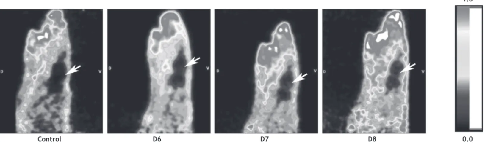
reactive astrocytes and microglia, has been successfully im-aged recently in a HSV-1-induced encephalitis rat model using 18F-FEAnGA68 (Figure 4). Also, the imidazoline2 Binding Site (I2BS), I(2)R, has been shown to be altered in several brain disorders including ND.69 Several ligands for I
2BS, including
deprenyl, are able to inhibit monoamine oxidase (MAO),70
whose activity is increased in AD human brain astrocytosis (or astrogliosis, i.e. an abnormal increase in the number of
astrocytes due to neurotoxicity or brain injury) measured
by [11C]DED71 (see below). Thus, imaging I
2BS is a promising
tool for the study of neuroinlammation in NDs. Recently,
the ligand [11C]FTIMD was evaluated with an improved
ultra-high speciic activity which afforded the detection of
small changes in I(2)R expression in the rat brain.72 Another
potentially useful ligand is BU99008, which demonstrates better in vivo brain uptake and speciicity in comparison
with [11C]FTIMD.73
Finally, cyclooxigenase (COX) enzymes are widely known as key molecular targets for anti-inflammatory drugs. Recently, [11C]ketoprofen methyl ester was pre-clinically
evaluated and proved to be eficient in quantifying COX-1 expression in both neuroinlammation and neurodegenera -tion models. In addi-tion, it afforded better results than [11C]
PK11195 in quantifying neuroinlammation. Therefore, [11C]
ketoprofen methyl ester demonstrated to be sensitive for
neuroinlammatory processes targeting COX-1 in activated
microglia and macrophages.74
Moreover, as astrocytosis is a commonly observed
phe-nomenon involved in neuroinlammation and neurodegenera -tion, this may also be evaluated in the human brain using PET tracers. The most relevant radioligand evaluated for this purpose to date is 11C-deuterium- L-deprenyl or [11C]DED, as
mentioned before. Recent studies demonstrated an increase in [11C]DED binding throughout the brain of AD patients that
also display high levels of [11C]PIB uptake, suggesting that
astrocytosis is an early phenomenon in the development of AD, probably being an intermediate step between amyloidosis and neuronal loss.75,76
Finally, there are also promising results in the use of
SPECT tracers for neuroinlammation. For instance, [123I]
PK11195 has recently been used in a pilot SPECT study with AD patients.77 In addition, the 123I-radiolabeled compound
6-chloro-2-(4’iodophenyl)-3-(N,N-diethyl)-imidazo[1,2-a] pyridine-3-acetamide or [123I]CLINDE was also successfully
tested in preclinical studies. Using different animal models
of neuroinlammation, [¹²³I]CLINDE demonstrated good per -formance to assess TSPO changes related to both astroglial and microglial activation.78,79 There are also preliminary
investigations of [125I]DPA-713 in rats exposed to a
seizure-inducing neurotoxicant; these studies revealed increased brain radioactivity in neurotoxicant-treated rats compared with controls, which was completely blocked by administra-tion of PK11195.80
Figure 4 Sagittal view of the head of a control rat (control) and a rat infected with HSV-1 (day 6(D6); day 7 (D7) and day 8
(D8) after virus inoculation). The images represent tracer uptake between 10 and 60 minutes after injection of [18F]FEAnGA.
Note the time-dependent microglial activation in the brain (arrows).
Control D6 D7 D8 0.0
Tracers for brain lipid metabolism
Arachidonic acid (AA) and docosahexaenoic acid (DHA), an omega-6 and omega-3 polyunsaturated fatty acid (PUFA), re-spectively, are very important constituents of phospholipids in cell membranes and contribute extensively to cell signal-ing in the brain. AA can be obtained from the conversion of its precursor linoleic acid obtained from the diet, whereas the brain concentration of DHA depends on dietary DHA
con-tent as well as liver synthesis from its precursor, α-linolenic
acid.81,82 The CNS response to injury and to the onset (and
progression) of neurodegeneration involves the release of
free DHA and AA along with the synthesis of stereospeciic
docosanoid derivatives and prostanoids, respectively.82,83
The release of AA in such conditions is mediated by speciic
phospholipases, e.g. PLA2, which contribute to the
conver-sion of AA into inlammatory molecules such as prostaglandin
E2 (PGE2) by the cyclooxygenase (COX) 1 and 2 enzymes. Interestingly, a recent study trying to understand the role of PLA2 in NDs demonstrated that the inhibition of PLA2 in rat brain leads to a decrease in total tau protein.84 On the
other hand, DHA has anti-inlammatory properties and their
docosanoid derivative (e.g. neuroprotectin D1) displays a neuroprotective bioactivity in the brain against various
insults, including oxidative injury, ischemia-reperfusion, and inlammation. In addition, low concentrations in the
brain have been detected in patients with brain disorders such as AD and depression.83,85,86,87 Importantly, measured
rates of AA and DHA incorporation into brain phospholipids represent their respective rates of metabolic consump-tion, because these PUFAs are not synthesized de novo or
converted signiicantly from their precursors in the brain82
(see below).
In recent PET investigations, an increase of 26% in the global brain incorporation of AA in AD patients compared with
healthy subjects was observed using [11C]arachidonic acid
([11C]AA).88 Such incorporation was particularly increased
in brain regions where neuroinlammation is thought to be
present in AD, and [11C] AA could thus be a novel marker of
activated microglia to be used in studies of neurodegenera-tive disorders. Further studies have evaluated the positron-labeled [1- (11)C]DHA tracer to map the incorporation of
unesteriied plasma DHA into the brain of healthy adult human volunteers. Values of incorporation coeficients k* for DHA
were higher in gray than white matter brain regions. For the entire human brain, the net DHA incorporation rate, Jin, the
product of k* and the unesteriied plasma DHA concentration,
equaled 3.8 ± 1.7 mg/day. The authors highlighted that this net rate, approximating the net rate of DHA consumption by brain, is less than the recommended human dietary DHA supplementation of 200 mg per day.89 Thus, with the use of
[1- (11)C]DHA, it is possible to quantify regional and global human brain DHA metabolism in relation to health and dis-ease.90 In addition, a more recent study demonstrated that
is possible to measure brain incorporation of plasma DHA in vivo. Thus, quantitative imaging of DHA incorporation from plasma into the brain can be used as an in vivo biomarker of brain DHA metabolism and neurotransmission.87 Importantly,
this may help to monitor DHA consumption in vivo in patients with disorders such as depression and AD, in which DHA supplementation may be helpful.91–93
Other new molecular targets for neuroimaging
in neurodegeneration
A potent and selective protein kinase C (PKC) inhibitor, Enzastaurin (LY317615), was recently labeled with 11C for PET imaging applications ([11C]Enzastaurin).94 PKC is an
enzyme involved in several cell biology mechanisms and is one of the most important initial elements involved in the
induction of the previously mentioned α-secretases,
ADAM-10 and 17, which are involved in neuroprotection.95 Also, a
sensitive myelin probe, [11C]MeDAS, was recently synthesized
and proved to be effective in detecting myelin changes in the brain. This radiotracer, which can be used as a myelin-imaging marker to early monitor myelin degeneration in vivo,96 is a
potentially useful development for the investigation of NDs, since degeneration of neurons, axons, and synapses is clearly present in AD as much as in multiple sclerosis.97
Heat shock proteins (HSP) also display important roles in neuroprotection. HSP70, for example, is a known protein found in aggresome of Lewy bodies and is mainly implicated in the degradation of aberrant proteins.98 Aggresome is a
general response of cells which occurs when the capacity of the proteasome (involved in degradation of unusable pro-teins) is exceeded by the production of aggregation-prone misfolded proteins.99 Similarly, cathepsins are also critical for
the degradation of enzymes that may be implicated in NDs.100
Thus, impaired function of these proteins may facilitate the progression of NDs. Interestingly, a PET reporter system (i.e. which uses reporter genes) for imaging gene expression in the intact brain was recently used to image and monitor the activation of the heat shock factor 1 (HSF1)/HSP70 transcription factor.101 In addition, another group recently
imaged the activity of the cysteine cathepsin using 64
Cu-Z-FK(DOTA)-AOMK and PET. Imaging of these proteins using the more widely available 18F radioisotope tracer may provide a
great tool in the future for early diagnosis and monitoring of disease progression of NDs.
Innate immune responses also play an important role in neurodegeneration. For example, it was recently found
that monocytes and microglia that are deicient for myeloid
differentiation factor 88 (MyD88) (involved in Toll-like re-ceptor signaling pathway) exhibit a functionally impaired
phagocytic reaction to Aβ.102 In addition, MyD88 is involved
in the dopaminergic neuronal degeneration induced by the neurotoxin MPTP in the enteric nervous system (ENS) of the mouse.103 However, this neurodegeneration is not a
MyD88-dependent mechanism.104 Thus, more knowledge is needed
before PET/SPECT imaging studies can be considered for this new target protein.
Oxidative stress (OS) leading to mitochondrial damage is
a major and early phenomenon triggering neurodegenera -tion.105,106 Interestingly, a tracer named [62Cu]ATSM, initially
designed for the study of tumor hypoxia,107 was recently used,
for the irst time, to assess OS in PD. This study demonstrated
that striatal OS was enhanced in PD patients compared with controls and increased with the progression of disease severity, particularly in the contralateral striatum.108 It was
further demonstrated that this tracer is very speciic for the
tracer for the study of brain disorders involving mitochondrial dysfunction, such as AD and PD.109
P-glycoprotein (P-gp) is a known BBB active efflux transporter involved in neuroprotection. Its dysfunction is considered one of the causes of the onset of PD110,111 and
AD.112 In addition, a correlation between aging and decreased
function of this transporter has also been established in vivo.113 Thus, developing methods for imaging P-gp in such
diseases is an important challenge nowadays. A number of studies have already been carried out using the radiolabelled P-gp substrate [11C]verapamil in PET studies. However, polar
radiolabelled metabolites are formed after injection of this
radioligand, and this may result in a non-P-gp-mediated signal as a confounding factor.114 Therefore, new P-gp tracers for
imaging P-gP function in the BBB are needed.
Concluding remarks
The studies reviewed in this article demonstrate the many opportunities to be explored using the already available molecular imaging tracers that map targets of known
rel-evance to NDs, including Aβ, α-synuclein, tau protein, and neuroinlammatory markers.
On the other hand, the brain mechanisms underlying NDs have not yet been fully elucidated and other targets of potential relevance to NDs emerge continuously.115 It is
clear the need to develop and use novel molecular imaging compounds for such targets, in addition to those related to
Aβ, α-synuclein and tau protein, in order to achieve a more
complete knowledge about the molecular basis of AD, PD and other NDs. Using such novel molecular imaging compounds, it is expected that PET and SPECT methods will help us to further understand the underlying pathological processes
and speciic molecular alterations that unfold during early
stages of NDs.
With an increasingly larger number of animal research facilities worldwide with access to micro-PET and SPECT tech-nology for preclinical studies, it is expected that pharmacol-ogy studies using novel radiotracers will help to identify and validate molecular processes as novel biomarkers to be used as therapeutic targets for treatment, and assess how new drugs are able to modify these biomarkers in animal models
of NDs. In this ield, one of the most promising strategies
should be the use of multi-tracer protocols for the simultane-ous evaluation of different molecular targets of relevance to ND. For instance, recent investigations using transgenic mice that express pathologies that characterize both dementia with Lewy bodies (DLB) and AD (DLB-AD mice) have revealed
that Aβ, tau, and α-synuclein act synergistically to promote
the aggregation, phosphorylation, and accumulation of each other, as well as leading to accelerated cognitive decline.116
Thus, multi-tracer protocols for these three molecular targets must be strongly considered in investigations of NDs.
Finally, it would be highly desirable to translate, in the
forthcoming years, some of the above novel indings into di
-rect and objective diagnostic applications in clinical practice.
With such developments, PET and SPECT imaging patterns might be used more incisively to improve diagnostic accuracy in doubtful cases, as well as to predict prognoses and treat-ment response in sufferers of NDs. The larger availability of SPECT and its lower costs may make it the method of choice for such future clinical applications, but a progressively
greater access to PET methods across a larger number of hospitals is also expected, particularly in regard to the use of 18F-labeled tracers. The use of any novel radiotracer for
clinical applications with PET or SPECT should be weighted against the availability of other promising biomarkers, such
as cerebrospinal luid (CSF) indices of Aβ, tau and other
pathologies.117 Large-scale clinical studies should
continu-ously be carried out to ascertain the comparative diagnostic
accuracy and cost-beneit of novel PET and SPECT imaging
probes and CSF markers, as well as the usefulness of employ-ing such measurements in combination.
Acknowledgements
We thank Janine Doorduin, PhD, Inês Farinha Antunes, PhD, and Andor Glaudemans, MD, for making available
the igures depicting [11C]PK11195, [18F]FEAnGA and PIB
PET Scans, respectively. We also thank the Coordenação de Aperfeiçoamento de Pessoal de Nível Superior (CAPES)
Brazilian agency for the inancial support to Marcel Benadiba.
Disclosures
Marcel Benadiba, PhD
Employment: Department of Nuclear Medicine and Molecular Imaging, University Medical Center Groningen, University of Groningen, Groningen, Netherlands; Laboratory of Psychiatric Neuroimaging (LIM 21), Department of Psychiatry, Faculdade de Medicina, Universidade de
São Paulo, Brazil. Other: Núcleo de Apoio à Pesquisa em Neurociência
Aplicada (NAPNA), Universidade de São Paulo, São Paulo, Brazil. Gert Luurtsema, PhD
Employment: Department of Nuclear Medicine and Molecular Imaging, University Medical Center Groningen, University of Groningen, Groningen,
Netherlands.
Lauro Wichert-Ana, PhD
Employment: Department of Internal Medicine, Faculdade de Medicina de Ribeirão Preto, Universidade de São Paulo, Ribeirão Preto, SP, Brazil. Other: Núcleo de Apoio à Pesquisa em Neurociência Aplicada (NAPNA),
Universidade de São Paulo, São Paulo, Brazil. Carlos Alberto Buchpigel, PhD
Employment: Center of Nuclear Medicine, Department of Radiology, Faculdade de Medicina da Universidade de São Paulo, São Paulo, SP, Brazil. Other: Núcleo de Apoio à Pesquisa em Neurociência Aplicada (NAPNA),
Universidade de São Paulo, São Paulo, Brazil. Geraldo Busatto Filho, PhD
Employment: Laboratory of Psychiatric Neuroimaging (LIM 21), Department of Psychiatry, Faculdade de Medicina, Universidade de São
Paulo, Brazil. Other: Núcleo de Apoio à Pesquisa em Neurociência Aplicada
(NAPNA), Universidade de São Paulo, São Paulo, Brazil.
* Modest
** Signiicant
*** Signiicant. Amounts given to the author’s institution or to a colleague for research in which the author has participation, not directly to the author.
References
1. Willmann JK, van Bruggen N, Dinkelborg LM, Gambhir SS. Molecular imaging in drug development. Nat Rev Drug Discov. 2008;7(7):591-607.
2. Pimlott SL, Sutherland A. Molecular tracers for the PET and SPECT imaging of disease. Chem Soc Rev. 2011;40(1):149-62. 3. Laruelle M, Slifstein M, Huang Y. Positron emission tomography:
imaging and quantiication of neurotransporter availability.
Methods. 2002;27(3):287-99.
4. Någren K, Halldin C, Rinne JO. Radiopharmaceuticals for positron emission tomography investigations of Alzheimer’s disease. Eur J Nucl Med Mol Imaging. 2010;37(8):1575-93. 5. Zhang Y-wu, Xu H. Molecular and cellular mechanisms for
6. Klunk WE, Engler H, Nordberg A, Wang Y, Blomqvist G, Holt DP et al. Imaging brain amyloid in Alzheimer’s disease with Pittsburgh Compound-B. Ann Neurol. 2004;55(3):306-19.
7. Kemppainen NM, Aalto S, Wilson IA, Någren K, Helin S, Brück A
et al. PET amyloid ligand [11C]PIB uptake is increased in mild
cognitive impairment. Neurology. 2007;68(19):1603-6. 8. Scheinin NM, Aalto S, Koikkalainen J, Lötjönen J, Karrasch
M, Kemppainen N et al. Follow-up of [11C]PIB uptake and
brain volume in patients with Alzheimer disease and controls. Neurology. 2009;73(15):1186-92.
9. Jack CR, Lowe VJ, Weigand SD, Wiste HJ, Senjem ML,
Knopman DS et al. Serial PIB and MRI in normal, mild cognitive impairment and Alzheimer’s disease: implications for sequence of pathological events in Alzheimer’s disease. Brain. 2009;132(Pt 5):1355-65.
10. Engler H, Forsberg A, Almkvist O, Blomquist G, Larsson E, Savitcheva I et al. Two-year follow-up of amyloid deposition in patients with Alzheimer’s disease. Brain.. 2006;129(Pt 11):2856-66.
11. Rowe CC, Ng S, Ackermann U, Gong SJ, Pike K, Savage G et al. Imaging beta-amyloid burden in aging and dementia. Neurology. 2007;68(20):1718-25.
12. Engler H, Santillo AF, Wang SX, Lindau M, Savitcheva I, Nordberg A et al. In vivo amyloid imaging with PET in frontotemporal dementia. Eur J Nucl Med Mol Imaging. 2008;35(1):100-6. 13. Petersen RC. Mild cognitive impairment as a diagnostic entity.
J Intern Med. 2004;256(3):183-94.
14. Mintun MA, Larossa GN, Sheline YI, Dence CS, Lee SY, Mach RH et al. [11C]PIB in a nondemented population: potential antecedent
marker of Alzheimer disease. Neurology. 2006;67(3):446-52. 15. Forsberg A, Engler H, Almkvist O, Blomquist G, Hagman G, Wall
A et al. PET imaging of amyloid deposition in patients with mild cognitive impairment. Neurobiol aging. 2008;29(10):1456–65. 16. Okello A, Koivunen J, Edison P, Archer HA, Turkheimer FE,
Någren K et al. Conversion of amyloid positive and negative MCI to AD over 3 years: an 11C-PIB PET study. Neurology. 2009;73(10):754-60.
17. Edison P, Rowe CC, Rinne JO, Ng S, Ahmed I, Kemppainen N et al. Amyloid load in Parkinson’s disease dementia and Lewy body dementia measured with [11C]PIB positron emission tomography.
J Neurol Neurosurg Psychiatry. 2008;79(12):1331-8.
18. Gomperts SN, Locascio JJ, Marquie M, Santarlasci AL, Rentz DM, Maye J et al. Brain amyloid and cognition in Lewy body diseases. Mov disord [Available from: http://www.ncbi.nlm.nih. gov/pubmed/22693110]. 2012 [cited 2012 Jun 24].
19. Fodero-Tavoletti MT, Smith DP, McLean C a, Adlard P a, Barnham KJ, Foster LE et al. In vitro characterization of Pittsburgh compound-B binding to Lewy bodies. J Neurosci. 2007;27(39):10365-71.
20. Kadir A, Andreasen N, Almkvist O, Wall A, Forsberg A, Engler H et al. Effect of phenserine treatment on brain functional activity and amyloid in Alzheimer’s disease. Ann Neurol. 2008;63(5):621-31.
21. Kung HF, Choi SR, Qu W, Zhang W, Skovronsky D. 18F stilbenes and styrylpyridines for PET imaging of A beta plaques in Alzheimer’s disease: a miniperspective. J Med Chem. 2010;53(3):933-41. 22. Vandenberghe R, Van Laere K, Ivanoiu A, Salmon E, Bastin C,
Triau E et al. 18F-lutemetamol amyloid imaging in Alzheimer
disease and mild cognitive impairment: a phase 2 trial. Ann Neurol. 2010;68(3):319-29.
23. Wong DF, Rosenberg PB, Zhou Y, Kumar A, Raymont V, Ravert HT, et al. In vivo imaging of amyloid deposition in Alzheimer
disease using the radioligand 18F-AV-45 (lorbetapir [corrected]
F 18). J Nucl Med. 2010;51(6):913-20.
24. Barthel H, Sabri O. Florbetaben to trace amyloid-β in
the Alzheimer brain by means of PET. J Alzheimers Dis. 2011;26(Suppl 3):117-21.
25. FDA Approves 18F-Florbetapir PET Agent. J Nucl Med. 2012;53(6):15N.
26. Cui M, Tang R, Li Z, Ren H, Liu B. 99mTc- and Re-labeled 6-dialkylamino-2-naphthylethylidene derivatives as imaging
probes for β-amyloid plaques. Bioorg Med Chem Lett.
2011;21(3):1064-8.
27. Hardy JA, Higgins GA. Alzheimer’s disease: the amyloid cascade hypothesis. Science. 1992;256(5054):184-5.
28. Mirra SS, Heyman A, McKeel D, Sumi SM, Crain BJ, Brownlee LM, et al. The Consortium to Establish a Registry for Alzheimer’s Disease (CERAD). Part II. Standardization of the neuropathologic assessment of Alzheimer’s disease. Neurology. 1991;41(4):479-86.
29. Arriagada PV, Growdon JH, Hedley-Whyte ET, Hyman BT.
Neuroibrillary tangles but not senile plaques parallel duration
and severity of Alzheimer’s disease. Neurology. 1992;42(3 Pt 1):631-9.
30. Gómez-Isla T, Hollister R, West H, Mui S, Growdon JH, Petersen RC, et al. Neuronal loss correlates with but exceeds
neuroibrillary tangles in Alzheimer’s disease. Ann Neurol.
1997;41(1):17-24.
31. Gómez-Isla T, Wasco W, Pettingell WP, Gurubhagavatula S, Schmidt SD, Jondro PD et al. A novel presenilin-1 mutation:
increased beta-amyloid and neuroibrillary changes. Ann Neurol.
1997;41(6):809-13.
32. Ono M, Saji H. Molecular Approaches to the Treatment,
Prophylaxis, and Diagnosis of Alzheimer’s Disease: Novel PET/ SPECT Imaging Probes for Diagnosis of Alzheimer’s Disease. J Pharmacol Sci. 2012;344(338):338-44.
33. Agdeppa ED, Kepe V, Liu J, Flores-Torres S, Satyamurthy N, Petric A et al. Binding characteristics of radiofluorinated 6-dialkylamino-2-naphthylethylidene derivatives as positron emission tomography imaging probes for beta-amyloid plaques in Alzheimer’s disease. J Neurosci. 2001;21(24):RC189. 34. Shoghi-Jadid K, Small GW, Agdeppa ED, Kepe V, Ercoli LM,
Siddarth P et al. Localization of neuroibrillary tangles and
beta-amyloid plaques in the brains of living patients with Alzheimer disease. Am J Geriatr Psychiatry. 2002;10(1):24-35.
35. Velasco A, Fraser G, Delobel P, Ghetti B, Lavenir I, Goedert
M. Detection of ilamentous tau inclusions by the luorescent Congo red derivative FSB
[(trans,trans)-1-luoro-2,5-bis(3-hydroxycarbonyl-4-hydroxy)styrylbenzene]. FEBS lett. 2008;582(6):901-6.
36. Mohorko N, Repovs G, Popović M, Kovacs GG, Bresjanac M. Curcumin labeling of neuronal ibrillar tau inclusions in human
brain samples. J Neuropathol Exp Neurol. 2010;69(4):405-14. 37. Small GW, Kepe V, Ercoli LM, Siddarth P, Bookheimer SY, Miller
KJ, et al. PET of brain amyloid and tau in mild cognitive impairment. N Engl J Med. 2006;355(25):2652-63.
38. Luurtsema G, Schuit RC, Takkenkamp K, Lubberink M, Hendrikse NH, Windhorst AD et al. Peripheral metabolism of [(18)F]FDDNP and cerebral uptake of its labelled metabolites. Nucl Med Biol. 2008;35(8):869-74.
39. Okamura N, Suemoto T, Furumoto S, Suzuki M, Shimadzu H, Akatsu H et al. Quinoline and benzimidazole derivatives: candidate probes for in vivo imaging of tau pathology in Alzheimer’s disease. J Neurosci. 2005;25(47):10857-62. 40. Fodero-Tavoletti MT, Okamura N, Furumoto S, Mulligan RS,
Connor AR, McLean CA, et al. 18F-THK523: a novel in vivo tau imaging ligand for Alzheimer’s disease. Brain. 2011;134(Pt 4):1089-100.
41. Ono M, Ishikawa M, Kimura H, Hayashi S, Matsumura K, Watanabe H et al. Development of dual functional SPECT/luorescent
42. Honson NS, Johnson RL, Huang W, Inglese J, Austin CP, Kuret J. Differentiating Alzheimer disease-associated aggregates with small molecules. Neurobiol Dis. 2007;28(3):251-60.
43. Jellinger KA. Interaction between α-synuclein and other proteins in neurodegenerative disorders. ScientiicWorldJournal.
2011;11:1893-907.
44. Liu G, Zhang C, Yin J, Li X, Cheng F, Li Y et al. alpha-Synuclein is differentially expressed in mitochondria from different rat brain regions and dose-dependently down-regulates complex I activity. Neurosci lett. 2009;454(3):187-92.
45. Bellucci A, Zaltieri M, Navarria L, Grigoletto J, Missale C, Spano
P. From α-synuclein to synaptic dysfunctions: New insights into
the pathophysiology of Parkinson’s disease. Brain Res [Available at: http://www.ncbi.nlm.nih.gov/pubmed/22560500]. 2012 [cited 2012 Jun 25].
46. Alim MA, Hossain MS, Arima K, Takeda K, Izumiyama Y, Nakamura M et al. Tubulin seeds alpha-synuclein ibril formation. J Biol
Chem. 2002;277(3):2112-7.
47. Alim MA, Ma Q-L, Takeda K, Aizawa T, Matsubara M, Nakamura M et al. Demonstration of a role for alpha-synuclein as a functional microtubule-associated protein. J Alzheimers Dis. 2004;6(4):435-42 [discussion 443-9].
48. Cooper AA, Gitler AD, Cashikar A, Haynes CM, Hill KJ, Bhullar B
et al. Alpha-synuclein blocks ER-Golgi trafic and Rab1 rescues
neuron loss in Parkinson’s models. Science. 2006;313(5785):324-8.
49. Uversky VN. Neuropathology, biochemistry, and biophysics of alpha-synuclein aggregation. J Neurochem. 2007;103(1):17-37. 50. Kudo Y, Okamura N, Furumoto S, Tashiro M, Furukawa K,
Maruyama M et al. 2-(2-[2-Dimethylaminothiazol-5-yl]
ethenyl)-6- (2-[luoro]ethoxy)benzoxazole: a novel PET agent
for in vivo detection of dense amyloid plaques in Alzheimer’s disease patients. J Nucl Med. 2007;48(4):553-61.
51. Fodero-Tavoletti MT, Mulligan RS, Okamura N, Furumoto S, Rowe CC, Kudo Y et al. In vitro characterisation of BF227 binding to alpha-synuclein/Lewy bodies. Eur J Pharmacol. 2009;617(1-3):54-8.
52. Kikuchi A, Takeda A, Okamura N, Tashiro M, Hasegawa T, Furumoto S et al. In vivo visualization of alpha-synuclein deposition by carbon-11-labelled
2-[2-(2-dimethylaminothiazol-5-yl)ethenyl]-6-[2-(luoro)ethoxy]benzoxazole positron emission
tomography in multiple system atrophy. Brain. 2010;133(Pt 6):1772-8.
53. Pizza V, Agresta A, D’Acunto CW, Festa M, Capasso A. Neuroinflammation and ageing: current theories and an overview of the data. Rev Recent Clin Trials. 2011;6(3):189-203. 54. Chen M-K, Guilarte TR. Translocator protein 18 kDa (TSPO):
molecular sensor of brain injury and repair. Pharmacol Ther.
2008;118(1):1-17.
55. Ouchi Y, Yoshikawa E, Sekine Y, Futatsubashi M, Kanno T, Ogusu T et al. Microglial activation and dopamine terminal loss in early Parkinson’s disease. Ann Neurol. 2005;57(2):168-75.
56. Yokokura M, Mori N, Yagi S, Yoshikawa E, Kikuchi M, Yoshihara Y et al. In vivo changes in microglial activation and amyloid deposits in brain regions with hypometabolism in Alzheimer’s disease. Eur J Nucl Med Mol Imaging. 2011;38(2):343-51. 57. Cagnin A, Brooks DJ, Kennedy AM, Gunn RN, Myers R, Turkheimer
FE et al. In-vivo measurement of activated microglia in dementia. Lancet. 2001;358(9280):461-7.
58. Cagnin A, Rossor M, Sampson EL, Mackinnon T, Banati RB. In vivo
detection of microglial activation in frontotemporal dementia. Ann Neurol. 2004;56(6):894-7.
59. Hickman SE, Allison EK, El Khoury J. Microglial dysfunction and defective beta-amyloid clearance pathways in aging Alzheimer’s disease mice. J Neurosci. 2008;28(33):8354-60.
60. Venneti S, Wiley CA, Koler J. Imaging microglial activation
during neuroinflammation and Alzheimer’s disease. J Neuroimmune Pharmacol. 2009;4(2):227-43.
61. Ching ASC, Kuhnast B, Damont A, Roeda D, Tavitian B, Dollé F. Current paradigm of the 18-kDa translocator protein (TSPO) as
a molecular target for PET imaging in neuroinlammation and
neurodegenerative diseases. Insights Imaging. 2011;3(1):111-9. 62. Doorduin J, Klein HC, Dierckx R a, James M, Kassiou M, de
Vries EFJ. [11C]-DPA-713 and [18F]-DPA-714 as new PET tracers
for TSPO: a comparison with [11C]-(R)-PK11195 in a rat model
of herpes encephalitis. Mol Imaging Biol. 2009;11(6):386-98. 63. Boutin H, Chauveau F, Thominiaux C, Grégoire M-C, James
ML, Trebossen R, et al. 11C-DPA-713: a novel peripheral benzodiazepine receptor PET ligand for in vivo imaging of
neuroinlammation. J Nucl Med. 2007;48(4):573-81.
64. Van Camp N, Boisgard R, Kuhnast B, Thézé B, Viel T, Grégoire M-C, et al. In vivo imaging of neuroinlammation: a comparative
study between [(18)F]PBR111, [ (11)C]CLINME and [ (11)C] PK11195 in an acute rodent model. Eur J Nucl Med Mol Imaging. 2010;37(5):962-72.
65. Abourbeh G, Thézé B, Maroy R, Dubois A, Brulon V, Fontyn Y et al. Imaging Microglial/Macrophage Activation in Spinal Cords of Experimental Autoimmune Encephalomyelitis Rats by Positron Emission Tomography Using the Mitochondrial 18 kDa Translocator Protein Radioligand [18F]DPA-714. J Neurosci.
2012;32(17):5728-36.
66. Tang D, Hight MR, McKinley ET, Fu A, Buck JR, Smith RA et al. Quantitative preclinical imaging of TSPO expression in glioma using N,N-diethyl-2-(2-(4-(2-18F-fluoroethoxy)phenyl)-5,7-dimethylpyrazolo[1,5-a]pyrimidin-3-yl)acetamide. J Nucl Med. 2012;53(2):287-94.
67. Winkeler A, Boisgard R, Awde AR, Dubois A, Thézé B, Zheng J
et al. The translocator protein ligand [(18)F]DPA-714 images glioma and activated microglia in vivo. Eur J Nucl Med Mol Imaging. 2012;39(5):811-23.
68. Antunes IF, Doorduin J, Haisma HJ, Elsinga PH, van Waarde A, Willemsen ATM et al. 18F-FEAnGA for PET of β-glucuronidase activity in neuroinlammation. J Nucl Med. 2012;53(3):451-8.
69. García-Sevilla JA, Escribá PV, Guimón J. Imidazoline receptors and human brain disorders. Ann N Y Acad Sci. 1999;881:392-409. 70. Lalies MD, Hibell A, Hudson AL, Nutt DJ. Inhibition of central monoamine oxidase by imidazoline2 site-selective ligands. Ann N Y Acad Sci. 1999;881:114-7.
71. Gulyás B, Pavlova E, Kása P, Gulya K, Bakota L, Várszegi S
et al. Activated MAO-B in the brain of Alzheimer patients, demonstrated by [11C]-L-deprenyl using whole hemisphere
autoradiography. Neurochem Int. 2011;58(1):60-8.
72. Kawamura K, Kimura Y, Yui J, Wakizaka H, Yamasaki T, Hatori A
et al. PET study using [11C]FTIMD with ultra-high speciic activity
to evaluate I2-imidazoline receptors binding in rat brains. Nucl Med Biol. 2012;39(2):199-206.
73. Tyacke RJ, Fisher A, Robinson ESJ, Grundt P, Turner EM, Husbands SM et al. Evaluation and initial in vitro and ex vivo characterization of the potential positron emission tomography ligand, BU99008 (2-(4,5-dihydro-1H-imidazol-2-yl)-1-
methyl-1H-indole), for the imidazoline binding site. Synapse.
2012;66(6):542-51.
74. Shukuri M, Takashima-Hirano M, Tokuda K, Takashima T, Matsumura K, Inoue O et al. In vivo expression of cyclooxygenase-1 in activated microglia and macrophages during
neuroinlammation visualized by PET with 11C-ketoprofen
methyl ester. J Nucl Med. 2011;52(7):1094-101.
75. Carter SF, Schöll M, Almkvist O, Wall A, Engler H, Långström B
76. Santillo AF, Gambini JP, Lannfelt L, Långström B, Ulla-Marja L,
Kilander L et al. In vivo imaging of astrocytosis in Alzheimer’s disease: an 11C-L-deuteriodeprenyl and PIB PET study. Eur J Nucl Med Mol Imaging. 2011;38(12):2202-8.
77. Versijpt JJ, Dumont F, Van Laere KJ, Decoo D, Santens P,
Audenaert K et al. Assessment of neuroinlammation and
microglial activation in Alzheimer’s disease with radiolabelled PK11195 and single photon emission computed tomography. A pilot study. Eur Neurol. 2003;50(1):39-47.
78. Arlicot N, Katsiis A, Garreau L, Mattner F, Vergote J, Duval S et al. Evaluation of CLINDE as potent translocator protein (18 kDa)
SPECT radiotracer relecting the degree of neuroinlammation
in a rat model of microglial activation. Eur J Nucl Med Mol Imaging. 2008;35(12):2203-11.
79. Mattner F, Bandin DL, Staykova M, Berghofer P, Gregoire MC, Ballantyne P et al. Evaluation of [123I]-CLINDE as a potent SPECT radiotracer to assess the degree of astroglia activation
in cuprizone-induced neuroinlammation. Eur J Nucl Med Mol
Imaging. 2011;38(8):1516-28.
80. Wang H, Pullambhatla M, Guilarte TR, Mease RC, Pomper MG. Synthesis of [(125)I]iodoDPA-713: a new probe for imaging
inlammation. Biochem Biophys Res Commun. 2009;389(1):80-3.
81. Gao F, Kiesewetter D, Chang L, Ma K, Bell JM, Rapoport SI et al. Whole-body synthesis-secretion rates of long-chain n-3
PUFAs from circulating unesteriied alpha-linolenic acid in
unanesthetized rats. J Lipid Res. 2009;50(4):749-58.
82. Rapoport SI. Arachidonic acid and the brain. J Nutr. 2008;138(12):2515-20.
83. Zhang C, Bazan NG. Lipid-mediated cell signaling protects against
injury and neurodegeneration. J Nutr. 2010;140(4):858-63.
84. Schaeffer EL, De-Paula VJ, da Silva ER, de A Novaes B, Skaf HD, Forlenza OV et al. Inhibition of phospholipase A(2) in rat brain decreases the levels of total Tau protein. J Neural Transm. 2011;118(9):1273-9.
85. Bazan NG, Musto AE, Knott EJ. Endogenous signaling by omega-3 docosahexaenoic acid-derived mediators sustains homeostatic synaptic and circuitry integrity. Mol Neurobiol. 2011;44(2):216-22.
86. Bazan NG, Molina MF, Gordon WC. Docosahexaenoic
acid signalolipidomics in nutrition: signiicance in aging, neuroinlammation, macular degeneration, Alzheimer’s, and
other neurodegenerative diseases. Annu Rev Nutr. 2011;31:321-51.
87. Rapoport SI, Ramadan E, Basselin M. Docosahexaenoic acid (DHA) incorporation into the brain from plasma, as an in vivo
biomarker of brain DHA metabolism and neurotransmission. Prostaglandins Other Lipid Mediat. 2011;96(1-4):109-13. 88. Esposito G, Giovacchini G, Liow J-S, Bhattacharjee AK,
Greenstein D, Schapiro M et al. Imaging neuroinlammation in
Alzheimer’s disease with radiolabeled arachidonic acid and PET. J Nucl Med. 2008;49(9):1414-21.
89. Kris-Etherton PM, Taylor DS, Yu-Poth S, Huth P, Moriarty K, Fishell V et al. Polyunsaturated fatty acids in the food chain in the United States. Am J Clin Nutr. 2000;71(1 Suppl):179S-88S. 90. Umhau JC, Zhou W, Carson RE, Rapoport SI, Polozova A, Demar J et al. Imaging incorporation of circulating docosahexaenoic acid into the human brain using positron emission tomography. J Lipid Res. 2009;50(7):1259-68.
91. Conquer JA, Tierney MC, Zecevic J, Bettger WJ, Fisher RH. Fatty acid analysis of blood plasma of patients with Alzheimer’s disease, other types of dementia, and cognitive impairment. Lipids. 2000;35(12):1305-12.
92. Quinn JF, Raman R, Thomas RG, Yurko-Mauro K, Nelson EB, Van Dyck C et al. Docosahexaenoic acid supplementation and cognitive decline in Alzheimer disease: a randomized trial. JAMA. 2010;304(17):1903-11.
93. Hibbeln JR. Fish consumption and major depression. Lancet.
1998;351(9110):1213.
94. Wang M, Xu L, Gao M, Miller KD, Sledge GW, Zheng Q-H. [11C]
enzastaurin, the first design and radiosynthesis of a new potential PET agent for imaging of protein kinase C. Bioorg Med Chem Lett. 2011;21(6):1649-53.
95. Kozikowski AP, Chen Y, Subhasish T, Lewin NE, Blumberg PM, Zhong Z et al. Searching for disease modiiers-PKC activation and
HDAC inhibition - a dual drug approach to Alzheimer’s disease that decreases Abeta production while blocking oxidative stress. ChemMedChem. 2009;4(7):1095-105.
96. Wu C, Wang C, Popescu DC, Zhu W, Somoza EA, Zhu J et al. A novel PET marker for in vivo quantiication of myelination.
Bioorg Med Chem. 2010;18(24):8592-9.
97. Lassmann H. Mechanisms of neurodegeneration shared between multiple sclerosis and Alzheimer’s disease. J Neural Transm. 2011;118(5):747-52.
98. Witt SN. Hsp70 molecular chaperones and Parkinson’s disease. Biopolymers. 2010;93(3):218-28.
99. Johnston JA, Ward CL, Kopito RR. Aggresomes: a cellular response to misfolded proteins. J Cell Biol. 1998;143(7):1883-98. 100. Bhutani N, Piccirillo R, Hourez R, Venkatraman P, Goldberg AL. Cathepsins L and Z Are Critical in Degrading Polyglutamine-containing Proteins within Lysosomes. J Biol Chem. 2012;287(21):17471-82.
101. Doubrovin M, Che JT, Serganova I, Moroz E, Solit DB, Ageyeva L et al. Monitoring the induction of heat shock factor 1/ heat shock protein 70 expression following 17-allylamino-demethoxygeldanamycin treatment by positron emission tomography and optical reporter gene imaging. Mol Imaging. 2012;11(1):67-76.
102. Michaud J-P, Richard KL, Rivest S. Hematopoietic MyD88-adaptor Protein Acts as a Natural Defense Mechanism for Cognitive
Deicits in Alzheimer’s Disease. Stem Cell Rev [Available at:
http://www.ncbi.nlm.nih.gov/pubmed/22374079]. 2012 [cited 2012 May 17].
103. Côté M, Drouin-Ouellet J, Cicchetti F, Soulet D. The critical role
of the MyD88-dependent pathway in non-CNS MPTP-mediated toxicity. Brain Behav Immun. 2011;25(6):1143-52.
104. Drouin-Ouellet J, Gibrat C, Bousquet M, Calon F, Kriz J, Cicchetti F. The role of the MYD88-dependent pathway in MPTP-induced brain dopaminergic degeneration. J Neuroinflammation. 2011;8:137.
105. Martin LJ. Biology of mitochondria in neurodegenerative diseases. Prog Mol Biol Transl Sci. 2012;107:355-415.
106. Ray PD, Huang B-W, Tsuji Y. Reactive oxygen species (ROS)
homeostasis and redox regulation in cellular signaling. Cell Signal. 2012;24(5):981-90.
107. Kurihara H, Honda N, Kono Y, Arai Y. Radiolabelled Agents for PET Imaging of Tumor Hypoxia. Curr Med Chem [Available at: http://www.ncbi.nlm.nih.gov/pubmed/22664246]. 2012 [cited 2012 Jun 11].
108. Ikawa M, Okazawa H, Kudo T, Kuriyama M, Fujibayashi Y,
Yoneda M. Evaluation of striatal oxidative stress in patients with Parkinson’s disease using [62Cu]ATSM PET. Nucl Med Biol. 2011;38(7):945-51.
109. Yoshii Y, Yoneda M, Ikawa M, Furukawa T, Kiyono Y, Mori T et al. Radiolabeled Cu-ATSM as a novel indicator of overreduced intracellular state due to mitochondrial dysfunction: studies
with mitochondrial DNA-less ρ0 cells and cybrids carrying MELAS
mitochondrial DNA mutation. Nucl Med Biol. 2012;39(2):177-85. 110. Droździk M, Białecka M, Myśliwiec K, Honczarenko K,
111. Kortekaas R, Leenders KL, van Oostrom JCH, Vaalburg W, Bart J, Willemsen ATM et al. Blood-brain barrier dysfunction in parkinsonian midbrain in vivo. Ann Neurol. 2005;57(2):176-9. 112. Vogelgesang S, Cascorbi I, Schroeder E, Pahnke J, Kroemer
HK, Siegmund W et al. Deposition of Alzheimer’s beta-amyloid is inversely correlated with P-glycoprotein expression in the brains of elderly non-demented humans. Pharmacogenetics. 2002;12(7):535-41.
113. Bartels AL, Kortekaas R, Bart J, Willemsen ATM, de Klerk OL, de Vries JJ et al. Blood-brain barrier P-glycoprotein function
decreases in speciic brain regions with aging: a possible
role in progressive neurodegeneration. Neurobiol Aging. 2009;30(11):1818-24.
114. Luurtsema G, Molthoff CFM, Schuit RC, Windhorst AD, Lammertsma AA, Franssen EJF. Evaluation of (R)-[11C]verapamil
as PET tracer of P-glycoprotein function in the blood-brain barrier: kinetics and metabolism in the rat. Nucl Med Biol. 2005;32(1):87-93.
115. Donovan LE, Higginbotham L, Dammer EB, Gearing M, Rees HD, Xia Q et al. Analysis of a membrane-enriched proteome from postmortem human brain tissue in Alzheimer’s disease. Proteomics Clin Appl. 2012;6(3-4):201-11.
116. Clinton LK, Blurton-Jones M, Myczek K, Trojanowski JQ, LaFerla
FM. Synergistic Interactions between Abeta, tau, and alpha-synuclein: acceleration of neuropathology and cognitive decline. J Neurosci. 2010;30(21):7281-9.

![Figure 3 Representative PET parametric image of [ 11 C]](https://thumb-eu.123doks.com/thumbv2/123dok_br/19085026.495174/5.980.502.900.839.1072/figure-representative-pet-parametric-image-c.webp)