Pontifícia Universidade Católica do Rio Grande do Sul Faculdade de Odontologia
Programa de Pós-Graduação em Odontologia Mestrado em Odontologia
Área de Concentração em Estomatologia Clínica
Felipe Leal Martins
Contribuição da ultrassonografia como exame complementar no estabelecimento do diagnóstico de lesões nodulares submucosas e subcutâneas da região bucomaxilofacial
Felipe Leal Martins
PONTIFÍCIA UNIVERSIDADE CATÓLICA DO RIO GRANDE DO SUL
FACULDADE DE ODONTOLOGIA
CONTRIBUIÇÃO DA ULTRASSONOGRAFIA COMO EXAME COMPLEMENTAR NO ESTABELECIMENTO DO DIAGNÓSTICO DE LESÕES NODULARES SUBMUCOSAS E SUBCUTÂNEAS DA REGIÃO
BUCOMAXILOFACIAL
PORTO ALEGRE
Dados Internacionais de Catalogação na Publicação (CIP)
M386c Martins, Felipe Leal
Contribuição da ultrassonografia como exame complementar no estabelecimento do diagnóstico de lesões nodulares submucosas e subcutâneas da região bucomaxilofacial / Felipe Leal Martins. – Porto Alegre, 2014.
106 f. : il.
Diss. (Mestrado) – Faculdade de Odontologia, PUCRS. Orientador: Profª. Drª. Maria Antonia Zancanaro de Figueiredo.
1. Odontologia. 2. Patologia Bucal. 3. Ultrassonografia. 4. Diagnóstico por Imagem. 5. Lesões Nodulares. I. Figueiredo, Maria Antonia Zancanaro de. II. Título.
CDD 617.607 NLM WU 140
Ficha Catalográfica elaborada por Vanessa Pinent
Dissertação apresentada como parte dos requisitos para obtenção do grau de Mestre pelo Programa de Pós-Graduação da Faculdade de Odontologia da Pontifícia Universidade Católica do Rio Grande do Sul – Área de Concentração em EstomatologiaClínica.
FELIPE LEAL MARTINS
Contribuição da ultrassonografia como exame complementar no estabelecimento
do diagnóstico de lesões nodulares submucosas e subcutâneas da região
bucomaxilofacial
Linha de Pesquisa: Métodos de diagnóstico em Estomatologia
Aprovada em: 28 de Março de 2014.
Banca Examinadora:
_______________________________________________________ Prof. Dra. Helena Willhelm de Oliveira – PUCRS
_______________________________________________________ Prof. Dra. Heloísa Emília Dias da Silveira – UFRGS
________________________________________________________ Prof. Dra. Maria Antonia Zancanaro de Figueiredo – PUCRS (Orientadora)
________________________________________________________ Prof. Dra. Fernanda Gonçalves Salum – PUCRS (Suplente)
―Devemos gerar coragem igual ao tamanho dasdificuldades que enfrentamos‖
À minha mãe, Graça, agradeço por servir de inspiração na profissão, e principalmente, a sempre incentivar a aprimorar os conhecimentos.
À Magali, agradeço por me incentivar e confortar pacientemente, durante a realização deste estudo.
À minha irmã, Karen, por sempre acreditar que tudo vai dar certo.
Agradeço à minha querida orientadora, Profa. Dra. Maria Antonia Zancanaro de Figueiredo, por todo o seu conhecimento, inspiração e, principalmente, amizade. Por sempre transmitir tranquilidade, segurança e apoiar nos momentos difíceis.
Ao Dr. Roberto Oliveira por todo o seu conhecimento e acolhimento, além de ter se disponibilizado a realizar todos exames ultrassonográficos.
Às minhas colegas e amigas de ambulatório, Maria Noel Petruzzi e Ruchielli Borghetti, por sempre estarem disponíveis a ajudar na realização do estudo.
Às professoras Fernanda Salum, Karen Cherubini e Liliane Yurgel por todo conhecimento a mim transmitido durante a convivência no ambulatório de Estomatologia do Hospital São Lucas – PUCRS.
RESUMO
ABSTRACT
Ultrasonography is an innocuous noninvasive diagnostic tool, which is easy to perform and low-cost, and it is widely used in the medical field, indicated especially in the exam of soft tissues. Despite this, it is not usually requested by dental surgeons in the diagnostic investigation of intraoral lesions. The aim of this study was to determine how much this resource contributes to establishing the final diagnosis of nodules located in the oral and maxillofacial region. Accordingly, we evaluated the indication of use and reliability of the procedure in Dentistry, considering the usefulness to reach the definitive diagnosis of these alterations. Sixty-five patients with submucosal or subcutaneous nodules were recruited for the study. They had been indicated for and subjected to regional ultrasonography, since it was not possible to establish a conclusive diagnosis of the lesions only by clinical examination. Ultrasonography was carried out in an imaging diagnostic center utilizing the same apparatus Toshiba – Aplio 80 – Japan. The imaging reports were prepared by a single examiner, with wide experience in the interpretation of images of the oral and maxillofacial region. Of the total patients, 43 had a biopsy for histopathologic confirmation of the final diagnosis. In 22 individuals, it was not necessary to perform surgery, since imaging allowed the establishment of the diagnosis and management of the patient. Two investigators evaluated the results obtained with ultrasonography and estimated the scores, which were zero (ultrasonographic diagnosis differed from the final), 1 (contributed to the management of the patient) and 2 (defined the definitive diagnosis). The zero score accounted for 12.3% of the examinations performed, while 41.5 and 46,1% of patients had scores of 1 and 2, respectively, totaling 87.6% of cases where imaging was helpful. The procedure facilitated the diagnosis of vascular lesions in 93.3%, helped with the management of 87.5% of patients with both malignant and benign neoplasms, and had a role in establishing the final diagnosis in 76.5% of cases of mucus retention phenomena. The results obtained were in accordance to findings in the literature, which demonstrated that ultrasonography is an effective resource in determining the final diagnosis or in the management of patients with nonspecific nodular lesions in the soft tissues of the oral and maxillofacial region.
SUMÁRIO
1. INTRODUÇÃO 16
2. ARTIGO CIENTÍFICO I 22
3. ARTIGO CIENTÍFICO II 39
4. DISCUSSÃO GERAL 58
5. REFERÊNCIAS 66
6. ANEXOS 75
INTRODUÇÃO
O diagnóstico das inúmeras patologias que acometem a cavidade bucal é parte
essencial da prática odontológica (NTOMOUCHTSIS, KARAKINARIS,
POULOLPOULOS et al., 2010). A mucosa da boca tem sido considerada um espelho
da saúde geral e pode ser afetada por uma grande diversidade de doenças, tanto de
origem local quanto sistêmica. Temos como exemplo as lesões reacionais inflamatórias,
infecciosas, císticas, cancerizáveis, além das neoplasias, que podem, em determinados
casos, desencadear relevantes distúrbios no organismo do paciente. Enquanto a maioria
das enfermidades orais são auto-limitantes e inócuas, outras são consideradas
agressivas, podendo resultar em severas consequências, ocasionando, por vezes, a morte
do indivíduo. As neoplasias malignas da cavidade oral apresentam características
invasivas no sítio de sua localização e, se diagnosticadas tardiamente ou abandonadas
ao próprio curso, são associadas com altas taxas de morbidade e mortalidade (ALI M,
DEVIPRIYA, 2012).
O estabelecimento do diagnóstico das patologias da região bucomaxilofacial é
um processo que deve ser realizado de maneira minuciosa, seguindo todas as etapas
preconizadas, para que nenhuma informação seja omitida. A realização de uma
cuidadosa anamnese juntamente com apurado exame físico, possibilitam ao
cirurgião-dentista (CD) determinar o diagnóstico clínico da doença. Contudo, em alguns casos, o
profissional precisa lançar mão de exames complementares, buscando através deles,
subsídios que permitam definir o mesmo.
As lesões proliferativas que ocorrem na superfície da mucosa bucal são
extremamente comuns e costumam ser de fácil diagnóstico. Já, as nodulares
subcutâneas e submucosas dificultam o seu estabelecimento, uma vez que podem
al.(2011) mencionaram que a avaliação clínica, nestes casos, é muito questionada. Isto se deve à variabilidade da localização das lesões, além da possibilidade das mesmas, em
seus estágios iniciais, passarem por vezes desapercebidas em virtude das pequenas
dimensões. Jinbu et al. (2003) descreveram que o mucocele, por exemplo, quando
localizado próximo a superfície da mucosa é de fácil diagnóstico, apresentando
características clínicas definidas. Contudo, quando situado na intimidade dos tecidos,
torna-se de difícil identificação, uma vez que pode mimetizar outras patologias como
hemangioma, linfangioma, lipoma, abscessos, entre outras (HODDER, EVANS,
PATTON et al., 2000; ANAND, CHAUNDHARY, MITTAL et al., 2007). Desta
maneira, desfavorece a determinação da etiologia do quadro, que pode ser de natureza
inflamatória, infecciosa, neoplásica, sendo fundamental esta informação para definir o
tratamento e o prognóstico do portador (PELEG, HEYMAN, ARDEKIAN et al., 1998;
BAURMASH, WORTH, 1999; THIRUCHELVAM, SONGRA, 2002).
Os exames complementares desempenham um importante papel no adequado
manejo do paciente. Na rotina dos CDs, as radiografias são os exames mais solicitados,
seguidas pela tomografia computadorizada (TC) e ressonância magnética (RM).
Entretanto outras alternativas, também podem ser utilizadas, uma vez que fornecem
informações relevantes ou até mesmo conclusivas, em relação ao tipo de enfermidade
apresentada pelo paciente. Os recursos da medicina nuclear, bem como os exames
hematológicos e a ultrassonografia (US) dos tecidos bucais, são ferramentas solicitadas
com menor frequência, embora possam contribuir de forma significativa na condução
do caso avaliado.
Na tentativa de expor os pacientes a menores taxas de radiação ionizante,
sugere-se que o CD realize exames com qualidade suficiente e reduzido índice da
al.(2013), afirmaram que na seleção do método a ser utilizado, as vantagens devem ser maiores que os riscos relacionados ao uso da radiação. Entretanto as radiografias orais
são alguns dos procedimentos imaginológicos mais realizados, tendo o exame periapical
completo e a radiografia panorâmica como padrão inicial durante a investigação de
alterações da região bucomaxilofacial (ADIBI, ZHANG, SERVOS et al., 2012;
BAHREYNI TOOSI, AKBARI, BAYANI ROODI, 2012).
Atualmente, a TC Cone Beam também faz parte do cotidiano dos CDs. Por
apresentar imagens tridimensionais, ao contrário da radiografia, onde são demonstradas
em apenas 2 dimensões, é superior na localização e análise da extensão das patologias,
quantidade e qualidade óssea, bem como na relação espacial com estruturas anatômicas
adjacentes. Em contrapartida, se for utilizada para avaliar tecidos moles, a mesma não
fornece informações suficientes (ADIBI, ZHANG, SERVOS et al., 2012). Sendo assim,
para a visualização das enfermidades nestes tecidos através dos métodos, tanto a TC
médica, quanto na radiografia exigem a prévia ingestão de contraste endovenoso a base
de iodo e em ambiente hospitalar. Alerta-se em relação a possibilidade de ocorrerem
reações à droga utilizada (CHRISTIANSEN, 2002). As alergias ao contraste são mais
predisponentes em indivíduos com fatores de risco, como os que já desenvolveram
reações anteriormente, idosos, portadores de asma, cardiopatia, insuficiência renal e
usuários de medicações, tais como b-bloqueadores, metformina, agentes nefrotóxicos,
entre outros (BEDOLLA-BARAJAS, HERNÁNDEZ-COLÍN, MORALES-ROMERO
et al., 2013).
Entretanto, embora a US seja considerada por alguns autores como o método
imaginológico de escolha na análise dos tecidos moles da face, a maioria dos CDs não o
utiliza dentro da sua rotina (CAVALCANTI, 2008; SALMON, LE DENMAT et al.,
recurso não invasivo, indolor e bem tolerado pelos pacientes. Utiliza ondas sonoras em
vez de radiação ionizante, não gerando desta maneira, efeitos prejudiciais aos tecidos
examinados. Apresenta custo inferior em relação à TC e RM, rápida e fácil execução,
podendo assim ser repetida quantas vezes forem necessárias (ZAJKOWSKI,
JAKUBOWSKI, BIALEK et al., 2000; POZZA, SOARES, OLIVEIRA, 2005;
CAVALCANTI, 2008; WONG, LEE, KING et al., 2008; CHANDAK, DEGWEKAR,
BHOWTE et al., 2011; PALLAGATTI, SHEIKH, PURI et al., 2012; NISHA,
PARTHIBAN, SANTANA et al., 2013). Pode ser indicada para determinar a extensão,
tamanho e localização das infecções nos tecidos moles intra-orais e dos espaços fasciais
da região maxilofacial (NISHA, PARTHIBAN, SANTANA et al., 2013). Permite
diferenciar lesões císticas de sólidas (CAVALCANTI, 2008), identificar linfonodos
metastáticos diminutos (BORGEMEESTER, VAN DEN BREKEL, VAN TINTEREN,
2008), guiar a biópsia por agulha fina (HODDER, EVANS, PATTON et al., 2000;
BORGEMEESTER, VAN DEN BREKEL, VAN TINTEREN, 2008; CAVALCANTI,
2008; BAHN, LEE, KWON et al., 2011; PFEIFFER, RIDDER, 2012) e contribuir
efetivamente no diagnóstico das patologias relacionadas com as glândulas salivares
(ZAJKOWSKI, JAKUBOWSKI, BIALEK et al., 2000; CAVALCANTI, 2008;
KOVACEVIC, FABINAVIC, 2010; SODHI, BARTLETT, PRABHU, 2011;
PFEIFFER, RIDDER, 2012).
A US é uma ferramenta de diagnóstico onipresente na área médica, porém pouco
requisitada na rotina da prática odontológica. De maneira geral, o termo ―ultrassom‖,
quando utilizado pelos CDs, costuma se referir às ponteiras indicadas para o tratamento
periodontal e não ao recurso de diagnóstico por imagem. Por ser um exame pouco
difundido na odontologia, apresenta escassez de profissionais qualificados para a sua
PALLAGATTI, SHEIKH, PURI, et al., 2012). Além disto, o conhecimento das
estruturas anatômicas da região da cabeça e pescoço é complexo. Deste modo, a técnica
por si só, exige um operador experiente para analisar as imagens obtidas
(CAVALCANTI, 2008). Outro fator que provavelmente influencia o seu pouco uso,
relaciona-se ao fato de que, quando arquivadas, as imagens são de difícil compreensão,
ao contrário da TC e RM, o que as fazem serem, na maioria das vezes, preteridas em
relação à US (OEPPEN, GIBSON, BRENNAN, 2010).
Mediante a frequente presença de nódulos nos tecidos moles da região
bucomaxilofacial e em virtude de alguns se apresentarem na intimidade tecidual,
dificultando seu diagnóstico, buscou-se avaliar a possibilidade do uso de um exame
complementar que não faz parte do rotina dos profissionais da área, mas que pode
auxíliar no estabelecimento do diagnóstico e favorecer a correta condução do caso.
A presente dissertação compreende 2 artigos científicos. O primeiro deles faz
uma revisão de literatura sobre o tema em questão e tem por objetivo fundamentar, com
base científica, o uso da US em tecidos moles da região bucomaxilofacial. O segundo,
descreve os resultados da pesquisa, a qual buscou avaliar o grau de contribuição da US
ARTIGO I
Use of ultrasonography by the oral surgeon as auxiliary method in diagnosis of
lesions of oral soft tissues
Felipe Leal Martins1, Fernanda Gonçalves Salum2, Karen Cherubini3, Maria Antonia
Zancanaro de Figueiredo3
1 MSc student, Oral Medicine, Dentistry School, Pontifical Catholic University of Rio
Grande do Sul, Porto Alegre, RS, Brazil
2 DDS, MSc, PhD, Oral Medicine, Dentistry School, Pontifical Catholic University of
Rio Grande do Sul, Porto Alegre, RS, Brazil
3 DDS, MSc, PhD, Professor, Oral Medicine, Dentistry School, Pontifical Catholic
University of Rio Grande do Sul, Porto Alegre, RS, Brazil
Corresponding author:
Professor Maria Antonia Zancanaro de Figueiredo
Address: Serviço de Estomatologia do Hospital São Lucas, PUCRS, Pontifícia
Universidade Católica do Rio Grande do Sul
Av. Ipiranga, 6690 – 2º andar/sala 231, Porto Alegre, RS. Brasil. CEP 90610-000
Phone/Fax.: +55 51 3320.3254 E-mail: [email protected]
Keywords: intraoral lesions, ultrasonography, diagnosis by imaging, oral soft tissue
ABSTRACT
A review was made of the literature about the use of ultrasonography as
complementary examination in the area of odontology during the diagnosis of changes
in soft tissues of the oral and maxillofacial region. A survey was conducted using the
PubMed database, prioritizing scientific articles that reported the indications,
advantages and limitations of the use of echography in the oromaxillofacial region.
Although ultrasonography is rarely requested by the oral surgeon, the results
demonstrated its use as a complementary examination that helps in establishing the
differential and/or conclusive diagnosis of innumerable diseases. It can demonstrate the
presence of mucous, submucous, cutaneous or subcutaneous nodules, their respective
diameter and precise localization. It determines if lesions are solid or contain liquid,
differentiates edemas from abscesses, and identifies cysts, pathologies of salivary
glands, and vascular lesions, besides contributing to the staging of cervical metastases.
Like the available imaging examinations, ultrasonography has specific indications and
limitations. However, compared to other methods, it demonstrates widely favorable
results for its utilization. It is suggested that this resource be promoted in Dentistry as
another alternative for obtaining a clinical diagnosis in oral and maxillofacial changes.
Ultrasonography is an innocuous, non-invasive examination, which is readily available
and low-cost, with eminent potential of helping in the diagnosis of diseases in soft
tissues. However, to date, it is little utilized by the dentist, who is often not aware of its
effective indications and advantages in relation to classic methods of diagnosis.
INTRODUCTION
Ultrasonography (US) or echography is a complementary examination utilized in
considered a tool for diagnosis routinely used by professionals, which began to happen
when grayscale mapping was introduced [1].
Echography reproduces images that not only complement the diagnosis by
radiography, but also has numerous advantages in relation to computed tomography
(CT) and magnetic resonance imaging (MRI). It utilizes a non-ionized form of radiation
and is thus considered safe and with low cost in relation to similar methods. This
examination has the capacity of reproducing images in real time, showing millimeter
resolution, and giving vascular information when combined with doppler, besides using
portable and easily transportable equipment [2,3]. Particularly, this method is utilized for
examinations of superficial structures where a spatial resolution is obtained that is better
than that of other techniques. Its use at high frequencies (7.5 – 12 MHz) reproduces
images with excellent definition and when combined with biopsy techniques, such as
fine-needle aspiration [4], it is characterized as having high sensitivity in lymph nodes
and salivary gland tumors [3].
Ultrasonographic examination has specific terminology for the description of
images, where they are classified as hypoechoic, hyperechoic and anechoic. The intense
reflection of sound wave results in a white image called hyperechoic, for example with
salivary calculi. The term hypoechoic is utilized when there is moderate to low
reflection, where the visualization of pathology detected is less intense in relation to
adjacent tissues and is represented in gray tones. Meanwhile, the anechoic expression,
represents no sound reflection, making the image of the lesion appear black, as in the
case of cystic lesions [5,6]. In virtue of being an accessible examination that is readily
accepted by the patient, it is currently the second most utilized modality in diagnosis by
imaging in the medical area [1]. It is a truly widespread method, which is utilized, for
the pre-natal evaluation in pregnancy. It is also of great help in the diagnosis of patients
with biliary or renal calculi, uterine myomas and endometrial polyps, mammography
and detection of lesions suspected of malignancy, when the biopsies are usually guided
by ultrasound. Furthermore, it can be utilized in areas of orthopedics, angiology,
vascular surgery, neurology, ophthalmology, endocrinology and oncology, among
others [7].
However, in soft tissues of the oral and maxillofacial region, US is not a
routinely requested examination. This is likely due to a gap in academic training,
without due training of professionals in the use and interpretation of this resource [8]. In
many situations, it is believed that the utilization of this method could effectively
contribute to the establishment of the clinical diagnosis, and as a consequence, to the
appropriate management and treatment of the patient.
DIFFERENTIATION BETWEEN ABSCESS AND FACIAL CELLULITIS
US can be utilized by oral surgeons in many different situations, for example, to
distinguish an abscess from facial cellulitis. The first represents a lesion that is
circumscribed or confined to a specific area. However, facial cellulitis is a diffuse
infection in soft tissues [9], which shows rapid progression where it can result in severe
tissue destruction or risk of death due to bacteremia [10,11], determining the need for
incision and drainage. In this way, high resolution US is an effective preoperative tool
to confirm the presence of fluids or abscesses in superficial spaces of the face [2] and to
distinguish the stage of infection [12], since the presence of pus is determined difficultly
by clinical examination [13]. Besides, it also provides the exact dimensions of the
cavity, in width as well as depth, and consequently prevents a blind incision, giving
Echography can also differentiate edema originating from infectious conditions,
localizing the increase in volume in the deepest planes of soft tissues [14]. Thus, some
authors consider it an ideal imaging examination for differentiating superficial
infections, which show lower risk than those deeper and consequently more serious,
such as Ludwig‘s angina. This is a severe infection of oropharyngeal cavity that can
rapidly progress and compromise upper airways [15]. Evidence that suggests the
presence of purulent exudate in edemas in the oral and maxillofacial region includes in
acoustic accentuation, which indicates the presence of fluid inside the lesion [10].
Another form of evidence is uneven distribution denoted by the exam, which can
suggest a stage of advanced facial cellulitis with the formation of purulent exudate [11].
This allows assessing the severity of pus or inflammatory area [10]. Besides this, tissue
destruction correlates with the duration and severity of clinical symptoms, such as fever
and erythema, together with information from laboratory findings, for example in case
of leukocytosis [11]. Various authors have found values close to 97% sensitivity and
88% specificity, when evaluating the use of US in determining the presence of purulent
exudate in increased volume in the oral and maxillofacial region [6,13,15,16]. In the
other hand, another study demonstrated a sensitivity of 65% and specificity of 88% in
determining the presence of pus [17].
DETERMINATION OF METASTATIC LYMPH NODES
In the treatment of patients with squamous cell carcinoma (SCC), the aim of the
surgeon is to cure patient by resection of the pathological tissue. The size of the primary
lesion is part of a series of information necessary for establishing the correct plan of
treatment. The site of neoplasia, the result of the histopathologic examination and
presence or absence of cervical lymph node metastasis are also of utmost importance
metastasis to the cervical lymph nodes [19]. One of the reasons for this is the lack of a
precise analysis by the surgeon, with regard to the tumor extent and invasion, as well as
to its relation to anatomic structures. This is possibly favored by virtue of the surgical
margins being determined in most cases only by palpation [20].
The identification of metastases in the lymph nodes of cervical region shows a
great effect on the prognosis and treatment of head and neck cancer. A survival rate of
approximately 5 years is reduced by 50% when the presence of cervical nodules is
identified. Therefore, staging of cervical lymphadenopathies is an important
consideration in the management of the oral cancer patient, since it favors the
determination of the patient‘s prognosis [21-23]. Its objective is to increase the quality
of life as well as the survival rate of patients. [24]. However, the detection of cervical
lymph nodes is hindered when they have a diameter less than 1 cm, where they can
usually go undetected on physical examination. Studies have demonstrated that clinical
examination in the cervical region shows low sensitivity and specificity for detecting
the presence of regional lymph nodes [18,23,25].
In dealing with the problem of occult metastases, the surgeon can choose
elective surgical treatment of the neck or clinical follow up of cervical region when
routine physical examination does not indicate metastatic lymph nodes in the neck
region. When follow up of the cervical region is chosen, the primary tumor is surgically
removed, while the neck is given a thorough evaluation. When any metastatic cervical
lymph node is identified, it is very important to begin oncological therapy immediately
[26]. US usually provides good images of the extent of neoplastic disease in soft tissues
of the floor of the mouth and tongue. It also allows the accurate analysis of the
compromise of muscle and bone tissue, as well the correlation with vessels of the
presence of metastasis in cervical lymph nodes, finding respective accuracies of 85.9%
and 83% [25]. The use of ultrasound-guided aspiration cytology to identify metastatic
lymph nodes in the neck region has been popular in Europe since the end of the 1980s
and in the United States. The major advantage of the combination of these diagnostic
techniques is that echography detects minute lymph nodes, while aspiration cytology
determines safely and conclusively the presence of metastasis at the site biopsied [26].
In this way, relevant information is obtained for ideal treatment to be carried out, mainly
in the early stages of cancer.
SALIVARY GLANDS
A diverse group of pathologies can compromise the structure of the major or
accessory salivary glands. Inflammatory lesions, mucus retention phenomena, and
malignant or benign neoplasms can develop in these anatomic regions. Neoplasms
rarely occur in the salivary glands, corresponding to less than 3% of lesions in the head
and neck region [28]. However, despite showing a low frequency, when they occur, 70
to 80% of cases are benign in nature. Pleomorphic adenoma and Warthin‘s tumor are
the most commonly diagnosed [29]. Some lesions of salivary glands can be treated in a
conservative manner, but surgical resection is the treatment of choice for neoplasms.
The behavior of these pathologies varies significantly, because it depends on type,
phase and degree of tumor, where the histopathologic diagnosis is considered the
determinant for defining the type of treatment [28]. Therefore, US is the standard
examination, which is conclusive for the majority of diagnoses of salivary gland
diseases. It has a high resolution, is done in real time and obtains multiplanar images
[28-31]. When utilized in the region of the salivary glands, the examination shows the
identifying precisely, for example, cases of sialolithiasis localized close to the ducts or
salivary gland itself [4,32].
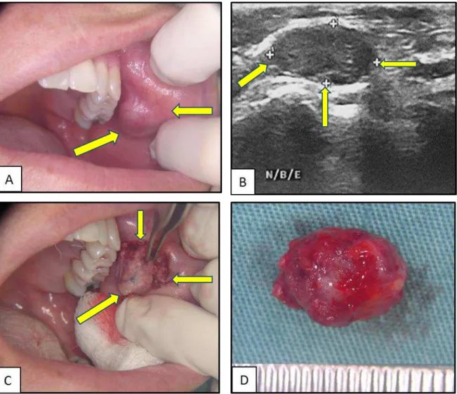
Among the mucus retention phenomena, plunging ranula is an uncommon lesion
that requires greater care, since it can reach large proportions. A definitive diagnosis is
difficult, even with the help of imaging resources such as CT or MRI. This condition
can mimic other diseases, such as lymphangioma and hemangioma [33]. The
differential diagnosis for clinical symptoms related to painless increase in volume
present in the submandibular and/or sublingual region, can include abscesses, cystic
hygroma/lymphangioma, thyroglossal duct cyst and lipoma [34]. Since the treatment of
the lesions mentioned can be different from that utilized for plunging ranula, it is
essential to determine the diagnosis, establishing the type of lesion the patient has [33].
Therefore, US becomes the tool of choice for evaluating abnormalities in the
submandibular region, since it allows the visualization of adjacent superficial
structures, confirming the presence of cystic lesions in submandibular triangle [35] and
demonstrating when there is communication of lesion with the mylohyoid muscle
(pathognomonic sign of plunging ranula). Considering the importance of visualizing
this invasion in mylohyoid muscle, the examination is appropriate for patients that do
not know the origin of the edema, since it is reliable, non-invasive and low-cost [33].
The efficacy of this diagnostic resource increases when combined with fine needle
aspiration biopsy [36]. In the study by Jain, Morton and Ahmad in 2010, the authors
reported limitations presented by MRI and CT, these being attributed to the fact that
images obtained by these methods are static. These examinations were not capable of
demonstrating the herniation of the sublingual gland, or evaluating the extent on the
sublingual gland in subjacent tissues, as well as the extent of spatial involvement with
the lesion [35].
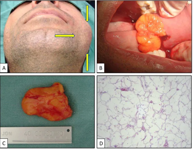
Mucocele is also a mucus retention phenomenon and is characterized by the
involvement of the minor salivary glands, especially those of the lower lip. In contrast
to plunging ranula, it is a common lesion whose presence is usually associated with
local trauma. This pathology is clinically characterized by an increase in a superficial,
translucent and well-defined volume, with 75% of lesions having a diameter of up to 1
cm. However, when the lesion is deep, it can show coloration similar to that of normal
mucosa, or by the presence of edema in the area, mucocele can give a reddish
appearance or even look vascular. Thus, it can mimic other lesions, such as
hemangioma, lymphangioma, lipoma and abscess in soft tissues [37]. When the lesion is
superficial, the history of the lesion and clinical evaluation can lead to a definitive
diagnosis, but when it is deep in tissue planes, an imaging examination is necessary to
determine type of lesion to be treated. In this way, US has been utilized to evaluate
these lesions, because through high-frequency transducers it is capable of demonstrating
the internal structures of the lesion more clearly than CT and MRI. Therefore, US can
identify the type of lesion and determine its treatment [38].
Neoplasias of salivary glands can be completely delineated by echography,
especially when they are localized in the superficial lobe of the parotid [32]. However,
this method is incapable of identifying reliably lesions and masses theinvolve the deep
lobe of the parotid gland, since it is obscured by the mandible [28]. Still, it allows a
dependable differentiation between intra- and extraglandular masses, cystic from solid
lesions, and presence of vascularization intraglandular and in adjacent structures [33].
Histopathology is considered a determinant in the choice of treatment of the
by ultrasound is effective in making a definitive diagnosis. According to a study by
Kovacevic and Fabijanic [30] who detected malignant lesions in the salivary glands, US
showed a specificity varying from 86 to 96% and sensitivity of 82 to 85%. Since it is a
minimally invasive, precise and low cost technique, ultrasound-guided fine needle
aspiration biopsy has been considered the ―gold standard‖ for preoperative evaluation of
salivary gland diseases [30]. Despite showing various advantages over other diagnostic
methods, it is essential to point out the importance of the skill and adequate training of
the professional in obtaining and interpreting the material. Otherwise, the information
obtained can impair the use of US and cause variation in the specificity and sensitivity
of the method [28].
VASCULAR LESIONS
Hemangioma is a benign vascular tumor which is more common in infants.
When present in the head and neck region, it is more easily visualized in the region of
lips, where this location is an important esthetic and functional part of the face [34]. A
10-year retrospective study revealed that hemangioma is the lip tumor of greatest
prevalence (19.28%) among all benign lesions in the region [35].
Another alteration of vascular origin is caliber persistent labial artery (CPLA),
which is an anomaly in which a main artery penetrates the submucous tissue without
division or loss of caliber. On physical examination, it appears as an asymptomatic
papular or nodular lesion, sometimes ulcerated, generally in the lower lip. It can be
confused with a neoplasm and during biopsy, cause severe hemorrhage [36].
In vascular lesions of the head and neck region, US has also been shown as an
important resource, mainly with the use of doppler, where it can, in particular cases,
essential for evaluating diseases of vascular origin, since it determines the origin of the
flow in the lesion (arterial or venous), as well as its velocity (cm/s). It allows a direct
visualization in real time of this vascular alteration, without the necessity of intravenous
contrast agents. Thus, it avoids biopsies and unnecessary surgical procedures, besides
being a non-invasive imaging technique that is well accepted by patients [36].
CONCLUSION
Like all available imaging examinations, US shows specific indications and
limitations in its use. However, compared to other methods, it demonstrates widely
favorable results for its utilization as an auxiliary resource in the diagnoses in soft
tissues of oral and maxillofacial region. With technological advances, ultrasound
instruments are made considering a higher frequency, which resulta in increased
definition of images. They utilize small, compact probes, allowing their introduction
inside the oral cavity.
As it is an innocuous, non-invasive complementary method, of easy access and
low cost, with eminent potential of helping in the diagnosis of diseases in soft tissues,
it is suggested that this resource be promoted in Dentistry as an alternative for
obtaining clinical diagnosis of diseases and appropriate management of patients.
REFERENCES
1) Shung, K K. Diagnostic ultrasound: imaging and blood flow measurements. 3rd Edition. Florida: CRC Press; 2005.
2) Thiruchelvam JK, Songra AK. Intraoperative ultrasound imaging to AID abscess drainage – a technical note. Int J Oral Maxillofac Surg. 2002;31:442–43.
3) Koischwitz D, Gritzmann N. Ultrasound of the neck. Radiol Clinics North Am. 2000;38:1029-1045.
4) Oeppen RS, Gibson D, Brennan PA. An update on the use of ultrasound imaging in oral and maxillofacial surgery. Br J Oral and Maxillofac Surg. 2010;48:412–18.
5) Papaleo EC. O uso da ultra-sonografia na odontologia [Monograph]. Porto Alegre (RS): Universidade Federal do Rio Grande do Sul; 2010.
6) Pallagatti S, Sheikh S, Puri N, Mittal A, Singh B. To evaluate the efficacy of ultrasonography compared to clinical diagnostic radiography and histopathological findings in the diagnosis of maxillofacial swellings. Eur J Radiol. 2012;81:1821–27. 7) Ultrasound [internet]. San Clemente (CA): eMedicine Health. c2003 – [cited 2012 June
25]. Available from: http://www.emedicinehealth.com/ultrasound/article_em.htm. 8) Sniezek JC. Head and Neck ultrasound: why now? Otolaryngol Clin North Am.
2010;43:1143–47.
9) Gaspari R, Dayno M, Briones J, Blehar D. Comparison of computerized tomography and ultrasound for diagnosing soft tissue abscesses. Crit Ultrasound J. 2012;17:1–7.
10)Srinivas K, Sumanth KN, Chopra SS. Ultrasonographic evaluation of inflammatory swellings of buccal space. Indian J Dent Res. 2009;20:458–62.
11)Chao HC, Lin SJ, Huang YC, Lin TY. Sonographic evaluation of cellulitis in children. J Ultrasound Med. 2000;19:743–49.
12)Baurmash HD, Worth L. Ultrasonography in the diagnosis and treatment of facial abscess. J Oral Maxillofac Surg. 1999;57:635–36.
13)Mallorie CN, Jones SD, Drage NA, Shepherd J. The reliability of high resolution ultrasound in the identification of pus collections in head and neck swellings. J Oral Maxillofac Surg. 2012; 41:252–55.
16)Chandak R, Degwekar S, Bhowte RR, Motwani M, Banode P, Chandak M, et al. An evaluation of efficacy of ultrasonography in the diagnosis of head and neck swellings. Dentomaxillofac Radiol. 2011;40:213–21.
17)Douglas AS, Jennings S, Owen VMF, Elliot S, Parker D. Is ultrasound useful for evaluating paediatric inflammatory neck masses? Clin Otalaryngol. 2006;30:526–29. 18)Douglas AS, Jennings S, Owen VMF, Elliot S, Parker D. Ultrasound and fine needle
aspiration cytology in the staging of neck lymph nodes in oral squamous cell carcinoma. Br J Oral Maxillofac Surg. 2000;38:430–36.
19)Natori T, Koga M, Anegawa E, Nakashima Y, Tetsuka M, Yoh J, et al. Usefulness of intra-oral ultrasonography to predict neck metastasis in patients with tongue carcinoma. Oral Dis. 2008;14: 591–99.
20)Ota Y, Aoki T, Karakida K, Otsuru M, Kurabayashi H, Sasaki M, et al. Determination of deep surgical margin based on anatomical architecture for local control of squamous cell carcinoma of the buccal mucosa. Oral Oncol. 2009;45:605–9.
21)Yuasa K, Kawazu T, Nagata T, Kanda S, Ohishi M, Shirasuna K. Computed tomography and ultrasonography of metastatic cervical lymph nodes in oral squamous cell carcinoma. Dentomaxillofac Radiol. 2000;29:238–44.
22)Di Martino E, Nowak B, Hassan HA, Hausmann R, Adam G, Buell U, et al. Diagnosis and staging of head and neck cancer: a comparison of modern imaging modalities (positron emission tomography, computed tomography, color-coded duplex sonography) with panendoscopic and histopathologic findings. Arch Otolaryngol Head Neck Surg. 2000;126:1457–61.
23)van den Breckel MW, Stel HV, Castelijns JA, Croll GJ, Snow GB. Lymph node staging in patients with clinically negative neck examinations by ultrasound and ultrasound-guided aspiration cytology. Am J Surg. 1991;162:362–66.
24)Hohlweg-Majert B, Metzger MC, Voss PJ, Hölzle F, Wolff KD, Schulze D. Preoperative cervical lymph node size evaluation in patients with malignant head/neck tumors: comparison between ultrasound and computer tomography. J Cancer Res Clin Oncol. 2009;135:753–59.
25)Anand N, Chaundhary N, Mittal MK, Prasad R. Comparison of the efficacy of clinical examination, ultrasound neck and computed tomography in detection and staging of cervical lymph node metastasis in the head and neck. India J Otolaryngol Head and Neck Surg 2007;59:19–23 .
27)Millesi W, Prayer L, Helmer M, Gritzmann N. Diagnostic imaging of tumor invasion of the mandible. Int J Oral Maxillofac Surg. 1990;19:294–98.
28)Pfeiffer J, Ridder GJ. Diagnostic value of ultrasound-guided core needle biopsy in patients with salivary gland masses. Int J Oral Maxillofac Surg. 2012;41:437–43. 29)Zajkowski P, Jakubowski W, Bialek EJ, Wysocki M, Osmólski A, Serafin-Król M.
Pleomorphic adenoma and adenolymphoma in ultrasonography. Eur J Ultrasound. 2000;12:23–29.
30)Kovacevic DO, Fabinavic I. Sonographic diagnosis of parotid gland lesions: correlation with the results of sonographically guided fine-needle aspiration biopsy. J Clin Ultrasound. 2010;38:294–98.
31)Sodhi KS, Bartlett M, Prabhu NK. Role of high resolution ultrasound in parotid lesions in children. Int J Pediatr Otorhinolaryngol. 2011;75:1353–358.
32)Gritzmann N, Rettenbacher T, Hollerweger A, Macheiner P, Hübner E. Sonography of the salivary glands. Eur Radiol. 2003;13:964–75.
33)O´Conner R, McGurk M. The plunging ranula: diagnostic difficulties and a less invasive approach to treatment. Int J Oral Maxillofac Surg http://dx.doi.org/10.1016/j.ijom.2013.03.019
34)Jain R, Morton RP, Ahmad Z. Diagnostic difficulties of plunging ranula: case series. J Laryngol Otol. 2012;126:506–10.
35)Jain P, Jain R, Morton RP, Ahmad Z. Plunging ranulas: high-resolution ultrasound for diagnosis and surgical management. Eur Radiol. 2010;20:1442–449.
36)Zhi K, Wen Y, Zhou H. Management of the pediatric plunging ranula: results of 15 years´clinical experience. Oral Surg Oral Med Oral Pathol Oral Radiol Endod. 2009;107:499–502.
37)Jinbu Y, Kusama M, Itoh H, Matsumoto K, Wang J, Noguchi T. Mucocele of the glands of Blandin-Nuhn: clinical and histopathologic analysis of 26 cases. Oral Surg Oral Med Oral Pathol Oral Radiol Endod. 2003;95:467–70.
38)Senthilkumar B, Mahabob MN. Mucocele: an usual presentation of the minor salivar gland lesion. J Pharm Bioallied Sci. 2012;4:180–82.
39)Pozza DH, Soares LP, Oliveira MG. Exames complementares por imagens no diagnóstico e no planejamento cirúrgico de patologias em glândulas salivares. Rev Bras Patol Oral. 2005;4:156–61.
41)Ntomouchtsis A, Karakinaris G, Poulolpoulos A, Kechagias N, Kittikidou K, Tsompanidou C, et al. Benign lip lesions a 10-year retrospective study. Oral Maxillofac Surg. 2010;14:115–18.
42)Wortsman X, Calderón P, Arellano J, Orellana Y. High-resolution color Doppler ultrasound of a caliber-persistent artery of the lip, a simulator variant of a dermatologic disease: case report and sonographic findings. Int J Dermatol. 2009;48:830–33.
43)Oates CP, Wilson AW, Ward-Booth RP, Williams ED. Combined use of Doppler and conventional ultrasound for diagnosis of vascular and other lesions in the head and neck. Int J Oral Maxillofac Surg. 1990;19:235–39.
ARTIGO II
Contribution of ultrasonography to the diagnosis of submucosal and subcutaneous nodular lesions of the oral and maxillofacial region – analysis of cases.
Felipe Leal Martins, Fernanda Gonçalves Salum, Karen Cherubini, Roberto Oliveira, Maria Antonia Zancanaro de Figueiredo
F. L. Martins, F. G. Salum, K. Cherubini, M. A. Z. de Figueiredo
Dentistry School of the Pontifical Catholic University of Rio Grande do Sul (PUCRS) Oral Medicine Unit of São Lucas Hospital - Pontifical Catholic University of Rio Grande do Sul (PUCRS)
Av. Ipiranga, 6690 – 2º andar/sala 231, Porto Alegre, RS. Brasil. CEP 90610-000 R. Oliveira
Specialist in Radiology and Imaging Diagnosis by the Brazilian College of Radiology.
Medical Director of the Clinical Radiology Unit and Dean of the Radiology and Ecography Fundation of Rio Grande do Sul, Brazil
Corresponding author:
Professor Maria Antonia Zancanaro de Figueiredo
Address: Serviço de Estomatologia do Hospital São Lucas, PUCRS, Pontifícia Universidade Católica do Rio Grande do Sul
Av. Ipiranga, 6690 – 2º andar/sala 231, Porto Alegre, RS. Brasil. CEP 90610-000 Phone/Fax.: +55 51 3320.3254 E-mail: [email protected]
Objective. The aim of this study was evaluate the contribution of ultrasonography in the establishment of the diagnosis of nonspecific nodular lesions of
the soft tissues of the oral and maxillofacial region. We determined the indication of use
and reliability of ultrasonography in the dental field.
Materials and Methods. We recruited 65 patients from the Oral Medicine Unit of São Lucas Hospital, who had nonspecific nodules located in the submucosa or
subcutaneously. They were subjected to a doppler ultrasonography, carried out with
standardization of the protocol and equipment. The ultrasonography report was made by
an experienced professional. Another 2 calibrated examiners analyzed the data,
comparing the ultrasonographic report with the final diagnosis. Accordingly, we used
established scoring, where zero corresponded to no contribution to the final diagnosis,
1, helped in its determination, and 2, estabilished the diagnosis.
Results. Zero was obtained for 12.3% of the examinations performed, and 1 and 2 accounted for respectively 41.5% and 46.1%, totaling a contribution about 88%.
Ultrasonography was of value in the diagnosis of vascular lesions in 93.3% and of
neoplasms in 87.5%. In the salivary gland diseases, it contributed in 75% of cases.
Conclusions. The results obtained demonstrated that ultrasonography is an effective tool in the determination of the definitive diagnosis of nonspecific nodular
lesions of the soft tissues of the oral and maxillofacial region.
Clinical relevance. Ultrasonography is an innocuous noninvasive examination, easy to perform and has low cost, which is effective in aiding to the diagnosis of
INTRODUCTION
Ultrasonography (US) was more used as a diagnostic tool by health care professionals during the 1970s, when a mapping of gray levels was introduced [1]. While it is widely used in the medical area, it is a resource that is rarely requested by the dentist, who is often unaware of its indications, limitations and advantages [2]. Echography utilizes a nonionized form of radiation and is thus considered safe, where it can be repeated whenever necessary. It shows few artifacts, and it is of low cost in relation to similar methods and painless, being well tolerated by patients. This exam has the capacity to reproduce images in real time and to provide millimetrical resolution and vascular information, if doppler is utilized [3–6].
However, US of the head and neck region is considered one of the most complex examinations to be interpreted, requiring an experienced professional, who should have wide knowledge of the regional anatomy, be informed about the clinical condition of the patient and technically qualified to interpret abnormalities detected [4]. Since it is a dynamic test, the images obtained are better understood live. When archived and reproduced in planes, they can be difficult to interpret, especially if compared with computed tomography and magnetic resonance. It is believed that this is one of the reasons the makes echography an examination less requested than others that also utilize sectioned imaging [5].
MATERIALS AND METHODS
We selected 65 patients with nonspecific nodular lesions localized in the oral
and maxillofacial region, seen at the Oral Medicine Unit at the São Lucas Hospital
(HSL) of the Pontifical Catholic University of Rio Grande do Sul (PUCRS), Brazil,
during 18 months.
All patients included in the study had been indicated for US and were subjected
to the examination, since it was not possible to establish a definitive diagnosis through
clinical examination. US was performed in a diagnostic imaging center utilizing the
same apparatus Toshiba Japan – Aplio 80 with linear probe for examination of soft
tissues and frequencies of 7.5 to 12 MHz with a color doppler system for high
definition. US was carried out by a single examiner with ample experience in the
interpretation of images of the oral and maxillofacial region.
The study was conducted after approval at the Committee of Ethics in Research
of PUCRS. Patient information was collected and annotated from medical charts which
contained data with regard to clinical characteristics of the lesion, descriptive US report
and the final diagnosis, including the anatomopathological examination, whenever
needed. All data obtained were kept confidential. The information was tabulated in
spreadsheets developed specifically for this study, which included the diagnosis from
US and the final diagnosis. Accordingly, each lesion was stratified into scores on the
basis of the following criteria:
- Score zero: when the diagnosis obtained with US differed from the final
diagnosis.
- Score 1: when the diagnosis obtained with US contributed to establishing the
- Score 2: when the diagnosis obtained with US defined the final diagnosis.
Later, 2 calibrated examiners performed the data analysis, comparing the US
diagnosis with the final diagnosis (confirmed by histopathologic examination, if
necessary), so that the scores could be defined.
The pathologies included in the statistical analysis were those that affected 10 or
more individuals. The small number of cases for the others made it impossible to carry
out mathematical calculations. On the basis of the data obtained, percentages were
determined for the 3 scores of impact of US on the final diagnosis of the subcutaneous
and submucosal nodular lesions of the oral and maxillofacial region. After obtaining
these results, a global Fisher exact test was performed to determine the p values that
revealed if the method showed a statistically significant difference in the definition of
the final diagnosis between the diseases detected in the study. Afterwards, for
determination of p between all lesions, post-hoc comparisons were carried out with
RESULTS
Of the patients evaluated, 29 were males and 36 females, aged between 4 and 84
years. The individuals who had a histopathologic examination to determine the final
diagnosis of the lesion, represented 66.1% (n=43) of the total. Of the 22 remaining
patients, 33.8% did not need a biopsy to confirm the definitive diagnosis, since the US
descriptive report determined how the patient would be treated (Table 1).
Table 1 Distribution of patients with nonspecific submucosal or subcutaneous nodules underwent surgery and histological examination, and those who were not required biopsy of lesions
PATIENTS TOTAL %
CONTAINED PATHOLOGIC EVALUATION
43 66,1
CONTAINED NO PATHOLOGIC EVALUATION
22 33,8
65 99,9
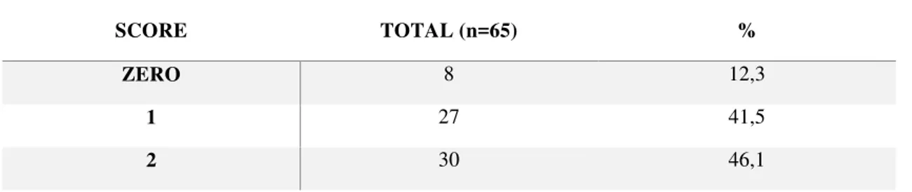
The zero score, which meant no contribution of echography in the establishment
of the final diagnosis accounted for 12.3% of the examinations performed. But the
scores 1, indicating US helped in the management of the case, and 2, where it was
possible to define the diagnosis through the image, represented respectively 41.5% and
46.1%, totaling 87.6% of cases where US was of value. This demonstrates that of the
total of patients analyzed (n=65), in 57 cases US contributed in a significant manner to
the appropriate management of the patient (Table 2).
Table 2 Distribution of scores set by the examiners, as well as the percentage that each of them represented in aid of determining the final diagnosis of patients
SCORE TOTAL (n=65) %
ZERO 8 12,3
1 27 41,5
The diseases of the salivary glands (sialadenitis, sialolithiasis and mucus
retention phenomena) represented 26.1% (n=17) of the alterations presented by the
patients. The neoplastic lesions (malignant and benign) corresponded to 24.6% (n=16)
and the vascular, blood and lymphatic, were diagnosed in 23.1% (n=15) of the
individuals. The inflammatory reaction processes made up 21.5% (n=14) of the cases
evaluated. The cystic lesions and lymph retention phenomena accounted for only 3.1
(n=2) and 1.5% (n=1), respectively (Table 3).
Table 3 Classification, absolute and percentage distribution of subcutaneous and submucosal nodular lesions
LESIONS TOTAL (n=65) %
VASCULAR LESIONS 15 23,1
NEOPLASMS 16 24,6
SALIVARY GLAND DISEASES
17 26,1
INFLAMMATORY PROCESSES REACTION
14 21,5
CYSTIC LESIONS 2 3,1
RETENTION LYMPH 1 1,5
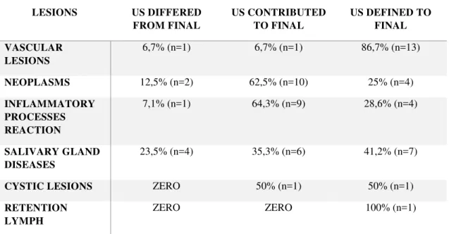
According to the scores determined by the examiners (Table 4), the method
contributed to the diagnosis of the vascular lesions in 93.3%. In the neoplasms,
ultrasonographiy was useful in the management of the lesions, the malignant as well as
the benign ones, in 87.5%. With regard to mucus retention phenomena, US had a role
in the establishment of the final diagnosis in 76.5%. The other alterations described
were represented by limited numbers of patients, making effective analysis of these data
The rates of the contribution of US were not the same for the different types of
lesions (p=0.0017). It was possible to see that in the pathologies of vascular nature, US
demonstrated a statistically greater contribution than in the others. This means that the
vascular lesions, when compared with salivary gland diseases, inflammatory reaction
processes and neoplasms, showed a statistically significant difference (p=0.024,
p=0.009, p=0.06). However, the study did not have appropriate conditions to differentiate the others, despite the nominal values of the neoplasms being less than that
of the others, because comparisons of the diagnostic method did not show statistically
significant differences.
US was able to determine the contents of the lesions in 80% of the examinations.
It indicated the vascular nature of the nodules in 23.1%, the presence of thick fluid or
mucus in the vicinity of the lesion in 21.5% and if the nodule was solid in 35.4%.
However, some ultrasonographic reports were not considered clear in relation to the
contents of the lesions.
Table 4 Distribution of lesions diagnosed in related score contribution of ultrasonography study to establish the final diagnosis
LESIONS US DIFFERED
FROM FINAL
US CONTRIBUTED TO FINAL
US DEFINED TO FINAL
VASCULAR LESIONS
6,7% (n=1) 6,7% (n=1) 86,7% (n=13)
NEOPLASMS 12,5% (n=2) 62,5% (n=10) 25% (n=4)
INFLAMMATORY PROCESSES REACTION
7,1% (n=1) 64,3% (n=9) 28,6% (n=4)
SALIVARY GLAND DISEASES
23,5% (n=4) 35,3% (n=6) 41,2% (n=7)
CYSTIC LESIONS ZERO 50% (n=1) 50% (n=1)
RETENTION LYMPH
DISCUSSION
High frequency US is a diagnostic tool that is noninvasive, low cost and
painless, which is commonly utilized in the medical area, where it is particularly
indicated in the examination of soft tissues. However, it is not usually utilized for
diagnostic investigation of intraoral lesions [6]. In this study, we evaluated the
contribution of US as an auxiliary method in the diagnosis of submucosal or
subcutaneous nodular lesions in the soft tissues of the oral and maxillofacial region.
This study did not consider the obligation of performing an anatomopathologic
examination as the gold standard in all cases, because in some lesions, especially
vascular, ultrasonographic diagnosis was considered definitive for the treatment of the
patients. The histopathologic diagnosis was established only in the lesions with surgical
indication.
Studies evaluating the use of US have been conducted in different lesions in the
facial soft tissues, and despite extremely satisfactory results, they did not emphasize its
minimal utilization as a diagnostic method by oral surgeons. This is probably due to an
academic gap, lacking due training of the professionals in the use and interpretation of
this resource [2].
The result obtained for the extent of the contribution that US showed as a
diagnostic tool, in all patients, was 87.6%. In the diseases of possible inflammatory
cause, we demonstrated that US was effective in 92.8%. This percentage is in contrast
to that obtained by various authors who analyzed nonspecific swelling in orofacial soft
tissues. They evaluated lesions of probable inflammatory nature and found a sensitivity
of 96%, that is, the fraction of the patients who showed a positive response in the
ultrasonographic examination among those individuals who had the disease [8 - 12]. On
same line of research, Pallagatti et al. [14] and Nisha et al. [12] respectively determined
an accuracy of 88.9% and 97.1% for US.
According to some authors, US is a complementary examination that helps
confirm the diagnosis of abscesses and delineate their anatomic location, differentiating
them from of cellulitis [11,15], and thereby demonstrating the presence of fluids or
abscesses in the superficial spaces of the face [3] distinguishing the stage of the
infection [16].
Among the vascular alterations, the hemangiomas were the most prevalent,
representing 23.1% of the lesions studied. The result obtained in this study is similar to
that of Ntomouchtsis et al. [17] who studied benign lesions in the lips of 420 patients.
The authors found that the hemangiomas were the most common, corresponding to
19.3%. When these lesions are present on the tissue surface, the clinical diagnosis can
be favored, especially through a semitechnical procedure called diascopy. However, in
deeper lesions, that is, located in the submucosa of the orofacial soft tissues, sufficient
information generally cannot be obtained from a clinical diagnosis to determine the
vascular origin of the pathology evaluated. For this, we must introduce available
imaging resources, including echography. Vascular alterations of the lesions visualized
by US using the doppler system were determinant and defined the management of the
patient. This means that in 13 (86.7%) of the 15 vascular alterations present in the
study, US defined the final diagnosis. The use of the doppler system was essential for
evaluation of the diseases of vascular origin, since it determines the origin of the flow in
the lesion (arterial or venous), as well as its velocity (cm/s). The examination allows a
direct visualization of this alteration in real time, without the need of intravenous
Salivary gland diseases encompass a wide range of pathologies. Various authors
have described imaging methods that can help in making the final diagnosis. A near
unanimity of authors consider US the first choice among all available imaging resources
in the detection of diseases of the salivary glands [5,7,20-26]. However, the majority of
the scholars only refer to its indications, advantages and disadvantages in relation to
other imaging examinations. In the present study, the extent that US contributed
determined by summing the scores of definition and aid in the management of the
patient, was 76.5% of the total cases.
Of the 16 cases of neoplasms, US helped in the management of the patient in
87.5% of cases. The malignant and benign lesions were grouped together, since
according to Zengel et al. [27] tumors smaller than 2 cm in diameter usually have a
homogeneous structure and show defined borders and can be consequently diagnosed
mistakenly as benign lesions. Gritzmann [20] analyzed 302 patients and found a
sensitivity of 100% for the examination in the identification of neoplasms. The results
of the study were in accordance with those reported by Millesi et al. [28] and Hodder et
al. [29], who found values over 90%. Other authors obtained different results; Haberal
et al. [30] and Anand et al. [31] found rates over 70%, but Hohlweg-Majert et al. [32]
described a sensitivity of 24.5% for US.
In bone tissue, according to Millesi et al. [28], US is capable of evaluating the
tissue destruction caused by malignant lesions in the oral and maxillofacial region, with
the exception of the lingual side of the mandibular ramus. Compared with other
techniques, US can be considered of great value, with a high contribution in particular
regions. Ng et al. [33] described a case of mandibular osteosarcoma evaluated by US
and concluded that the use of this resource was essential for the visualization of signs of
stages of these pathologies. McCann et al. [34] also mentioned US as an effective tool
in the initial analysis of fractures of the orbital-zygomatic complex, thereby reducing
the number of X-rays required for the patients. It is known that although echography
can be used in the analysis of bony structures, some authors believe that its use is not
recommended for diagnosing temporomandibular disorders in a conclusive manner
[35].
Some limitations that occurred during the course of the present study should be
mentioned. A limited number of patients made up of some groups of analyzed lesions,
making it difficult to make comparisons between the groups of nodular lesions and also
with other studies previously conducted in this area. However, it was our purpose to
bring together a larger number of patients with nonspecific swelling, where the clinical
diagnosis represented similar conditions. For this reason, the neoplasms, malignant and
benign, including those originating from the salivary glands, as well as metastatic
lymph nodes, were grouped together. But in the diseases associated with the salivary
glands, they were put in the group exclusively for sialadenitis, sialolithiasis and
Sjögren‘s syndrome.
In the literature, there are still few studies that have evaluated the use of US in
investigating nodular lesions in nonspecific clinical diagnosis and have indicated it for
professionals in dentistry. In contrast, in the medical area, it is a widely used in the most
diverse specialties. Currently, more studies are being conducted on the use of US for
evaluation of intraoral lesions and structures, due to the emergence of new technologies,
such as the use of higher frequencies and smaller transducers, making it possible to
introduce them inside the oral cavity.
The results obtained in this study demonstrated that US supplies quality
management of the submucosal or subcutaneous nodular lesions. This is a recurring
clinical situation in dental practice, and through this examination, the dentist is more
able to rapidly and adequately manage the patient. Thus, this resource can be indicated
as a viable method in the routine evaluation of the nonspecific swelling of the soft
tissues in the oral and maxillofacial region, since it is well tolerated by the patient, has
low cost and does not expose to ionizing radiation.
CONFLICT OF INTEREST
REFERENCES
1. Shung, K K (2005) Diagnostic ultrasound: imaging and blood flow measurements. CRC Press, Florida.
2. Sniezek JC. Head and neck ultrasound: why now? Otolaryngol Clin North Am (2010): 43(6):1143-1147.
3. Thiruchelvam JK, Songra AK (2002) Intraoperative ultrasound imaging to AID abscess drainage – a technical note. Int J Oral Maxillofac Surg 31(4):442-443
4. Koischwitz D, Gritzmann N (2000) Ultrasound of the neck. Radiol Clinics North Am 38(5): 1029-1045
5. Oeppen RS, Gibson D, Brennan PA (2010) An update on the use of ultrasound imaging in oral and maxillofacial surgery. Br J Oral and Maxillofac Surg 48(6):412-418.
6. Salomon B, Le Denmat D (2012) Intraoral ultrasonography: development of a specific high-frequency probe and clinical pilot study. Clin Oral Invest 16(2):643-649
7. Pfeiffer J, Ridder GJ (2012) Diagnostic value of ultrasound-guided core needle biopsy in patients with salivary gland masses. Int J Oral Maxillofac Surg 41(4):437-443
8. Srinivas K, Sumanth KN, Chopra SS (2009) Ultrasonographic evaluation of inflammatory swellings of buccal space. Indian J Dent Res 20(4):458-462
9. Chandak R, Degwekar S, Bhowte RR, Motwani M, Banode P, Chandak M, Rawlani S (2011) An evaluation of efficacy of ultrasonography in the diagnosis of head and neck swellings. Dentomaxillofac Radiol 40(2):213-221.
10.Mallorie CN, Jones SD, Drage NA, Shepherd J (2012) The reliability of high resolution ultrasound in the identification of pus collections in head and neck swellings. J Oral Maxillofac Surg 41(2):252-255
11.Gaspari R, Dayno M, Briones J, Blehar D (2012) Comparison of computerized tomography and ultrasound for diagnosing soft tissue abscesses. Crit Ultrasound J 17(4):1-7