(1) Departamento de Fonoaudiologia da Universidade Federal de Santa Maria, UFSM, Santa Maria (RS), Brasil. (2) Universidade Anhanguera de São Paulo,
UNIAN, São Paulo (SP), Brasil. (3) Universidade Federal de Santa Maria,
UFSM, Santa Maria (RS), Brasil. (4) Pontifícia Universidade Católica de São
Paulo, PUC, São Paulo (SP), Brasil. Source Feature: CnPq
Conlict of interest: non-existent
Electroacoustic evaluation of the olivocochlear efferent pathway
in subjects with tinnitus complaint
Avaliação eletroacústica da via eferente olivococlear
em indivíduos com queixa de zumbido
Valdete Alves Valentins dos Santos Filha(1) Fátima Cristina Alves Branco-Barreiro(2) Ariane Macedo Gomes(3) Teresa Maria Momensohn dos Santos(4)
Received on: December 30, 2015 Accepted on: May 11, 2016
Mailing address:
Valdete Alves Valentins dos Santos Filha Rua Riachuelo, 417 AP. 902 Bairro Centro – Santa Maria – RS CEP: 97050-011
E-mail: [email protected]
ABSTRACT
Purpose: to investigate the suppressive effect of transient-evoked otoacoustic emissions in subjects with
tinnitus complaint and normal audiometry and to analyze the relation to age, gender, laterality of tinnitus and its degree of discomfort.
Methods: we assessed 60 subjects, 14 males and 46 females, aged between 20 and 59 years, 30 with
tinnitus (experimental group) and 30 without tinnitus complaint (control group). The suppression of tran-sient-evoked otoacoustic emissions was investigated with contralateral white noise at 50 dBHL at the frequency bands of 700, 1000, 1400, 2000, 2800 and 4000Hz.
Results: the mean value for the suppression of transient-evoked otoacoustic emissions in the
experimen-tal group ranged from 2.14 to 4.38. In the control group, the mean value for suppression of transient
--evoked otoacoustic emissions ranged from 2.27 to 4.88.
Conclusion: suppression values of otoacoustic emissions were similar in subjects with and without
tinni-tus, although the results of the tinnitus group were lower, suggesting worse performance of the Superior Olivary Complex.
Keywords: Tinnitus; Hearing; Hearing Tests; Hair Cells, Auditory, Outer; Signal-To-Noise Ratio
RESUMO
Objetivo: investigar o efeito supressor das emissões otoacústicas por estímulos transientes em indiví
-duos com queixa zumbido e audiometria normal e analisar sua relação com as variáveis idade, sexo,
lateralidade do zumbido e grau de incômodo.
Métodos: foram avaliados 60 sujeitos, 14 do gênero masculino e 46 do gênero feminino, entre 20 e
59 anos de idade, sendo 30 com queixa de zumbido (grupo experimental) e 30 sem zumbido (grupo
controle). Foi realizada a pesquisa da supressão das emissões otoacústicas por estímulos transientes,
para ruído branco de 50 dBNA, na condição contralateral nas bandas de frequência de 700, 1000, 1400, 2000, 2800 and 4000Hz. Resultado: no grupo experimental, a supressão das emissões otoacústicas
transientes média variou de 2,14 a 4,38. No grupo controle o valor médio da supressão das emissões otoacústicas transientes variou de 2,27 a 4,88.
Conclusão: os valores de supressão das emissões otoacústicas foram semelhantes nos indivíduos com e sem zumbido, embora o grupo com o sintoma tenha tido resultados menores, sugerindo pior desempe
-nho do Complexo Olivar Superior.
INTRODUCTION
Tinnitus is deined as the perception of a sound in one or both ears, as well as in the head, generated in the absence of an external sound stimulus, and it may be the irst indication of a number of diseases that endanger health and well-being of individuals1,2.
Also called acouphène, this symptom may or may not be associated with hearing alterations, varying in intensity and quality from person to person and can be perceived as a high-pitched sound, like a bell, or even a low-pitched sound, such as an engine3. It ranges from a mild discomfort to a complete incapacity1,4.
This complaint is still unclear in many aspects, especially concerning its origin and production. According to researchers5, the lack of knowledge about the pathophysiology of tinnitus conducted to the development of many theories that attempt to explain the origin of this symptom. It is known that there is the possibility of involvement of more than one mechanism in the same individual.
One of the hypotheses for the occurrence of tinnitus would be a dysfunction in the efferent auditory system, speciically in the medial superior olivary complex (MSOC) region.
The medial olivocochlear tract modulates the movement of the outer hair cells by the release of acetylcholine in the synaptic cleft, causing a hyper-polarization, which is opposed to the depolarization induced by sound stimuli. This hyperpolarization can be quantiied by the decrease in the amplitude of otoacoustic emissions in the presence of noise in contralateral ear6,7.
Some studies have shown a decrease in the amplitude of otoacoustic emissions in the presence of contralateral noise, which is called suppression effect, in individuals with tinnitus complaints8,9. However, other
studies have found similar indings when comparing the suppression effect of otoacoustic emissions in individuals with and without tinnitus10,11.
Although otoacoustic emissions are useful to inves-tigate the medial olivocochlear tract, methodological problems affect the interpretation of academic indings. Many studies use a probe stimulus to produce the otoacoustic emission and an eliciting stimulus to evoke the efferent activity, changing, therefore, the otoacoustic emissions. Little attention has been given to the possi-bility of the probe stimulus evoke the efferent activity. Besides, many studies use contralateral elicitors and do not include measures to discard the inluence of stapedius muscle contractions12,13.
The clariication of the mechanisms involved in the production of tinnitus is necessary and essential to propose effective measures, which aim to the permanent relief of this symptom14.
Knowing that the efferent system plays a key role in the modulation of active cochlear process, is there a decrease of noise suppression effect on otoacoustic emissions in individuals with complaint of tinnitus?
Therefore, this study aimed to investigate the suppression effect of transient-evoked otoacoustic emissions (TEOAEs) in individuals with complaint of tinnitus, as well as analyze its relation to the variables age, gender, laterality of tinnitus and level of discomfort.
METHODS
This study was approved by the Research Ethics Committee of the Postgraduate Studies Program in Speech Therapy at Pontiical Catholic University of São Paulo (PUC/SP) REC/PUC-SP.
, under the number 0015/2004 and all individuals signed an informed consent to participate in this study. The same proceedings were done in a private specialized clinic in the municipality of São Paulo.
This is a descriptive, experimental and comparison group study, and quantitative analysis were adopted.
The convenience sample was composed of 60 individuals, including male and female genders, with a mean age of approximately 37 years, ranging from 20 to 59 years. Both the experimental group (EG) and the control group (CG) were formed by 30 individuals, 23 women and seven men. Individuals who reported tinnitus composed the EG, while those who did not report this symptom remained in CG.
Inclusion criteria consisted in: complaint of constant or intermittent tinnitus, unilateral or bilateral for EG; hearing thresholds within the normal range, i.e., less than or equal to 25 dB HL in the frequencies of 250 kHz up to 8 kHz and TEOAEs in both ears for EG and CG. We excluded individuals who have reported outer and/ or middle ear alterations and neurological changes, as well as those who have already undergone treatment with ototoxic medications and/or are in drug treatment for tinnitus.
All individuals were submitted to: audiological anamnesis, questionnaire about tinnitus charac-teristics15, pure tone audiometry, transient-evoked otoacoustic emissions (TEOAEs) and research of TEOAEs suppression.
(μs), with rareied polarity, presented in a series of 260 stimuli in eight clicks blocks each, with pulse repetition frequency of 50 cycles per second. Concerning the spectrum of emissions, the standard stimulus contains energy distributed in the frequency bands of 0.7; 1.0; 1.4; 2.0; 2.8 and 4 kHz16.
The collection of emissions started with the right ear. For the measurement of transient-evoked otoacoustic emissions (TEOAEs), it was used the cochlear analyzer Ero Scan, produced by Maico Diagnostics Company, in a soundproof booth.
The technique used to record the suppression of TEOAEs followed the aforementioned procedures, added to the presence of white noise17 in the opposite ear. The noise was provided by the audiometer Interacoustic AC 40, through TDH 39 phone with intensity of 50 dB HL18. This measure has always been performed after recording without contralateral stimulation for both ears, in order to not change the placement of the probe during the two measurements16. Therefore, the measurement was initiated in the right ear, after collection without contralateral stimulation, and after the left ear was evaluated with and without contralateral stimulation.
In the analysis of results, the otoacoustic emissions were classiied as present or absent according to the following criteria: emissions were considered present when the value of S/N (signal-to-noise ratio) was greater than or equal to 7 dB in at least three frequencies.
The following criteria17 were used to classify TEOAEs suppression as present or absent: the suppression value of the olivocochlear system was obtained from the measurement of the difference of the values obtained in the conditions with and without contra-lateral stimulation in each ear. It was considered that the suppression occurred when the value was positive (greater than or equal to 1) and it was considered that there was no suppression of the amplitude of TEOAEs when the value was zero or negative.
In order to compare the results in the suppression of transient-evoked otoacoustic emissions (TEOAEs) concerning the variable age (if it inluences or not), the experimental group was divided by age – less than or
equal to 37 years and over 37 years, and the division parameter was the average age (paired between groups).
The severity of tinnitus was analyzed by visual analogue scale (VAS). Following this method, we ask individuals to give a score from 1 to 10 concerning tinnitus, considering that 1 would be a mild tinnitus while 10, the worst tinnitus they could imagine. The scores were classiied as the following: 1 to 3 - mild tinnitus; 4 to 6 - moderate tinnitus, and 7 to 10 - severe tinnitus19.
After analyzing the scores attributed to the severity of tinnitus, the individuals were divided into three subgroups: Group 1 - mild tinnitus; Group 2 - moderate tinnitus; Group 3 - severe tinnitus.
In the analysis of results, the Mann-Whitney Test and the Kruskal-Wallis Test were used with signiicance level of p <0.05; as well, it was performed a descriptive critical analysis of the variables: age, sex, severity and location of tinnitus.
RESULTS
The study sample consisted of 30 individuals in each group studied, 23 (76.7%) women and seven (23.3%) men, with an average age of 37 years for the experi-mental group and 36.2 years for the control group. There was no statistically signiicant difference between the groups regarding average age (p = 0.753).
In relation to the severity of tinnitus, there was an occurrence of 86.65% for moderate or severe tinnitus, while 13.35% for mild tinnitus.
Concerning the symptom laterality, 56.65% of the individuals reported bilateral tinnitus, whereas 43.35% informed unilateral tinnitus, considering that 23.35% reported this complaint in the right ear and 20% in the left ear.
Table 1. Descriptive analysis of TEOAEs amplitude suppression, by frequency on the right ear, in the Control Group (CG) (n=30) and in
the Experimental Group (EG) (n=30).
Frequency / Group TEOAEs amplitude suppression p
Average SD Median Minimum Maximum
700 Hz
CG (n=8) 4,88 3,09 4,5 1 9 0,062
EG (n=7) 2,14 1,46 2,0 1 5
1000 Hz
CG (n=17) 4,41 2,53 5 1 8 0,983
EG (n=13) 4,38 2,02 5 1 8
1400 Hz
CG (n=14) 3,21 1,48 3,5 1 5 0,676
EG (n=12) 3,67 1,92 3,0 1 7
2000 Hz
CG (n=11) 3,09 1,64 3 1 6 0,209
EG (n=13) 2,31 1,49 2 1 5
2800 Hz
CG (n=11) 2,27 1,10 2,0 1 4 0,613
EG (n=12) 2,75 1,71 2,5 1 6
4000Hz
CG (n=7) 3,14 1,77 3 1 6 0,650
EG (n=10) 2,80 1,62 3 1 6
Mann-Whitney Test
Legend: TEOAEs– Transient-evoked otoacoustic emissions CG – control group; EG – experimental group SD – standard deviation; n –number of individuals Hz - Hertz
Table 2. Descriptive analysis of TEOAEs amplitude suppression, by frequency on the left ear, in the Control Group (CG) (n=30) and in the
Experimental Group (EG) (n=30).
Frequency / Group TEOAEs amplitude suppression p
Average SD Median Minimum Maximum
700Hz
CG (n=7) 4,57 3,46 5 1 9
0,872
EG (n=9) 4,11 3,02 3 1 9
1000Hz
CG (n=15) 2,93 1,71 3 1 6
0,655
EG (n=13) 4,15 3,81 2 1 13
1400HZ
CG (n=16) 2,44 1,83 2,0 1 8
0,735
EG (n=12) 2,92 2,31 2,5 1 8
2000Hz
CG (n=12) 2,92 2,02 2,5 1 8
0,515
EG (n=9) 3,67 2,45 3,0 1 8
2800Hz
CG (n=13) 3,15 2,12 3 1 8
0,063
EG (n=12) 2,17 1,90 1 1 6
4000Hz
CG (n=5) 3,00 1,00 3 2 4
0,377
EG (n=4) 2,25 1,50 2 1 4
Mann-Whitney test
Legend: TEOAEs– Transient-evoked otoacoustic emissions CG – control group EG – experimental group SD – standard deviation
In tables 4 and 5, there is the presence of TEOAEs suppression by frequency and by ear, according to the variable severity (mild, moderate and severe) and laterality of tinnitus (right unilateral, left unilateral left or bilateral) respectively. It was observed that there were no signiicant differences in the comparisons performed using the Kruskal-Wallis test.
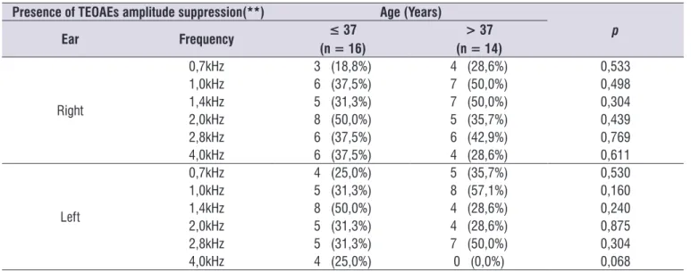
The distribution of the presence of TEOAEs suppression, by frequency and by ear, in individuals in the experimental group according to age (less than or equal to 37 and greater than 37 years) showed no signiicant differences using Mann-Whitney test (Table 3).
Table 3. Presence of TEOAEs suppression by frequency and by ear, according to the variable age (years), in the experimental group
(n=30).
Presence of TEOAEs amplitude suppression(**) Age (Years)
p ≤ 37
(n = 16)
> 37 (n = 14)
Ear Frequency
Right
0,7kHz 3 (18,8%) 4 (28,6%) 0,533
1,0kHz 6 (37,5%) 7 (50,0%) 0,498
1,4kHz 5 (31,3%) 7 (50,0%) 0,304
2,0kHz 8 (50,0%) 5 (35,7%) 0,439
2,8kHz 6 (37,5%) 6 (42,9%) 0,769
4,0kHz 6 (37,5%) 4 (28,6%) 0,611
Left
0,7kHz 4 (25,0%) 5 (35,7%) 0,530
1,0kHz 5 (31,3%) 8 (57,1%) 0,160
1,4kHz 8 (50,0%) 4 (28,6%) 0,240
2,0kHz 5 (31,3%) 4 (28,6%) 0,875
2,8kHz 5 (31,3%) 7 (50,0%) 0,304
4,0kHz 4 (25,0%) 0 (0,0%) 0,068
Mann-Whitney test
(**) It was considered that the suppression occurred when the value was positive (greater than or equal to 1 dB).
Legend: TEOAEs– Transient-evoked otoacoustic emissions n –number of individuals kHz – kilo Hertz
Table 4. Presence of TEOAEs suppression by frequency and by ear, according to the variable severity of tinnitus (mild, moderate and
severe) (n = 30).
Presence of TEOAEs suppression Severity of tinnitus
p
Ear Frequency Mild
(n = 4)
Moderate (n = 17)
Severe (n = 9)
Right
0,7 kHz 1 (25,0%) 4 (23,5%) 2 (22,2%) 0,994
1,0 kHz 1 ( 25,0%) 9 (52,9%) 3 (33,3%) 0,472
1,4 kHz 1 (25,0%) 8 (47,1%) 3 (33,3%) 0,649
2,0 kHz 0 ( 0,0%) 8 (47,1%) 5 (55,6%) 0,167
2,8 kHz 0 (0,0%) 9 (52,9%) 3 (33,3%) 0,143
4,0 kHz 0 (0,0%) 7 (41,2%) 3 (33,3%) 0,303
Left
0,7 kHz 0 (0,0%) 6 (35,3%) 3 (33,3%) 0,382
1,0 kHz 1 (25,0%) 9 (52,9%) 3 (33,3%) 0,472
1,4 kHz 1 (25,0%) 9 (52,9%) 2 (22,2%) 0,265
2,0 kHz 2 (50,0%) 5 (29,4%) 2 (22,2%) 0,610
2,8 kHz 1 (25,0%) 8 (47,1%) 3 (33,3%) 0,649
4,0 kHz 0 ( 0,0%) 2 (11,8%) 2 (22,2%) 0,542
Kruskal-Wallis test
(**) It was considered that the suppression occurred when the value was positive (greater than or equal to 1 dB).
The mean amplitude of TEOAEs suppression in this study ranged from 2.14 to 4.38 dB in the right ear and 2.17 to 4.15 dB in the left ear for the EG, and from 2.27 to 4.88 dB in the right ear and 2.44 to 4.57 dB in the left ear for the CG (Tables 1 and 2). Such values were higher than those found in the literature consulted (1.28 dB in the right ear and 1.25 dB in the left ear26 and 1.29 dB in the right ear and 1.26 dB in the left ear)4.
Therefore, despite the lack of a statistically signif-icant difference, the mean amplitude of TEOAEs suppression were lower in patients with tinnitus (EG) than in individuals without the symptom (CG), similarly to another study that also compared the suppression effect between groups9. Although the difference is not
statistically signiicant, this result suggests lower effec -tiveness of the medial efferent olivocochlear system concerning the EG7,13..
Numerical comparisons between studies are dificult, since there are methodological differences used to measure the suppression of TEOAEs, such as type and intensity of suppressive noise, intensity and polarity of the click, and the ear in which the masking was presented (contralateral/ipsilateral/bilateral) and the equipment used27.
This study did not show statistically signiicant differ -ences between the presence of suppression and age in the EG individuals (Table 3), although previous studies have demonstrated a reduction in the suppression effect according to age increases28, 29.
DISCUSSION
The study sample consisted mostly of females (76.7%). Some authors1,20 reported that women present a higher prevalence of tinnitus complaint. On the other hand, a national study21 did not identify differences between the sexes; moreover, it was characterized by an average age of young adults (37 years), similar to the study of Fernandes and Santos22, whose average age was 37.8 years.
In relation to the severity of tinnitus, it was evidenced similar results to the Brazilian academic literature in identifying the occurrence of moderate tinnitus in 86.65%20; 61.8%13 and 57%23 of cases. However, studies have reported that 72%1 and 53.2%24 of the population they have studied presented mild to moderate tinnitus. Perhaps the difference in values observed in these studies is due to the method used for data collection and analysis, as well as the difference between the populations studied.
Regarding the laterality of tinnitus, we veriied 56.65% of bilateral and 43.35% of unilateral complaints, being 23.3% in the right ear and 20% in the left ear, similarly to the studies that reported 70%; 67% and 60% of patients with bilateral tinnitus and 30%; 33% and 25% with unilateral tinnitus, respectively13,23,25. However, other authors have reported higher incidence of unilateral tinnitus (left ear) (65%) in individuals with normal pure tone audiometryl22.
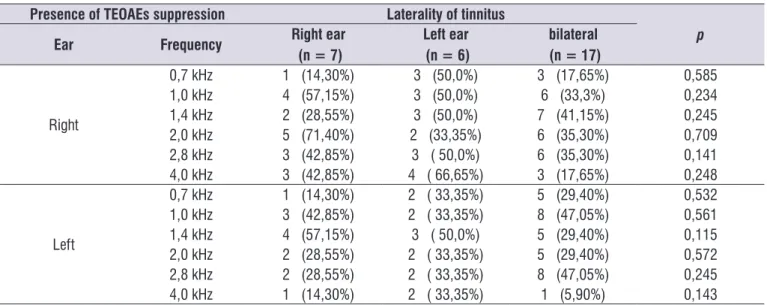
Table 5. Presence of TEOAEs suppression by frequency and by ear, according to the variable laterality of tinnitus (right unilateral, left
unilateral left or bilateral) (n = 30).
Presence of TEOAEs suppression Laterality of tinnitus
p
Right ear (n = 7)
Left ear (n = 6)
bilateral (n = 17)
Ear Frequency
Right
0,7 kHz 1 (14,30%) 3 (50,0%) 3 (17,65%) 0,585
1,0 kHz 4 (57,15%) 3 (50,0%) 6 (33,3%) 0,234
1,4 kHz 2 (28,55%) 3 (50,0%) 7 (41,15%) 0,245
2,0 kHz 5 (71,40%) 2 (33,35%) 6 (35,30%) 0,709
2,8 kHz 3 (42,85%) 3 ( 50,0%) 6 (35,30%) 0,141
4,0 kHz 3 (42,85%) 4 ( 66,65%) 3 (17,65%) 0,248
Left
0,7 kHz 1 (14,30%) 2 ( 33,35%) 5 (29,40%) 0,532
1,0 kHz 3 (42,85%) 2 ( 33,35%) 8 (47,05%) 0,561
1,4 kHz 4 (57,15%) 3 ( 50,0%) 5 (29,40%) 0,115
2,0 kHz 2 (28,55%) 2 ( 33,35%) 5 (29,40%) 0,572
2,8 kHz 2 (28,55%) 2 ( 33,35%) 8 (47,05%) 0,245
4,0 kHz 1 (14,30%) 2 ( 33,35%) 1 (5,90%) 0,143
Kruskal-Wallis test
(**) It was considered that the suppression occurred when the value was positive (greater than or equal to 1 dB).
REFERENCES
1. Pinto PC, Sanchez TG, Tomita S. Avaliação da relação entre severidade do zumbido e perda auditiva, sexo e idade do paciente. Braz J Otorhinolaryngol. 2010;76(1):18-24.
2. Sogebi AO. Characterization of tinnitus in Nigeria. Auris Nasus Larynx. 2013;40(4):356-60.
3. American Tinnitus Association. Information About Tinnitus. Portland, ATA, 1997.
4. Urnau D, Tochetto TM. Occurrence and suppression effect of otoacoustic emissions in normal hearing adults with tinnitus and hyperacusis. Braz. J. Otorhinolaryngol. 2012;78(1):87-94.
5. Han BIH, Lee HW, Kim TY, Lim JS, Shin KS. Tinnitus: characteristics, causes, mechanisms, and treatments. J Clin Neurol. 2009;5(1):11-9.
6. Fávero ML, Sanchez TG, Bento RF, Nascimento AF. Contralateral suppression of otoacoustic emission in patients with tinnitus. Rev. Bras. Otorrinolaringol. [Internet]. 2006 Apr[cited 2015 Aug 01];72(2):223-6. Available from: http://www.scielo. br/scielo.php?script=sci_arttext&pid=S0034-72992006000200012&lng=en. http://dx.doi. org/10.1590/S0034-72992006000200012.
7. Sztuka A, Pospiech L, Gawron W, Dudek K. DPOAE. In: Estimation of the function of the cochlea in tinnitus patients with normal hearing. Auris Nasus Larynx. 2010;37(1):55-60.
8. Chéry-Croze S, Collet L, Morgon A. Medial olivo-cochlear system and tinnitus. Acta Otolaryngol. (Stockh). 1993;113(3):285-90.
9. Paglialonga A, Del Bo L, Ravazzani P, Tognola G. Quantitative analysis of cochlear active mechanisms in tinnitus subjets with normal hearing sensitivity: multiparametric recording of evoked otoacustic emissions and contralateral suppression. Auris nasus Larynx. 2010;37(3):527-38.
10. Chéry-Croze S, Truy E, Morgon A. Contralateral suppression of transiently evoked otoacoustic emissions and tinnitus. Br J Audiol. 1994;28(4-5):255-66.
11. Geven LI, Kleine E, Free RH, Dijk PV. Contralateral suppression of otoacoustic emissions in tinnitus patients. Otol Neurotol. 2011;32(2):315-21.
12. Guinan Jr JJ, Backus BC, Lilaonitkul W, Aharonson V. Medial olivocochlear efferent relex in humans: otoacoustic emission (OAE) measurement issues and the advantages of stimulus frequency OAEs. J Assoc Res Otolaryngol. 2003;4(4):521-40.
There were also no differences in TEOAEs suppression according to the severity of tinnitus (Table 4). In the literature consulted, there were no studies that associate the presence of TEOAEs suppression to the variable severity of tinnitus in individuals with this symptom.
The association between laterality of tinnitus and the presence of TEOAEs suppression was also not signiicant (Table 5), in agreement with the interna -tional academic literature, a less eficient functioning of the MSOC in one ear does not necessarily imply that tinnitus is present in that ear, as some cases in which it is lateralized in the opposite side10. On the other hand, some studies have shown that the function of the MSOC is reduced in the tinnitus side4,6,23.
Therefore, although the results related to the suppression of TEOAEs were similar in individuals with and without tinnitus, we observed a tendency to less suppression effect in patients with tinnitus. Thereby, further studies may support the under-standing of the role of efferent pathways in cases of tinnitus and, especially, the assessment of the hypothesis of MSOC relation10. We suggest that these studies attempt to control confounding variables such as manual preference, since the efferent auditory system operates under lateral conditions, following the standards of hemispheric dominance and, thus, does not have the same suppression effects for the right and left ears in people right-handed and left-handed6 and with tolerance to noise, since hyperacusis, which accompanies many cases of tinnitus, may increase the suppression effect of otoacoustic emissions30.
In this context, even considering the inexistence of statistical signiicance, it is supposed the importance of the TEOAEs suppression for the tinnitus topographic diagnosis. However, it is necessary to standardize procedures and also normative values for clinical applicability.
CONCLUSION
temporomandibular. Braz J Otorhinolaryngol. 2012;78(2):59-65.
26. Aita ADC. Capacidade e autopercepção auditivas: um estudo em hiperacúsicos [tese]. São Paulo: Universidade Federal de São Paulo. Escola Paulista de Medicina; 2002.
27. Oliveira JR, Fernandes CF, Costa Filho AO. Study on suppression of otoacoustic emissions: lateral domain. Braz J Otorhinolaryngol. 2011; 77(5):547-54.
28. Parthasarathy TK. Aging and contralateral
suppression effects on transient evoked otoacoustic emissions. J Am Acad Audiol. 2001;12(2):80-5. 29. Yilmaz ST, Sennaroğlu G, Sennaroğlu L, Köse
SK. Effect of age on speech recognition in noise and on contralateral transient evoked otoacoustic emission suppression. J Laryngol Otol. 2007;121(11):1029-34.
30. Knudson IM, Shera CA, Melcher JR. Increased contralateral suppression of otoacoustic emissions indicates a hyperresponsive medial olivocochlear system in humans with tinnitusand hyperacusis. J Neurophysiol. 2014;112(12):3197-208.
13. Serra LSM. Estudo da supressão das emissões otoacústicas evocadas e a relação com o incômodo do zumbido em indivíduos com limiares auditivos normais [dissertação]. Brasília/DF: Universidade de Brasília, Faculdade de Ciências da Saúde; 2014. 14. Samelli AG. Hipóteses atuais sobre a geração
do zumbido. In: Samelli AG. Zumbido: avaliação diagnóstico e reabilitação – abordagens atuais. São Paulo. 2004. p.23-35.
15. Branco FCA. Zumbido em adultos ouvintes
normais: Um estudo sobre o processamento auditivo central e o handicap [Dissertação]. São Paulo: Pontifícia Universidade Católica de São Paulo (PUC); 1998.
16. Kemp DT, Ryan S, Bray P. A guide to the effective use of otoacoustic emissions. Ear Hear. 1990;11(2):93-105.
17. Ryan S, Kemp DT. The inluence of evoking stimulus level on the neural suppression of transient evoked otoacoustic emissions. Hear Res. 1996;94(1-2):140-7.
18. Hood LJ, Berlin CI, Hurley A, Cecola RP, Bell B. Contralateral suppression of transient-evoked otoacoustic emissions in human: intensity effects. Hear Res. 1996;101(1-2):113-8.
19. Valente JP, Pinheiro LA, Carvalho GM, Guimarães AC, Mezzalira R, Stoler G et al. Evaluation of factors related to the tinnitus disturbance. Int Tinnitus J. 2012;17(1):21-5.
20. Gomes SJV, Barbosa RM, Santos TMM. A
incidência de zumbido numa amostra aleatória na cidade de Salvador. Rev. CEFAC. 2004;6(1):94-100. 21. Mondelli MFCG, Rocha AB. Correlação entre
os achados audiológicos e incômodo com zumbido. Arq. Int. Otorrinolaringol. / Intl. Arch. Otorhinolaryngol. 2011;15(2)172-80.
22. Fernandes LC, Santos TMM dos. Tinnitus
and normal hearing: a study on the transiente otoacoustic emissions suppression. Braz. J Otorhinolaryngol. 2009;75(3):414-9.
23. Santos Filha VAV, Samelli AG, Matas CG. Noise-induced tinnitus: auditory evoked potential in symptomatic and asymptomatic patients. Clinics. 2014;69(7):487-90.
24. Gois RO, Gois BO, Pereira MCCS, Taguchi CK. Estado Mental e Impacto do Zumbido em Idosos. Rev. CEFAC. 2014;16(3):798-809.