Rev Bras Med Esporte _ Vol. 10, Nº 5 – Set/Out, 2004
367
1. Coordinator of the “Latus Senso” Trauma and Orthopedic PhysicalTher-apy Program of the Brazilian Rehabilitation Medicine Institute, IBMR – RJ – CREFITO 2/689-F).
2. PhD Professor, Coordinator of the “Strictus Senso” Motricity Science Program – Castelo Branco University, UCB-RJ 0066/1).
3. MsC Professor, Coordinator of the Scientific Initiation Program in Phys-ical Education – Brasilia Catholic University – UCB – CREF 0155/7). 4. MsC Professor, Coordinator of the Strength Study Laboratory – LABEF
– Physical Education Course - Brasilia Catholic University – UCB – CREF 0159/7).
Received in 12/5/04. 2nd version received in 16/9/04. Approved in 20/9/04. Correspondence to: Claudionor Delgado, Clínica de Fisioterapia Claudio-nor Delgado, Av. Princesa Isabel, 323, sl. 412 – Copacabana – 22011-010 – Rio de Janeiro, RJ, Brazil. E-mail: [email protected]
Use of the sphygmomanometer in the evaluation of the
knee joint flexor and extensor muscle strength in militaries
Claudionor Delgado1, José Fernandes Filho2, Fernando Policarpo Barbosa3 and Hildeamo Bonifácio Oliveira4
O
RIGINALA
RTICLEKey words: Isometric strength. Sphygmomanometer. Knee joint. ENGLISH VERSION
ABSTRACT
Bases and objective: This is a comparative and descriptive study that aims at analyzing the strength for the different angles of the knee flexion and extension in militaries. The objective of this study was to evaluate the extensor and flexor muscles of the knee joint for different angles by means of the Modified Sphygmomanome-ter (MS) in healthy militaries. Methods: The sample was composed of 31 militaries as follows: 19 male and 12 female with average age of 26.5 ± 5.8 years; respective average height of 162.00 ± 0.06 (cm) and 175.00 ± 0.06 (cm) and average body mass of 56.83 ± 5.85 (kg) and 73.25 ± 10.46 (kg). The evaluation methodology was the one proposed by Helewa, Goldsmith and Smithe (1981) using Modified Sphygmomanometer (MS). The maximal isometric contractions at 30o of flexion and 30o/90o of extension were
ob-tained in the Make test, in the Inbaf flexion-extension table and recorded by the MS Tycos. The data was analyzed using the “t” Student-test to compare the averages, and the significance level adopted was p > 0.05. Results: In both the female and the male groups, significant difference was only observed between angles of 30 and 90 degrees of the right knee extension (p > 0.05). At angles of 90 degrees for the knee extension and of 30 degrees for knee flexion, no intra-groups significant differences were observed (p > 0.05). Conclusion: Militaries present strength differences be-tween knee joint anterior and posterior muscular groups at the different angles studied. The methodology used showed to be sat-isfactory for the strength qualitative evaluation.
INTRODUCTION
The evaluation of the muscular strength has been objective of study in different knowledge areas. It can be verified in the litera-ture that different subjective (perimetry and manual muscular test) and objective methods (portable dynamometer and isokinetic dy-namometer) have been used to measure this physical valence (board 1). In the history of physical therapy, the importance of the strength evaluation can be verified in the rehabilitation process of the body segments(1-3).
The hypotrophy and the strength unbalance between the ago-nistic and the antagoago-nistic musculature are factors that may influ-ence the muscular dysfunctions and alter the articular stability, lead-ing to new lesions. Thus, it has become priority for physical therapists to perform evaluations that enable prophylactic actions as well as the evolutive treatment of the articular lesions. Howev-er, there are controversies regarding to the application and validity of the methods employed, once the perimeter asymmetry does
not necessarily indicate strength asymmetry(1,4-8), the manual
mus-cular test presents a reliability of only 60-65%(2,6,9,10), the portable
dynamometer is not yet regularly manufactured in Brazil and
pre-sents different readings according to the manufacturer(3) and the
high-reliability isokinetic dynamometer has as primary limiting
fac-tor its high cost and the necessity of adequate physical space(11)
(board 2).
BOARD 1
Methods of muscular strength evaluation
Subjective methods Objective methods
1. Perimetry 1. Portable dynamometer 2. Manual muscular test (MMT) 2. Isokinetic dynamometer
BOARD 2
Summarized discussion on the methods of muscular strength evaluation: limitations, feasibility, reliability
Method Discussion
1. Perimetry The thigh perimeter asymmetry is frequently related to the torque decrease. The association between perimeter and torque is discussed. The perimeter asymmetry does not indicate strength asymmetry(1,4,5,6,7,10).
2. Muscular test Main muscular test for many decades. Beasley, for over than 30 years, already supported the application of more objective tests. The MMT may provide a submaximal re-sponse if the patient’s strength exceeds the physical therapist’s. William reported that the reliability of this test is of only 60 to 65%(2,6,9,11).
3. Portable Although having limitations, its application has been sup-dynamometer ported in the last decade. Not yet regularly manufactured in Brazil and with high cost, it presents maintenance diffi-culties and different readings according to model and manufacturer; these are factors that hinder its utilization in the evaluation routine(3).
4. Isokinetic This device has aided to overcome some difficulties in dynamometer the muscular tests. Presenting good reliability, has as pri-mary limiting factor high cost and the necessity of proper physical space(8).
368
Rev Bras Med Esporte _ Vol. 10, Nº 5 – Set/Out, 2004Thus, it was verified the necessity of a method of easy applica-bility and low cost for the strength evaluation with result reliabili-ty(12,13). Among the methods presented by literature, the method
proposed by Helewa et al.(14) has demonstrated that the
applica-tion of the Modified Sphygmomanometer (MS) was more sensible in the muscular evaluation than the methodology that uses free weights. They observed that the strength measure methodology with the MS provided qualitative and objective measures more sensible to the different strength standards. These authors con-cluded that the method presented good reproducibility when the results obtained by different appraisers were observed. Fernando
and Robertson(15) showed a difference of less than 2% between
the measures obtained by different appraisers using MS in the
manual pressure strength test. Helewa et al.(14) assured that the
MS yet presented good security level, being able to be applied in at least twenty-four muscular groups.
In the specialized literature, reports of two types of muscular test in which the MS may be used are found(12,16,17): a) Break Test –
It is a manual test where the MS is placed between the segment of the appraised and the hand of the appraiser, the appraiser’s strength overcomes the maximal muscular strength of the ap-praised and b) Make Test – it is a mechanical test where the MS is placed between the segment of the appraised and an object or a stationary device with appraised performing maximal isometric strength.
This study is justified by the possibility of providing a practical method for the muscular strength evaluation. The body segments, objects of this study, are the lower limbs, more specifically the knee, joint with specific stability features, function and importance that presents high incidence of lesions and dysfunctions especial-ly due to the deficiencies in the periarticular musculature mentioned above, responsible for its dynamic stabilization(18-21).
The objective of this study is to use the MS for the knee flexor and extensor muscle strength evaluation with the application of the Make Test for angles of 30/90 degrees and 30 degrees respec-tively in adult individuals, apparently healthy.
MATERIAL AND METHODS Methodology
This study presents a descriptive-comparative approach(22); the
evaluation methodology proposed by Helewa et al.(14) was used in
militaries from both genders and ages ranging from 19 and 31 years, apparently healthy.
Sample
The sample of the present study was intentionally, composed of 31 militaries from both genders distributed in 19 women and 12 men with ages ranging from 19 and 31 years and with an average of 26.5 ± 5.8 years. The volunteers had no knee lesions or anatom-ical alterations. All participants were informed about the risks in-volved in the experiment, being invited to fill and to sign a consent form, according to Brazilian law number 196/96. Data regarding to age, gender, height, body mass, physical activity practice level with regular practice and dominance side were also collected.
Material
Modified Sphygmomanometer – – – The device used to assess – – blood pressure had part of its Velcro tape removed. The inflatable bag was folded in three equal parts and fixed inside an inelastic
bag. In this prototype, the sphygmomanometer label Tycos® was
used. After modification performed, the device presented the fol-lowing dimensions: 9 cm width, 14 cm length, 2.5 cm thickness and the aerial tubes presented 48 cm extension (figure 1). The unit
was inflated and applied to the positions standardized by Reese(1),
Daniels and Worthingham(9) for the test of the muscles
investigat-ed. We employed the Make Test, which stabilization is mechani-cal, to avoid measurement errors.
“Inbaf”flexion-extension table – The equipment was set to its maximal load of 80 kg and for some volunteers who achieved dislocating this load, a manual stabilization strength performed by the appraiser at the device’s arm crowbar extremity was added. The following positions for the strength test in the Inbaf
equip-ment were adopted: extension from 90o and flexion from 30o.
Procedures
Before performing the strength test, the volunteer was informed of how to proceed, also being requested to perform the desired movement one single time in each segment for apprenticeship, before the MS was positioned and the strength test was performed. The MS was positioned at the distal extremity between the leg and the support point of the leg with the device’s arm crowbar. The test was composed of three repetitions in each limb for each position adopted and the volunteer was induced to perform maxi-mal effort through verbal stimuli and encouragement.
Calculation of the general values obtained – – – The test’s final – – values for each position adopted previously described were calcu-lated through the arithmetic average of the value obtained in each one of the two positions adopted.
Example: If, in the extension from 90o, the volunteer reached
values of 58, 50 and 52 for the right lower limb, the arithmetic average would be 53.3.
Calculation of the asymmetry percentile – – – – – Comparative cal-culations were performed to establish the right and left thigh
an-tagonistic-agonistic asymmetry percentage at positions of 90o of
extension and 30o of flexion. For each position, the highest
arith-metic average was subtracted by the lowest aritharith-metic average and the result obtained from the subtraction (X) was divided by the highest arithmetic average (B) and this result (Y) multiplied by 100, thus finding the specific percentile for each case.
B – A = X X/B = Y.100 = %
With the objective of making this evaluation method simpler and feasible in the daily clinics as evaluation control and treatment evo-lution, no correlation between pressure (mmHg) and strength (N) was performed. The values obtained were only used as reference unit in order to establish the asymmetry index.
Statistics
For data analysis, the descriptive analysis and the “t” Student test was used to compare the averages. The statistical significance level adopted was p > 0.05. To do so, the “Statistical Package for the Social Sciences” (SPSS 11.0) was used.
RESULTS
Sample characterization
Rev Bras Med Esporte _ Vol. 10, Nº 5 – Set/Out, 2004
369
age height of 175.00 ± 0.06 (cm) and body mass of 73.25 ± 10.46 was observed.
With regards to the regular practice of physical exercise, it was verified that from the total number of individuals, 54.8% did prac-tice regular physical exercise while 45.2% did not.
The average values and standard deviation (SD) for the angle analysis studied in the knee flexion and extension are presented in table 1.
In women (n = 19)
Right knee – In 78.9% of cases, the extensor group presented strength predominance in relation to the flexor group (ischiotibial), with average percentile index of 26% (figure 2). In 21.1% of cas-es, the flexor group presented strength predominance in relation to the extensor group with average percentile index of 9.5%.
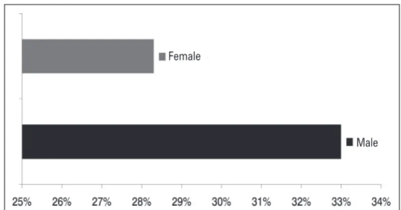
Left knee – In 73.7% of cases, the extensor group presented strength predominance in relation to the flexor group (ischiotibial) with average percentile index of 28.3% (figure 3). In 26.3% of cas-es, the flexor group presented strength predominance in relation to the extensor group with average percentile index of 9.2%.
The strength predominance of the extensor muscle group may be observed in 78% of men and 76.3% of women, with an asym-metry average index in relation to the flexion of 26.4% in the right knee and 33% in the left knee in men and 26% in the right knee and 28.3 in the left knee in women.
The flexors presented predominance in relation to the exten-sors in 21.9% of men and 23.7% of women, with an asymmetry average index of 18.8% in the right knee and 6% in the left, in men. In this group, which a large asymmetry difference between segments was observed, one may speculate that this fact might have occurred due to the type of physical activity practiced by the group. In women, the asymmetry index was of 9.5% in the right knee and 9.2 in the left knee.
Through the results obtained, one cannot corroborate the rela-tion between manual dominance and strength dominance in the lower limbs. This relation was observed in 31.5% of the total of individuals tested, which majority was right-handed, however, it was observed that the strength asymmetry average index between flexors and extensors was higher in the left knee, maybe due to the fact that it is not the support segment and the musculature is not properly used.
The data from the statistical analysis demonstrated that for knee extension in the female group, a significant difference between angles of 30o and 90o was observed in the right leg (p > 0.05) while
for the left leg, no significant difference was observed (p > 0.05); and the same behavior was observed for the male group.
For the Extension 30o x Flexion 30o in the female group, a
signif-icant difference was observed (p < 0.05) for both segments. The male group presented no significant difference (p > 0.05) in the same case observed.
In the Extension 90o x Flexion 30o analysis, both female and male
groups presented no significant difference for both segments (p < 0.05).
When the Extension 30o x Extension 90o of the right leg is
com-pared, a significant difference for both groups was observed (p < 0.05). In the left leg, no significant differences were observed be-tween groups (p > 0.05).
In the comparison of Flexion 30o x 30o, according to the analysis
of results, no significant differences were observed (p > 0.05) for segments both in the female group and for the male group.
The strength asymmetry average percentile index between the knee flexors and extensors (ischiotibial/quadriceps) found with the use of methodology described was:
In men (n = 12)
Right knee – In 83.3% of cases, the extensor group (quadriceps) presented strength predominance in relation to the flexor group (ischiotibial), with an average percentile index of 26.4% (figure 2). In 16.7% of cases, the flexor group presented strength predomi-nance in relation to the extensor group with average percentile index of 18.8%.
Left knee – In 66.6% of cases, the extensor group presented strength predominance in relation to the flexor group (ischiotibial) with an average percentile index of 33% (figure 3). In 26% of cas-es, the flexor group presented strength predominance in relation to the extensor group with average percentile index of 6%, and in one case, the extension strength was equivalent to the flexion strength.
TABLE 1
Descriptive values of the knee extension and flexion strength test in men and women for angles of 30/90 and 30 degrees
Variable Women Men
Extension Average ± SD Average ± SD
Right Left Right Left
knee knee knee knee
30o 73.76 ± 21.96* 75.16 ± 19.31* 085.46 ± 20.74 090.64 ± 22.03
90o 83.37 ± 19.84* 85.96 ± 13.35* 109.15 ± 22.04* 110.35 ± 35.87*
Flexion Average ± SD Average ± SD
Right Left Right Left
knee knee knee knee
30o 66.44 ± 16.28* 65.14 ± 16,90* 81.55 ± 12.50 76.91 ± 70.93
* (p < 0.05)
No correlation between pressure (mmHg) and strength (N) was performed. The values obtained were only used as reference unit in order to establish the asymmetry index.
Fig. 2 – Right knee. Strength asymmetry average percentile of the domi-nant extensor group (n = 25).
Female
Male
Fig. 3 – Left knee. Strength asymmetry average percentile of the dominant extensor group (n = 22).
Female
370
Rev Bras Med Esporte _ Vol. 10, Nº 5 – Set/Out, 2004DISCUSSION
We can emphasize as limiting factors for this study: 1) the num-ber of individuals evaluated and 2) in function of the strength lev-els variation observed, it is not possible to extrapolate the results in such way to create normative values.
The strength asymmetry between agonistic and antagonistic has already been object of discussion in some studies. The study per-formed by Safran et al.(23) affirms that athletes with strength
differ-ences of 60% in one leg when quadriceps and ischiotibial are com-pared have high chances of suffering a muscular lesion. Heiser et
al.(24) showed that a team of athletes presented an incidence of
7.7% of ischiotibial lesions with a recurrence rate of 31.7%. How-ever, after the muscular unbalance was recognized and corrected, the lesions incidence dropped down to 1.1%.
In function of these studies, one may suggest that the same could occur to non-athlete individuals, as in the sample investigat-ed in this study. This strength inequality between the knee joint agonistic and antagonistic musculature is favorable when we
ob-serve Heyward(25), who presents the studies of Golding, Meyers
and Sinning (1989), where the authors suggest a higher strength normal difference of the thigh anterior portion in relation to the
posterior portion of around 25%. Other authors(26-28) suggest that
this difference is of the order of 30 to 40%.
In the present study, part of the sample presented behavior sim-ilar to results found in literature(25). The order which the tests were
performed is also pointed as limiting factor in this study, once the agonistic/antagonistic action may have influenced on the loss of strength at maximal effort in function of the resistance generated
by the antagonistic, factor also known as Lombard(29,30) paradox.
Among other factors that may be considered as limiting factors, we emphasize the percentage of individuals who did not perform regular physical exercises or which the physical fitness level was
not measured. The effectiveness of the intramuscular coordina-tion and the coordinacoordina-tion between muscles is related to this exer-cise practice(31). Such factors are associated to the high incidence
of lesions in the thigh posterior muscular group(24,26-30,32-34). Thus, it
is observed that the posterior musculature must not present strength values close or similar to the anterior musculature.
The results obtained in this study demonstrated that the most participants presented strength equivalence suggested for the pro-phylaxis of muscular lesions.
We know that the pressure is proportional to the contact area and that this factor may have influenced the results when we es-tablished an asymmetry normal index, fact that requires further investigations.
CONCLUSION
Militaries present strength differences between anterior and posterior muscular groups of the knee joint at the different angles studied.
The use of the MS as a low-cost practical method for the strength evaluation between knee flexors and extensors was emphasized, being applicable as a comparison parameter when the prophylaxis of muscular lesions or the monitoring of a knee surgery recovery is the objective.
It is suggested that other studies be conducted with the objec-tive of investigating the strength relation obtained between the MS method and values standardized by isometric tests at the dif-ferent angles for the knee flexion and extension.
All the authors declared there is not any potential conflict of inter-ests regarding this article.
REFERENCES
1. Reese NB. Testes de função muscular e sensorial. Rio de Janeiro: Guanabara-Koogan, 2000.
2. Kendall FP, McCreary EK, Provance PG. Muscles – Testing and function. 4th ed.
Baltimore: Williams & Wilkins, 1993.
3. Williams M. Manual muscle testing – Development and current use. New York: World Confederation for Physical Therapy, 1956.
4. Allison S, Westpmol K, Finstuen K. Knee extension and flexion torque as func-tion of thigh asymmetry. JOSPT 1993;6:661-6.
5. Beasley WC. Influence of method on estimate of normal knee extensor force among normal and postpolio children. Physical Therapy Review 1956;86:21-44. 6. Bohannon RW, Lusardi MM. Modified sphygmomanometer versus strain gauge
hand-held dynamometer. Phys Ther 1991;72:911-4.
7. Brunnstrom S. Cinesiologia clínica. 4a ed. São Paulo: Manole, 1989.
11. Cooper M. Use and misuse of the tape measure as a mean of assessing muscle strength and power. Rheum and Rehab 1981;20:211-8.
9. Daniels L, Worthingham C. Provas de função muscular. 5a ed. Rio de Janeiro:
Guanabara, 1987.
8. Delorme TL. Restoration of muscle power by heavy resistance exercises. J Bone Joint Surg 1945;27:645-67.
10. Griffin JW, McClure MH, Bertorini TE. Sequential isometric and manual muscle testing in patients with neuro muscular disease. Phys Ther 1986;66:32-5. 12. Isherwood L, Lew L, Dean E. Indirect evidence for eccentric muscle contraction
during isometric muscle testing performed with a modified sphygmomanome-ter. Physiot Canada 1989;41:138-42.
13. Rice CL, Cunninghan DA, Peterson DH, Rechnitzer PA. Strength in an elderly population. Arch Phys Med Rehabil 1989;70:391-7.
14. Helewa A, Goldsmith CH, Smithe M. The modified sphygmomanometer – an instrument to measure muscle strength, a validation study. J Clin Epidemiol 1981;34:353-61.
15. Fernando UM, Robertson JC. Grip strength in the healthy. Rheum and Rehab 1982;21:179-81.
16. Smidt GL, Rogers MW. Factors contributing to regulation and clinical assess-ment of muscular strength. Phys Ther 1982;9:1283-90.
17. Bohannom RW. Make tests and break tests of elbow flexor muscle strength. Phys Ther 1988;2:193-4.
18. Kapandji AI. Fisiologia articular. 5a ed. São Paulo: Panamericana, 2000.
19. Podesta L, Magnusson J, Gillette T. Reconstrução do ligamento cruzado ante-rior. In: Maxey L, Magnusson J, editores. Reabilitação pós-cirúrgica para o pa-ciente ortopédico. Rio de Janeiro: Guanabara-Koogan, 2003;200-20.
20. Weber MD, Ware N. Reabilitação do joelho. In: Andrews JR, Harrelson GL, Wilk KE, editores. Reabilitação física das lesões desportivas. Rio de Janeiro: Guana-bara-Koogan, 2000; 235-94.
21. Wallace LA, Mangine RE, Malone TR. Joelho. In: Malone TR, McPoil TG, Nitz AJ, editores. Fisioterapia em ortopedia e medicina no esporte. 3a ed. São Paulo:
Santos, 2000;295-326.
22. Thomas JR, Nelson JK. Métodos de pesquisa em atividade física. Porto Alegre: Artmed, 2002.
23. Safran M, Seaber A, Garnett W. Warm-up and muscular injury prevention an update. Sports Med 1989;4:239-49.
24. Heiser TM, Weber J, Sullivan G, Clare P, Jacobs RR. Prophylaxis and manage-ment of hamstrings muscle injuries in intercollegiate football players. Am J Sports Med 1984;12:368-70.
25. Heyward VH. Advanced fitness – Assessment & exercise prescription. 3rd ed.
New York: Human Kinetics, 1998.
26. Arnheim DH, Prentice WE. Modern principles of athletic training. 2nd ed. St.
Louis: Mosby, 1993.
27. Agre JC. Hamstrings injuries: proposed etiological factors, prevention, treatment. Sports Med 1985;2:21-33.
28. Restron P, Kannus P, editors. Endurance in sport. London: Blackwell, 1992. 29. Distefano V. Functional anatomy and biomechanics of the knee. Athl Train 1978;
13:113-8.
30. Simão R. Fundamentos fisiológicos para o treinamento de força e potência. São Paulo: Phorte Editora, 2003.
31. Flek SJ, Kraemer WJ. Fundamentos do treinamento de força muscular. 2a ed.
Porto Alegre: Artes Médicas, 1999.
32. Liemhon W. Factors related to hamstrings strains. J Sports Med Phys Fitness 1978;18:71-75.
33. Fahey TD. Athletic training: principles and practice. Palo Alto: Mayfield, 1986. 34. Gallaspy JB. Reabilitação dos músculos isquiotibiais, quadríceps e virilha. In: