Heterochromatin Plasticity in
Saccharomyces cerevisiae
Bo O. Zhou1, Shan-Shan Wang1, Yang Zhang1, Xiao-Hong Fu1, Wei Dang1, Brian A. Lenzmeier2, Jin-Qiu Zhou1*
1State Key Laboratory of Molecular Biology, Institute of Biochemistry and Cell Biology, Shanghai Institutes for Biological Sciences, Chinese Academy of Sciences, Shanghai, China,2School of Science, Buena Vista University, Storm Lake, Iowa, United States of America
Abstract
Recent studies have established that the highly condensed and transcriptionally silent heterochromatic domains in budding yeast are virtually dynamic structures. The underlying mechanisms for heterochromatin dynamics, however, remain obscure. In this study, we show that histones are dynamically acetylated on H4K12 at telomeric heterochromatin, and this acetylation regulates several of the dynamic telomere properties. Using ade novoheterochromatin formation assay, we surprisingly found that acetylated H4K12 survived the formation of telomeric heterochromatin. Consistently, the histone acetyltransferase complex NuA4 bound to silenced telomeric regions and acetylated H4K12. H4K12 acetylation prevented the over-accumulation of Sir proteins at telomeric heterochromatin and elimination of this acetylation caused defects in multiple telomere-related processes, including transcription, telomere replication, and recombination. Together, these data shed light on a potential histone acetylation mark within telomeric heterochromatin that contributes to telomere plasticity.
Citation:Zhou BO, Wang S-S, Zhang Y, Fu X-H, Dang W, et al. (2011) Histone H4 Lysine 12 Acetylation Regulates Telomeric Heterochromatin Plasticity in
Saccharomyces cerevisiae. PLoS Genet 7(1): e1001272. doi:10.1371/journal.pgen.1001272
Editor:Hiten D. Madhani, University of California San Francisco, United States of America
ReceivedJune 10, 2010;AcceptedDecember 8, 2010;PublishedJanuary 13, 2011
Copyright:ß2011 Zhou et al. This is an open-access article distributed under the terms of the Creative Commons Attribution License, which permits unrestricted use, distribution, and reproduction in any medium, provided the original author and source are credited.
Funding:This work is supported by grants from Ministry of Science and Technology of China (2005CB522402) and National Natural Science Foundation of China (90919027). The funders had no role in study design, data collection and analysis, decision to publish, or preparation of the manuscript.
Competing Interests:The authors have declared that no competing interests exist. * E-mail: jqzhou@sibs.ac.cn
Introduction
Saccharomyces cerevisiaetelomeres and mating type loci (HMRand
HML) are well-characterized transcriptionally silenced domains. Heterochromatin tends to initiate at a specific DNA element and propagates along a chromatin fiber to repress the expression of nearby genes [1]. The major structural components of hetero-chromatin in yeast are known as Sir (silent information regulators) proteins, including Sir2, Sir3 and Sir4 [1]. For telomeric heterochromatin, the double-stranded telomeric DNA-binding protein Rap1 interacts with Sir4 and thereby recruits the whole Sir complex to the telomeres [2]. Once recruited to telomeres, Sir2 deacetylates a critical K16 acetyl mark on histone H4 [3], a process required for Sir proteins to bind throughout the subtelomeric regions of ,3 kb proximal to the terminal ,
350-bp TG-tracts [4–8].
The spreading of heterochromatin must be restricted by boundaries between silent and active chromatin [9]. Since histone H4 amino acid residues 16 to 29 are primarily required for Sir3 binding [4], histone H4K16 acetylation dominantly prevents Sir-mediated heterochromatin spreading [6,7]. Genetic and biochemical data also show that unacetylated H4 K5, 8, 12 together can substitute for the mutation of H4K16 in Sir3 binding [4,5]. Therefore, it has been generally believed that the acetylation of histone H4 at K5, 8 and 12, catalyzed by NuA4 complex [6,10], cumulatively antago-nizes heterochromatin spreading. However, it remains unclear whether H4 K5, 8 and 12 contribute equally to this process.
Yeast heterochromatin possesses the ability to exchange chromatin-bound Sir3 for soluble unbound protein throughout
the cell cycle [11]. In addition, heterochromatin is surprisingly permissive to activators, co-activators and transcriptional pre-initiation-complex (PIC) as well [12–14]. Moreover, heterochro-matin doesn’t prohibit active base-pair substitutions [15]. These lines of evidence have revealed the dynamic aspect of silent- and hetero-chromatin. One outstanding question of concern is what contributes to the dynamics of heterochromatin. Accordingly, it would be beneficial to know if altering the dynamics of heterochromatin structure has biological consequences.
In this study, we show that the heterochromatin in yeast harbors a dynamic H4K12 acetylation mark which suppresses Sir-mediated aberrant condensation of telomeric heterochromatin and promotes telomere-related processes, including telomere transcription, replication and recombination. Thus, histone acetylation may provide a scheme for yeast cells to maintain partially flexible telomeric heterochromatin that allows for normal changes in DNA metabolism.
Results
A distinct system for studyingde novotelomeric heterochromatin formation in yeast
was not affected. Technically, we used a galactose-inducible HO endonuclease to cut a chromosome to expose a pre-inserted 81-bp telomeric ‘‘seed’’ for further telomere addition [16] (Figure 1A). Chromosome ‘‘healing’’ was hardly observed in the first 6 hours upon HO induction. Later, the telomere seed was gradually extended in a telomerase dependent manner ([16] and Figure 1B). The recruitment of Rap1 and Sir proteins onto regions 100-bp to 5.0-kb from the telomere seed upon HO induction was then monitored by chromatin immunoprecipitation (ChIP). High occupancy of Rap1 was observed at the 81-bp telomere seed prior to HO induction (Figure 1C), indicating that the 81-bp telomeric DNA tract is sufficient for Rap1 recruitment. Upon HO induction, the Rap1 occupancy at the newly formed telomere was barely altered in the first 6 hours and then gradually increased along with telomere elongation (Figure 1B and 1C). Notably, upon HO induction, Sir proteins were recruited to the new telomere within the first 6 hours in an extremely efficient manner (Figure 1D–1F). Once occupying a newly formed telomere, Sir proteins began to spread into the lateral regions up to 3 kb proximal to the newly formed telomeres (Figure 1D–1F). Consistently, the expression of ADE2, a gene near the telomere seed, was gradually reduced upon HO induction (Figure 1G). Therefore, telomeric heterochromatin was quickly assembled at the 81-bp telomeric seed after HO induction.
Histone H4K5 and K12 acetylation partially survived the formation of telomeric heterochromatin
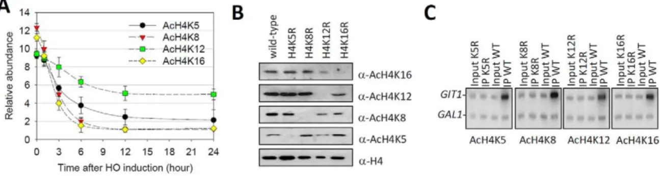
Since histone H4 hypoacetylation was required for the onset of telomere silencing [1,4,5] and K16 seemed to be the unique deacetylation target for Sir2 on H4 [3], we were curious about the deacetylation kinetics of different lysines on H4 (K5, K8, K12 and K16) during heterochromatin formation. To avoid recombination-induced histone acetylation, we deleted the RAD52gene in our strains [17]. The result showed that, prior to HO cleavage, all lysine acetylation was robust at the subtelomeric region (indicated asSubTelin Figure 1) (Figure 2A). Upon HO induction, H4K8 and K16 acetylation levels decreased most substantially and were largely eliminated by 6 hours (Figure 2A), a time point when Sir proteins saturatedSubTel(Figure 1D–1F). In contrast, H4K5 and K12 acetylation, especially K12 acetylation, decayed more slowly atSubTelduring HO induction, and were still detectable after 24-hour HO induction (Figure 2A). These observations raise a possibility that H4K5 and K12 acetylation may partially survive the formation of new telomeric heterochromatin.
To exclude the possibility that anti-acetyl-lysine antibodies cross-reacted with other subtelomeric factors, we compared the levels of H4 lysine acetylation in a wild-type strain to that of ‘‘unacetylated strains’’ where the corresponding lysines were mutated to arginines, respectively, thereby preventing anti-acetyl lysine antibodies from recognizing corresponding residues. In the western-blot experiment, we found that chromatin derived from strains with the lysine to arginine mutation was not detected by the corresponding anti-acetyl lysine antibodies (Figure 2B). In the ChIP experiment, we found that in the wild-type strain, all antibodies enrichedGIT1, a typical hyperacetylated region [18,19] (Figure 2C). In contrast, lysine to arginine mutations eliminated corresponding anti-acetyl lysine antibodies from enriching any chromatin fragments (Figure 2C). Additionally, increasing the amount of anti-acetyl-H4K12 antibody in the immunoprecipita-tion (IP) procedure couldn’t recover chromatin in the H4K12R strain (Figure S1). These data are consistent with previous report [20], and indicate that all anti-acetyl-H4 lysine antibodies perform appropriately in our ChIP experiments.
Histone H4K12 is modestly acetylated at telomeric heterochromatin
To determine if the acetylation of H4K5 and K12 was generally associated with native telomeres that were not undergoing a double-strand break response [21], we performed ChIP to examine the acetylation profile of H4 lysines at all yeast telomeres. Sir2 binding was firstly mapped (Figure 3, pink bars) so as to mark the silencing status of the individual telomeres. We found that the abundance of Sir2 at different telomeres varied (Figure 3). Low amounts of Sir2 were mostly observed at telomeres that contain Y’ elements (http://www.yeastgenome.org/ and Figure 3), support-ing an anti-silencsupport-ing role for these Y’ elements [22]. The acetylation levels at subtelomeric regions were normalized to an
HMRregion, where we detected lowest level of acetylation (Figure S3, region D). Figure 3 showed that H4K12 acetylation was observed at nearly all telomeres in a manner that was independent of Sir2 binding. In contrast, the relative amount of H4K5, K8 or K16 acetylation at telomeres seemed to correlate in an opposing manner to the level of Sir2 binding (Figure 3). For example, at the end of left arm of chromosome II where there was little Sir2 binding and all lysine acetylations were robust. In contrast, at the end of right arm of chromosome II where there was abundant Sir2, only H4K12 acetylation was detectable (Figure 3). Essentially identical results were also obtained when yeast were grown in galactose culture (Figure S2). Furthermore, H4K12 acetylation was also detected at some regions of yeastHMheterochromatin (Figure S3). Taken together, these data suggest that H4K12 acetylation exists within yeast heterochromatin.
Esa1 is responsible for H4K12 acetylation at telomeric heterochromatin
We next set out to ascertain how the subtelomeric H4K12 acetylation pattern was established. Genome-wide nucleosome H4K5, K8 and K12 acetylation in budding yeast is catalyzed by Piccolo NuA4 in a non-targeted manner [23]. We found that the subtelomeric H4K12 acetylation was sensitive to esa1-338
mutation (Figure 3, white bars). Since Esa1 is the catalytic subunit of NuA4 complex, these results support the idea that NuA4 is responsible for heterochromatic H4K12 acetylation. Interestingly, ChIP analysis revealed that Esa1 was specifically enriched at Sir2-abundant subtelomeres (Figure 3, red bars). Thus, heterochro-matic H4K12 was acetylated by NuA4 in a targeted manner while the euchromatic H4K12, as the case of other H4 lysines, was
Author Summary
Figure 1.De novo telomeric heterochromatin assembly on a short telomere.(A) Schematic representation of the de novotelomeric heterochromatin formation system [16]. TheADH4locus was replaced in a haploid yeast strain with a 6-kb fragment consisting of theADE2selection marker, 81-bp telomeric DNA sequence and the recognition site for the HO endonuclease. TheLYS2gene was placed approximately 10 kb from the natural telomere VII-L. HO was induced by the addition of galactose to the media.ADE2was a gene adjacent to the telomere seed. The black line indicated the primer set (SubTel) used for ChIP analysis. (B)De novotelomere addition analysis [16]. After HO induction, yeast cells were harvested at time points as indicated. Genomic DNA was isolated, digested withBstXI and subjected to Southern-blot analysis. The blots were hybrid with a telomere seed-proximal probe. (C–F) ChIP analyses of Rap1 (C), Sir2-myc (D), Sir3-myc (E) and Sir4-myc (F) in the time course of HO induction. Shown were the real-time Q-PCR results of the ChIP products. X axis indicated the distance from the 81-bp telomere seed. Y axis indicated the timed intervals after HO induction. Z axis indicated the enrichment values (IP/Input) at indicated regions relative toACT1. Values that were greater than 1 indicated more enrichment at indicated regions than background. (G) mRNA analysis ofADE2upon HO induction. The mRNA level ofADE2was normalized by
Figure 2. Histone H4K12 acetylation survives heterochromatin formation. (A) ChIP analyses of H4K5 acetylation, K8 acetylation, K12 acetylation and K16 acetylation in the time course of HO induction. Shown were the real-time Q-PCR results of the ChIP products. Error bars represented standard error of the mean for three independent experiments. The relative abundance indicated the enrichment value (IP/Input) atSubTelrelative to
HMR. Values that were greater than 1 indicated more enrichment at theSubTelthan background (HMR). (B) Whole-cell extracts from NSY429 strains expressing wild-type histone H4 (lane 1) and arginine substitution mutations of H4 Lys5 (lane 2), Lys8 (lane 3), Lys12 (lane 4) or Lys16 (lane 5) were subjected to western blot analysis with the specific antibodies indicated on the right. (C) ChIP assays were performed as in (A) except that chromatin from strains that contained either wild-type or H4 lysine substituted mutants was used. Q-PCR with32P-ATP incorporation was done with primer pairs
directed againstGIT1andGALregions. PCR products were separated by 6% TBE gel and product abundance was detected using a PhosphorImager. doi:10.1371/journal.pgen.1001272.g002
Figure 3. Acetylation of H4K12 at native telomeric heterochromatin. Relative abundance of H4K5 acetylation, K8 acetylation, K12 acetylation, K16 acetylation, histone H4, Sir2-myc and Esa1-myc at 32 telomeres. Real-time PCR was done with primer pairs against regions,0.5 kb
from each telomere. PCR data for acetylations were normalized to anHMRregion (region D in Figure S3); PCR data for Sir2-myc were normalized to
ACT1; PCR data for Esa1-myc were normalized to a subtelomeric region,4 kb from the end of chromosome III-R (Stel). The relative abundance of
acetylated by Piccolo NuA4 in a non-targeted manner [23]. We found that the binding of Esa1 at subtelomeric region was restricted to the distal region because there was no Esa1 binding in the subtelomeric domain 3.2 kb,7.6 kb from the end of
chromosome XIV-R (Figure S4A and S4B).
A previous study showed that Hat1, another lysine acetyl-transferase in yeast, also possesses H4K12 acetylation activity [24], however it is predominantly localized to the cytoplasm and thought to specifically acetylate free histone H4 [25]. Western-blot results showed thatHAT1deletion did not affect chromatic H4K12 acetylation (Figure S4D). ChIP analyses revealed that the H4K12 acetylation level at the subtelomeric region of ChrXIV-R was not reduced, but rather modestly increased in
hat1Dcells (Figure S4E). Therefore, these data suggest that Hat1 is likely not needed for dynamic H4K12 acetylation at telomeric heterochromatin.
Histone H4K12 acetylation regulates telomere replication
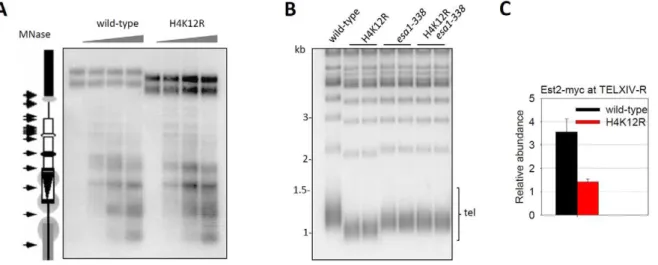
Histone acetylation is thought to regulate chromatin assembly [26]. To address whether H4K12R mutation affects subtelomeric nucleosomal organization, the chromatin from wild-type and H4K12R mutant cells was digested with increasing concentrations of micrococcal nuclease (MNase) and the chromatin structure of subtelomere III-L was analyzed by Southern-blot as previously reported [27]. The MNase digestion of the subtelomeric region at specific sites indicated the presence of a regular array of nucleosomes (Figure 4A). There was little difference in subtelo-meric nucleosome organization when comparing wild-type to H4K12R cells (Figure 4A), indicating that H4K12 does not influence the nucleosome organization of subtelomeric DNA on chromosome III-L. Surprisingly, the upper bands representing the undigested telomere-containing DNA, were much shorter in H4K12R cells when compared to wild-type cells (Figure. 4A), indicating that H4K12 acetylation affects the telomere length of chromosome III-L.
To investigate whether H4K12 acetylation regulates telomere length in a universal manner, we examined the average telomere length of wild-type and H4K12R mutant cells. Strikingly, Southern-blot analysis revealed that telomeres in H4K12R mutant cells were much shorter than that in wild-type cells (Figure 4B). Consistently, the ESA1 mutant esa1-338, which lost telomeric H4K12 acetylation (Figure 3), also had shorter telomeres (Figure 4B). However, the telomere length in H4K12Resa1-338
double mutation cells resembled that inesa1-338cells, rather than that in H4K12R cells (Figure 4B). Because H4K12R mutation didn’t affect Esa1-telomere association (Figure S4B), it remains possible that the shorter telomeres observed inesa1-338 mutant could not be solely attributed to a defect in H4K12 acetylation.
Histone H4K12R mutation reduces the accessibility of telomeres to telomerase
The major structural components of telomere heterochromatin are Sir2, Sir3 and Sir4 [1]. Previous studies have shown that deletion ofSIR3orSIR4caused telomere shortening, suggesting that disturbing telomere heterochromatin structure affects telo-mere replication [28]. Because the distal telotelo-meres are devoid of nucleosome structure [29], we proposed that the telomere length defect observed in H4K12R mutant was attributable to a change in telomere heterochromatin structure. To test this possibility and to elucidate the molecular mechanism by which histone H4K12 acetylation affects telomere length, we examined the structural change(s) of telomere heterochromatin in H4K12R mutant cells. Immuno-staining analysis of myc-tagged Sir3 (Sir3-myc) with monoclonal anti-myc antibody revealed that H4K12R mutation did not affect the sub-cellular localization of telomeres (Figure S5), excluding the possibility that H4K12 affects the perinuclear localization of telomeres, which potentially regulates telomerase activity [30].
A distinctive feature of telomeric heterochromatin involves high-order structure, such as a fold-back loop-like structure
Figure 4. Histone H4K12 acetylation regulates telomere replication.(A) Sensitivity of subtelomere III-L chromatin to MNase. Chromatin samples from wild-type and H4K12R cells were digested with increasing concentrations of MNase (0 U, 50 U, 150 U and 300 U) for 10 minutes and genomic DNA was purified, digested withBamHI and resolved in 1.2% agarose gel. Then the cutting profile was visualized after hybridization with an internal probe from Ty5-1. This probe abutted theBamHI site selected for the analyses and extended from position 1495 to 1725 ofS. cerevisiae
chromosome III. A scaled representation of the telomere III-L was shown on the left. Arrows pointed to the main cuts detected in chromatin. Gray circles represented translationally phased nucleosomes. The upper bands represented MNase-undigested telomere containing DNA. (B) Telomere-blot of wild-type and mutant cells. Theesa1-338mutation ofESA1was a point mutation at position 338 (Glu to Gln), which is the catalytic site of Esa1. Genomic DNA was digested withXhoI, separated by 1.0% agarose gel, transferred to a nitrocellulose membrane and hybridized with a probe for yeast telomeric DNA. Tel: terminal TG-tracts-containing DNA fragments. (C) ChIP experiment of Est2-myc in wild-type and H4K12R cells. The enrichment value represented the ratio (IP/Input) atTELXIV-R relative toACT1.
[31–33] and such structure plays a role in telomere length homeostasis [28]. It was possible that H4K12R mutation affected the access of telomerase machinery to telomere end and thereby impaired telomere replication. To test this idea, we performed a ChIP experiment to compare the telomere binding of Est2, the catalytic subunit of telomerase, in wild-type and H4K12R cells. Interestingly, the telomere association of Est2 was greatly reduced by H4K12R mutation (Figure 4C), supporting our idea that H4K12 influences the recruitment of telomerase to telomeres.
Histone H4K12 acetylation suppresses the aberrant accumulation of Sir proteins at telomeric
heterochromatin
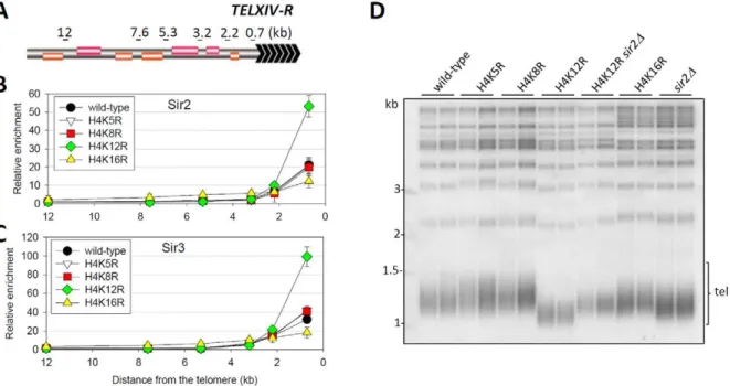
The formation of heterochromatin in yeast is dependent on Sir proteins [1]. To determine if H4K12 acetylation directly modulated the binding of Sir proteins, we carried out ChIP to compare the chromatin binding of Sir2 and Sir3 in wild-type and histone mutant cells at regions 0.7 kb,12 kb from the end of
chromosome XIV-R (TELXIV-R) (Figure 5A–5C). We found that Sir proteins in wild-type cells were present only in telomeric heterochromatin as far as 3 kb from the end of the chromosome. In H4K16R mutant cells, the abundance of Sir proteins was reduced within heterochromatin but was greatly increased at heterochromatin adjacent regions (Figure 5B and 5C), a result that is representative of a typical heterochromatin over-spreading phenotype [6,7]. Notably, compared to a H4K5R or H4K8R mutation that did not alter the binding of Sir proteins at TELXIV-R telomere (Figure 5B and 5C), the H4K12TELXIV-R mutation resulted in 2.52-fold more Sir2 binding and 3.09-fold more Sir3 binding 0.7 kb from the telomere end, but not at regions farther from the chromosome end. We also examined Sir2 binding at all other telomeres and found that the H4K12R mutation resulted in enhancement of Sir2 binding at most telomeres, especially at those where Sir2 was already abundant (Figure S6A). Because the H4K12R mutation does not perturb the transcriptional profile of
the genome [34], and the expression and nuclear distribution of Sir proteins were not affected by the H4K12R mutation (Figure S5 and Figure S7), we therefore favor a model where the acetylation status of H4K12 directly affects Sir protein binding. We also analyzed the heterochromatin structure in H4K12Q mutant. Presumably, a lysine to glutamine mimicked a hyperacetylated state of the corresponding lysine. However, we found that the abundance and distribution of Sir2 and Sir3 in H4K12Q cells were largely similar to that in H4K12R cells (Figure S6B and S6C). We proposed that glutamine does not accurately represent the hyperacetylated state of H4K12 in this case. Together, these data suggest that the H4K12 acetylation suppresses the over-congregation of Sir proteins at telomeric heterochromatin.
To investigate if the regulation of telomere replication by H4K12 acetylation is dependent on the telomeric heterochromatin structure, we detected the effect ofSIR2deletion on the telomere length in H4K12R mutant cells. As shown in Figure 5D,sir2Dcells exhibited a modest reduction of telomere length, which is much longer than that observed in H4K12R mutant cells. Interestingly, deletion ofSIR2efficiently rescued the severe telomere-shortening phenotype in H4K12R mutant cells (Figure 5D), indicating that H4K12 acetylation regulates telomere length though the Sir2 pathway. In addition, telomere length in H4K5R, K8R or K16R cells was indistinguishable from that in wild-type cells (Figure 5D), consistent with their already defined effect on the telomeric association of Sir proteins. Taken together, these data strongly supported the notion that H4K12 acetylation regulates telomere replication directly via modulating telomeric heterochromatin structure.
Histone H4K12 acetylation regulates telomere recombination
Telomeres are hotspots for recombination when they are deprotected. Telomerase-negative yeast cells could undergo homologous recombination on Y’ or TG1–3telomeric sequences,
thus generating Type I or Type II post-senescence survivors, respectively [35]. Since Type II survivors grew much faster than Type I, liquid culturing of post-senescent est2D cells yielded primarily Type II survivor cells [35] (Figure 6A). Interestingly, deletion of the catalytic subunit of telomerase EST2 in the H4K12R mutant background eventually led to the generation of a population of cells that contained amplified Y’ telomeric sequence (Figure 6A), a hallmark of Type I survivor cells. Therefore, we conclude that H4K12 acetylation suppresses homologous recom-bination of TG1–3 tracts during the creation of telomerase-null
post-senescent survivors.
Since histone H4K12 acetylation affected both telomere length and recombination, we wondered whether it regulated the senescence rate of telomerase inactive cells. Therefore, we compared the growth potential ofest2Dand H4K12Rest2Dcells. The result showed that H4K12R mutation greatly accelerated the senescence rate ofest2Dcells (Figure 6B). Further deletion ofSIR2
could suppress the accelerated senescence rate of H4K12Rest2D
cells (Figure 6B). These data suggested that H4K12 acetylation delays senescence driven by Sir-dependent telomere dysfunction.
Histone H4K12 acetylation facilitates basal transcription at telomeric heterochromatin
Previous work had shown that histone H4 mutations that led to increased telomere position effect (TPE) were usually associated with a dramatic decrease of K12 acetylation [36]. To further address this point, we carried out TPE assay [37] to evaluate the effects of histone mutations on the transcriptional state at heterochromatin. A URA3 gene was inserted into a locus that was proximal to the right telomeric TG-tracts of chromosome XIV. Strains were then tested for the relativeURA3expression in histone mutant strains compared with wild-type level. In agreement with a previous report [38], H4K16R mutation markedly reduced telomere silencing (Figure 7). Notably, H4K12R, but not K5R or K8R cells, had lessURA3expression than wild-type cells (Figure 7), suggesting that H4K12R mutation enhances telomere silencing. H4K12R mutation in sir2D back-ground had similarURA3expression to that insir2Dcells (Figure 7), indicating that the increased silencing by H4K12R mutation is
Sir-dependent. Therefore, we concluded that H4K12 acety-lation contributes to the basal transcription within telomeric heterochromatin.
Discussion
In this study, we have employed thede novotelomere addition assay [16] as a de novo telomeric heterochromatin formation system, to monitor chromatin dynamics occurred on a newly-formed telomere (Figure 1). Compared with the traditional Sir3-induction system [39–41], this system works in a more physiolog-ical condition, with normal Sir proteins level and much less transcriptional changes across the genome. Hence, the kinetics of multiple events in the course of telomeric heterochromatin formation can be more accurately followed.
The experimental observations we have made in this study establish histone H4K12 acetylation as an important component of yeast telomeric heterochromatin. We have provided phenotyp-ic, genetic and mechanistic evidence to support the presence of H4K12 acetylation inside telomeric heterochromatin. By using stringently controlled ChIP analyses, we detected H4K12 acetyla-tion at most telomeres (Figure 3). Compared with euchromatic H4K12 acetylation, the level of heterochromatic H4K12 acety-lation was relatively low but was greatly elevated bySIR2deletion (Figure S3), a phenomenon also observed in earlier reports [20,42]. Therefore, H4K12 acetylation, as is the case of acetyla-tion of other H4 lysines, is suppressed by heterochromatin structure. Genetically, the H4K12R mutation, which mimicked an unacetylated state of K12, increased Sir protein binding at telomeric heterochromatin and altered several dynamic telomere-related chromosomal processes (Figure 4, Figure 5, Figure 6, Figure 7). Heterochromatic H4K12 acetylation coincides with H4K12 as a memory mark for the heritable chromatin structure in yeast [36]. Mechanistically, Esa1, the catalytic subunit of NuA4 HAT, bound to silenced telomeres and was responsible for H4K12 acetylation (Figure 3). Since Arp4, another subunit of NuA4 complex, is also enriched at heterochromatic domains [43], it is possible that the whole NuA4 complex binds to telomeric heterochromatin and plays a direct role in the acetylation of H4K12. Earlier work suggested a phosphorylated H2A-dependent
Figure 6. Histone H4K12 acetylation regulates telomere recombination.(A) Telomere-blot ofest2Dandest2DH4K12R cells. Cells were continuously passaged for the indicated number of days and the genomic DNA was subjected to telomere-blot. Y’: Y’ elements. (B) Senescence rates were measured in liquid culture by serially passaging strains of the indicated genotypes.
mechanism for the recruitment of NuA4 during DNA damage repair [44]. However, due to the fact that normal telomeres are protected from being recognized as double-strand DNA break and phosphorylated H2A is absent from normal telomeres, we propose that yeast cells take a distinct strategy such as a Rap1-dependent mechanism to recruit NuA4 onto telomeric heterochromatin [45]. Esa1 preferentially acetylated nucleosome on both H4K5 and K12in vivo[46] andin vitro(Figure S4C). This raised a question of why H4K5 was hypoacetylated at telomeric heterochromatin. We suspected that another histone deacetylase besides Sir2 was involved in the establishment of the acetylation pattern at telomeric heterochromatin. Two candidates are Rpd3 and Hda1, which displayed the highestin vivoactivity toward acetylated H4K5 from among H4K5, K8, K12 and K16 [47]. Indeed, our recent study did reveal a genetic interaction between Rpd3L and H4K5 at subtelomeric regions [48]. Moreover, analysis of the CIDMS/MS spectra shows K12 to be the most highly acetylated site (54%) from among H4K5, K8 and K12, followed by K5 (32%), and K8 (24%) [49], suggesting that K12 acetylation covers a much wider range of yeast chromatin than that of K5 or K8 acetylation.
NuA4 complex also functions to prevent the Sir complex from spreading out of heterochromatic domains [19,50–52]. Therefore, mutation of ESA1 or other key subunits of the NuA4 complex resulted in gene silencing near heterochromatin [19,50,51] and a modest reduction of silencing within telomeric heterochromatin [53,54]. The histone H4K12 acetylationper sehas little effect on heterochromatin boundary activity (Figure 5B and 5C), however, since it is the uniquely acetylated site within heterochromatin, abolishment of histone H4K12 caused a increase of heterochro-matin silencing (Figure 7 and [36]).
Chromatin modifications have been implicated in telomere elongation in several organisms [55,56]. Recent study on H4K16 demonstrated the relationship between histone acetylation and telomere regulation [57]. The natural presence of H4K12 acetylation at telomeres and Sir-dependent regulation of telomere replication via H4K12 have provided additional direct evidence supporting the proposal that chromatin modifications affect telomere homeostasis. Elimination of H4K12 acetylation increased
subtelomeric binding of Sir proteins (Figure 5B and 5C), accelerated senescence inest2D cells (Figure 6B) and suppressed homologous recombination within TG-tracts (Figure 6A). By contrast, inactivation of Sas2, the acetyltransferase of H4K16 [38], decreased Sir3 binding at telomere ends and thereby delayed senescence in tlc1D cells through homologous recombination-dependent mechanism [57]. Therefore, H4K12 acetylation and K16 acetylation seem to play opposite roles in the Sir-dependent regulation of homologous recombination at telomeric heterochro-matin. It is quite interesting that H4K12 and K16 are in close proximity to each other but have opposing roles in regulating telomere dynamics through Sir-dependent mechanisms. Finally, a recent paper showed that Esa1 and Rpd3L controlled H4K12 acetylation, which is necessary for cell growth and viability [46]. Although H4K12R does not change the genome-wide transcrip-tion profile [34], it is still possible that H4K12 also fine-tunes the chromatin structure at sequences other than the telomeric heterochromatin.
In conclusion, heterochromatin is known as a highly condensed chromatin domain that is transcriptionally silent [1]. However, pioneering studies have recently revealed the dynamic aspect of yeast heterochromatin [11–13,15]. In this study, we have built on these pioneering studies and shown that the H4K12R mutation led to a more condensed telomeric heterochromatin structure (Figure 5) and more static telomere metabolism (Figure 4, Figure 5, Figure 6, Figure 7). Therefore, we propose that H4K12 provides a mechanism for yeast cells to maintain partial plasticity of their telomeric heterochromatin (Figure 8). Interestingly, histone H4K12 acetylation has also been observed at the chromocenter in fly [58]. Mst1, the orthologue of Esa1 in fission yeast, acetylates H3K4 at pericentric heterochromatin to regulate heterochromatin reassembly [59]. TIP60, the orthologue of NuA4 in mammals, physically interacts with Sirt1, the mammalian homologue of Sir2 [60], and associates with tri-methylated H3K9, a hallmark of heterochromatin [61]. Hence, it will be of great interest to determine if histone acetylation also plays a general role in heterochromatin dynamics in other eukaryotes.
Materials and Methods
Yeast strains and reagents
Antibodies, yeast strains and primers used in this study are listed in Tables S1, S2, S3, respectively.
Chromatin immuno-precipitation
ChIP assays were performed as described [18,19]. Most ChIP products were directly analyzed by real-time Q-PCR using SYBR green as a label (TOYOBO). Alternatively, pellet and whole-cell extract DNAs were analyzed by Q-PCR performed in a linear range with32P-dATP, electrophoresis through 6% PAGE in Tris-Borate-EDTA buffer, and phosphorimager quantification of radioactive bands in dried gels. The relative enrichment value represented the ratio (IPs/Input) at indicated loci relative to internal control. All ChIP experiments were performed in triplicate on paired isogenic wild-type and mutant strains.
HPLC analysis of DNA methylation status
After protein expression was induced by galactose for indicated time, total DNA was isolated from yeast cells by glass beads lysis, proteinase K digestion and extraction with QIAGEN Genomic DNA Kit, followed by RNaseA and RNaseT digestion, phenol/ chloroform extraction and re-precipitation. Finally, purified genomic DNA was digested into mononucleoside 59 -monophos-phates with Nuclease P1 (Sigma). Nucleosides were separated by an
Figure 7. Histone H4K12 acetylation regulates basal transcrip-tion at telomeric heterochromatin. URA3 gene was artificially inserted into a locus that is proximal to the telomeric TG-tracts of chromosome XIV-R. The mRNA ofURA3in wild-type and mutant strains were measured by real-time PCR and normalized toACT1. The relative mRNA level ofURA3/ACT1in wild-type strain was set as 1. Therefore, values above ‘‘1’’ indicated higherURA3expression.
AQ-C18 column (5mm, 4.66250 mm, Welch Materials Inc.) guarded by a precolumn (Phenomenex, Security Guard), using a Beckman device (SYSTEM GOLD 125 Solvent Module and SYSTEM GOLD 166 Detector). The eluate was obtained using a flow rate of 1 ml/min and 100% buffer A (10 mM KH2PO4/
H3PO4, pH 3.7) for 50 minutes, followed by a shift to 70% A/
30% methanol for 10 minutes, and then to 100% buffer A for 10 minutes. Peaks were quantified by measuring their heights with a 32 Karat software V7.0 after identity confirmation by comparison of spikes with different combinations of dCMP and 59-me-dCMP (Hongene Biotechnology Ltd. Shanghai). At least three indepen-dent yeast clones were assayed for each construct.
Southern blot
Southern blot was performed as described [62]. Yeast genomic DNAs were digested with restriction enzymes as indicated, separated by 1.2% agarose gel electrophoresis, and transferred to Hybond-N membrane (Amersham). The blot was hybridized with a 32P-dCTP incorporated probe as indicated. The radioactive signal was detected by phosphorimager.
Micrococcal nuclease sensitivity assay
Micrococcal nuclease (MNase) sensitivity assay was performed as described [27]. Briefly, Cells from 50-ml cultures were collected by centrifugation, treated with zymolyase, and digested with micrococcal nuclease (MNase). genomic DNA samples were purified, digested with BamHI, resolved in 1.2% agarose gels. Then the cutting profiles were visualized after hybridization with an internal probe from Ty5-1.
Immunofluorescence of yeast cells
Cells were grown in YPD medium overnight to a density of,1–
26107cells/ml and were fixed for 30 min by incubation with
3.7% formaldehyde. Next, cells were washed with 0.1 M potassium phosphate (pH 6.5) and P solution (1.2 M sorbitol, 1 M K2PO4), and re-suspended in P solution. Cells were
subsequently treated with 0.1 mg/ml Zymolyase (20T, MP Biomedicals) for 10 min, washed with P solution, spotted on Poly-L-Lysine pre-treated slides. After rinsing in PBS-T buffer (PBS containing 0.1% Triton X-100 and 1% BSA), slides were incubated overnight with anti-Myc, anti-Rap1 and anti-Nop1 antibody diluted in PBS containing 1% BSA. Slides were then washed with PBS-T and incubated with the appropriate secondary antibodies conjugated to Cy3 or fluorescein isothiocyanate (FITC). The DNA fluorescence signal was detected by DAPI (1mg/ml in Phosphate Buffered Saline (PBS) solution) staining. Slides were mounted with PBS containing 1 mg/ml p-phenylenediamine, 2.5mM NaOH, and 90% glycerol.
Confocal microscopy was performed on a Leica TCS SP2 microscope with a 636 lamda blue objective (oil). Image processing including similar filtration and threshold levels was standardized for all images.
Purification of Esa1 protein
N terminal 6xHis tagged Esa1 protein was overexpressed and purified inE. coliaccording to the manufacturer’s instructions (GE Healthcare).
Supporting Information
Figure S1 Specificity of the anti-acetyl-H4K12 antibody. ChIP assays were performed on chromatin from wild-type and H4K12R mutant strains. IPs were done with increasing amount of anti-acetyl-H4K12 antibody: 1, 1:200; 2, 1:200; 3, 1:50; 4, 1:25.
Found at: doi:10.1371/journal.pgen.1001272.s001 (0.12 MB PDF)
Figure 8. Model for the regulation of telomeric heterochromatin plasticity by histone H4K12 acetylation.In wild-type cells where subtelomeric histone H4K12 is partially acetylated, heterochromatin structure is plastic and accessible to proteins, such as the RNA polymerase machinery and the telomerase machinery. In H4K12 deacetylated strain, heterochromatin becomes aberrantly condensed and less accessible to proteins, thereby inhibiting chromosomal processes.
Figure S2 Acetylation of H4K12 at native telomeric hetero-chromatin in galactose condition. ChIP experiments were performed as Figure 3 except that the cells were cultured in galactose medium.
Found at: doi:10.1371/journal.pgen.1001272.s002 (0.06 MB PDF)
Figure S3 Histone N terminal lysine acetylation profile at HMR and HMR-proximal regions. (A) Schematic diagram of the HMR region and subtelomeric region of the right arm of chromosome III. Primer sets used for the following ChIP analysis were shown on the top. (B) Relative abundance of H4K5 acetylation, K8 acetylation, K12 acetylation and K16 acetylation at HMR and HMR-proximal regions. The relative abundance of lysine acetylation was corrected by H4 occupancy. Grey areas were predicted silenced chromatin.
Found at: doi:10.1371/journal.pgen.1001272.s003 (0.21 MB PDF)
Figure S4 Contribution of Esa1 and Hat1 to chromatic H4K12 acetylation (A) Schematic diagram of the subtelomeric region of the right arm of chromosome XIV (TELXIV-R). Primer sets used for the following ChIP analysis were shown on the top. (B) ChIP analysis of Esa1-myc at subtelomeric region of TELXIV-R. The locations of primer sets were depicted in (A). (C) Histone acetylation assay. Recombinant Esa1 was incubated with native nucleosomes derived from sas2Deaf4D cells. Reaction products were loaded onto 15% SDS-page gel and detected using indicated antibodies. (D) Western-blot of chromatin derived from wild-type and hat1Dcells. Antibodies used were indicated on the right. (E) ChIP of H4K12ac at subtelomere XIV-R in wild-type and hat1D
cells.
Found at: doi:10.1371/journal.pgen.1001272.s004 (0.25 MB PDF)
Figure S5 H4K12R mutation does not affect the perinuclear localization of telomeres. Sir3-myc (used to represent telomeres) and nuclear pores were stained by rabbit anti-myc antibody and mouse anti-mAb414 antibody, respectively.
Found at: doi:10.1371/journal.pgen.1001272.s005 (0.66 MB PDF)
Figure S6 H4K12 regulates subtelomeric binding of Sir proteins. (A) Relative abundance of Sir2-myc at all telomeres in isogenic wild-type and H4K12R strains. Regions at,0.5 kb from
each telomere were subjected to real-time PCR analysis. PCR data
were normalized to ACT1. (B) and (C) ChIP of Sir2-myc (B) and Sir3-myc (C) in wild-type and H4K12Q mutant cells.
Found at: doi:10.1371/journal.pgen.1001272.s006 (0.08 MB PDF)
Figure S7 Histone mutations do not affect the expression of silencing-related genes. (A) and (B) Western-blot of Sir2-myc (A) or Sir3-myc (B) in wild-type and histone mutants. Sir2 and Sir3 levels were detected using an anti-myc antibody and were normalized to anti-tubulin protein level. (C) Quantitative PCR of relative mRNA levels in a cluster of silencing-related genes in the indicated histone mutant strains. Shown are the average expression ratios (normalized by that of ACT1) relative to the wild-type. A log2 ratio less than zero indicates repression of transcription, whereas greater than zero indicates enhancement of transcription. Error bars represent standard error of the mean for three independent RNA purifications. (D) Immunolocalization of Sir2-myc and Nop1 in wild-type and H4K12R cells. Sir2-myc and Nop1 (indicating the nucleolus) were stained by rabbit anti-myc antibody and mouse anti-Nop1 antibody, respectively.
Found at: doi:10.1371/journal.pgen.1001272.s007 (1.04 MB PDF)
Table S1 Antibodies used in this study.
Found at: doi:10.1371/journal.pgen.1001272.s008 (0.04 MB DOC)
Table S2 Yeast strains used in this study.
Found at: doi:10.1371/journal.pgen.1001272.s009 (0.09 MB DOC)
Table S3 Primers used for real-time PCR.
Found at: doi:10.1371/journal.pgen.1001272.s010 (0.07 MB DOC)
Author Contributions
Conceived and designed the experiments: BOZ JQZ. Performed the experiments: BOZ XHF WD. Analyzed the data: BOZ XHF BAL JQZ. Contributed reagents/materials/analysis tools: BOZ SSW YZ. Wrote the paper: BAL JQZ. Assisted in constructing yeast strains, checking the specificity of antibodies, and detecting the expression, cellular localization, and telomere binding of Sir proteins: SSW. Assisted in strain construction: YZ.
References
1. Rusche LN, Kirchmaier AL, Rine J (2003) The establishment, inheritance, and function of silenced chromatin in Saccharomyces cerevisiae. Annu Rev Biochem 72: 481–516.
2. Luo K, Vega-Palas MA, Grunstein M (2002) Rap1-Sir4 binding independent of other Sir, yKu, or histone interactions initiates the assembly of telomeric heterochromatin in yeast. Genes Dev 16: 1528–1539.
3. Imai S, Armstrong CM, Kaeberlein M, Guarente L (2000) Transcriptional silencing and longevity protein Sir2 is an NAD-dependent histone deacetylase. Nature 403: 795–800.
4. Hecht A, Laroche T, Strahl-Bolsinger S, Gasser SM, Grunstein M (1995) Histone H3 and H4 N-termini interact with SIR3 and SIR4 proteins: a molecular model for the formation of heterochromatin in yeast. Cell 80: 583–592.
5. Carmen AA, Milne L, Grunstein M (2002) Acetylation of the yeast histone H4 N terminus regulates its binding to heterochromatin protein SIR3. J Biol Chem 277: 4778–4781.
6. Kimura A, Umehara T, Horikoshi M (2002) Chromosomal gradient of histone acetylation established by Sas2p and Sir2p functions as a shield against gene silencing. Nat Genet 32: 370–377.
7. Suka N, Luo K, Grunstein M (2002) Sir2p and Sas2p opposingly regulate acetylation of yeast histone H4 lysine16 and spreading of heterochromatin. Nat Genet 32: 378–383.
8. Katan-Khaykovich Y, Struhl K (2005) Heterochromatin formation involves changes in histone modifications over multiple cell generations. Embo J 24: 2138–2149.
9. Bi X, Broach JR (2001) Chromosomal boundaries in S. cerevisiae. Curr Opin Genet Dev 11: 199–204.
10. Smith ER, Eisen A, Gu W, Sattah M, Pannuti A, et al. (1998) ESA1 is a histone acetyltransferase that is essential for growth in yeast. Proc Natl Acad Sci U S A 95: 3561–3565.
11. Cheng TH, Gartenberg MR (2000) Yeast heterochromatin is a dynamic structure that requires silencers continuously. Genes Dev 14: 452–463. 12. Sekinger EA, Gross DS (2001) Silenced chromatin is permissive to activator
binding and PIC recruitment. Cell 105: 403–414.
13. Chen L, Widom J (2005) Mechanism of transcriptional silencing in yeast. Cell 120: 37–48.
14. Andrau JC, van de Pasch L, Lijnzaad P, Bijma T, Koerkamp MG, et al. (2006) Genome-wide location of the coactivator mediator: Binding without activation and transient Cdk8 interaction on DNA. Mol Cell 22: 179–192.
15. Teytelman L, Eisen MB, Rine J (2008) Silent but not static: accelerated base-pair substitution in silenced chromatin of budding yeasts. PLoS Genet 4: e1000247. doi:10.1371/journal.pgen.1000247.
16. Diede SJ, Gottschling DE (1999) Telomerase-mediated telomere addition in vivo requires DNA primase and DNA polymerases alpha and delta. Cell 99: 723–733.
17. Tamburini BA, Tyler JK (2005) Localized histone acetylation and deacetylation triggered by the homologous recombination pathway of double-strand DNA repair. Mol Cell Biol 25: 4903–4913.
18. Meneghini MD, Wu M, Madhani HD (2003) Conserved histone variant H2A.Z protects euchromatin from the ectopic spread of silent heterochromatin. Cell 112: 725–736.
20. Suka N, Suka Y, Carmen AA, Wu J, Grunstein M (2001) Highly specific antibodies determine histone acetylation site usage in yeast heterochromatin and euchromatin. Mol Cell 8: 473–479.
21. Negrini S, Ribaud V, Bianchi A, Shore D (2007) DNA breaks are masked by multiple Rap1 binding in yeast: implications for telomere capping and telomerase regulation. Genes Dev 21: 292–302.
22. Zhu X, Gustafsson CM (2009) Distinct differences in chromatin structure at subtelomeric X and Y’ elements in budding yeast. PLoS ONE 4: e6363. doi:10.1371/journal.pone.0006363.
23. Boudreault AA, Cronier D, Selleck W, Lacoste N, Utley RT, et al. (2003) Yeast enhancer of polycomb defines global Esa1-dependent acetylation of chromatin. Genes Dev 17: 1415–1428.
24. Kleff S, Andrulis ED, Anderson CW, Sternglanz R (1995) Identification of a gene encoding a yeast histone H4 acetyltransferase. J Biol Chem 270: 24674–24677.
25. Ruiz-Garcia AB, Sendra R, Galiana M, Pamblanco M, Perez-Ortin JE, et al. (1998) HAT1 and HAT2 proteins are components of a yeast nuclear histone acetyltransferase enzyme specific for free histone H4. J Biol Chem 273: 12599–12605.
26. Shahbazian MD, Grunstein M (2007) Functions of site-specific histone acetylation and deacetylation. Annu Rev Biochem 76: 75–100.
27. Vega-Palas MA, Venditti S, Di Mauro E (1998) Heterochromatin organization of a natural yeast telomere. Changes of nucleosome distribution driven by the absence of Sir3p. J Biol Chem 273: 9388–9392.
28. Palladino F, Laroche T, Gilson E, Axelrod A, Pillus L, et al. (1993) SIR3 and SIR4 proteins are required for the positioning and integrity of yeast telomeres. Cell 75: 543–555.
29. Wright JH, Gottschling DE, Zakian VA (1992) Saccharomyces telomeres assume a non-nucleosomal chromatin structure. Genes Dev 6: 197–210.
30. Schober H, Ferreira H, Kalck V, Gehlen LR, Gasser SM (2009) Yeast telomerase and the SUN domain protein Mps3 anchor telomeres and repress subtelomeric recombination. Genes Dev 23: 928–938.
31. de Bruin D, Kantrow SM, Liberatore RA, Zakian VA (2000) Telomere folding is required for the stable maintenance of telomere position effects in yeast. Mol Cell Biol 20: 7991–8000.
32. Strahl-Bolsinger S, Hecht A, Luo K, Grunstein M (1997) SIR2 and SIR4 interactions differ in core and extended telomeric heterochromatin in yeast. Genes Dev 11: 83–93.
33. de Bruin D, Zaman Z, Liberatore RA, Ptashne M (2001) Telomere looping permits gene activation by a downstream UAS in yeast. Nature 409: 109–113. 34. Dion MF, Altschuler SJ, Wu LF, Rando OJ (2005) Genomic characterization reveals a simple histone H4 acetylation code. Proc Natl Acad Sci U S A 102: 5501–5506.
35. Grandin N, Charbonneau M (2007) Control of the yeast telomeric senescence survival pathways of recombination by the Mec1 and Mec3 DNA damage sensors and RPA. Nucleic Acids Res 35: 822–838.
36. Smith CM, Haimberger ZW, Johnson CO, Wolf AJ, Gafken PR, et al. (2002) Heritable chromatin structure: mapping ‘‘memory’’ in histones H3 and H4. Proc Natl Acad Sci U S A 99 Suppl 4: 16454–16461.
37. Gottschling DE, Aparicio OM, Billington BL, Zakian VA (1990) Position effect at S. cerevisiae telomeres: reversible repression of Pol II transcription. Cell 63: 751–762.
38. Meijsing SH, Ehrenhofer-Murray AE (2001) The silencing complex SAS-I links histone acetylation to the assembly of repressed chromatin by CAF-I and Asf1 in Saccharomyces cerevisiae. Genes Dev 15: 3169–3182.
39. Li YC, Cheng TH, Gartenberg MR (2001) Establishment of transcriptional silencing in the absence of DNA replication. Science 291: 650–653. 40. Kirchmaier AL, Rine J (2001) DNA replication-independent silencing in S.
cerevisiae. Science 291: 646–650.
41. Hoppe GJ, Tanny JC, Rudner AD, Gerber SA, Danaie S, et al. (2002) Steps in assembly of silent chromatin in yeast: Sir3-independent binding of a Sir2/Sir4 complex to silencers and role for Sir2-dependent deacetylation. Mol Cell Biol 22: 4167–4180.
42. Braunstein M, Sobel RE, Allis CD, Turner BM, Broach JR (1996) Efficient transcriptional silencing in Saccharomyces cerevisiae requires a heterochromatin histone acetylation pattern. Mol Cell Biol 16: 4349–4356.
43. Ogiwara H, Ui A, Kawashima S, Kugou K, Onoda F, et al. (2007) Actin-related protein Arp4 functions in kinetochore assembly. Nucleic Acids Res 35: 3109–3117.
44. Bird AW, Yu DY, Pray-Grant MG, Qiu Q, Harmon KE, et al. (2002) Acetylation of histone H4 by Esa1 is required for DNA double-strand break repair. Nature 419: 411–415.
45. Reid JL, Iyer VR, Brown PO, Struhl K (2000) Coordinate regulation of yeast ribosomal protein genes is associated with targeted recruitment of Esa1 histone acetylase. Mol Cell 6: 1297–1307.
46. Chang CS, Pillus L (2009) Collaboration between the essential Esa1 acetyltransferase and the Rpd3 deacetylase is mediated by H4K12 histone acetylation in Saccharomyces cerevisiae. Genetics 183: 149–160.
47. Rundlett SE, Carmen AA, Kobayashi R, Bavykin S, Turner BM, et al. (1996) HDA1 and RPD3 are members of distinct yeast histone deacetylase complexes that regulate silencing and transcription. Proc Natl Acad Sci U S A 93: 14503–14508.
48. Zhou J, Zhou BO, Lenzmeier BA, Zhou JQ (2009) Histone deacetylase Rpd3 antagonizes Sir2-dependent silent chromatin propagation. Nucleic Acids Res 37: 3699–3713.
49. Smith CM, Gafken PR, Zhang Z, Gottschling DE, Smith JB, et al. (2003) Mass spectrometric quantification of acetylation at specific lysines within the amino-terminal tail of histone H4. Anal Biochem 316: 23–33.
50. Babiarz JE, Halley JE, Rine J (2006) Telomeric heterochromatin boundaries require NuA4-dependent acetylation of histone variant H2A.Z in Saccharomy-ces cerevisiae. Genes Dev 20: 700–710.
51. Chiu YH, Yu Q, Sandmeier JJ, Bi X (2003) A targeted histone acetyltransferase can create a sizable region of hyperacetylated chromatin and counteract the propagation of transcriptionally silent chromatin. Genetics 165: 115–125. 52. Altaf M, Auger A, Monnet-Saksouk J, Brodeur J, Piquet S, et al. (2010)
NuA4-dependent acetylation of nucleosomal histones H4 and H2A directly stimulates incorporation of H2A.Z by the SWR1 complex. J Biol Chem 285: 15966–15977. 53. Clarke AS, Samal E, Pillus L (2006) Distinct roles for the essential MYST family
HAT Esa1p in transcriptional silencing. Mol Biol Cell 17: 1744–1757. 54. Auger A, Galarneau L, Altaf M, Nourani A, Doyon Y, et al. (2008) Eaf1 is the
platform for NuA4 molecular assembly that evolutionarily links chromatin acetylation to ATP-dependent exchange of histone H2A variants. Mol Cell Biol 28: 2257–2270.
55. Blasco MA (2007) The epigenetic regulation of mammalian telomeres. Nat Rev Genet 8: 299–309.
56. Yu EY, Steinberg-Neifach O, Dandjinou AT, Kang F, Morrison AJ, et al. (2007) Regulation of telomere structure and functions by subunits of the INO80 chromatin remodeling complex. Mol Cell Biol 27: 5639–5649.
57. Kozak ML, Chavez A, Dang W, Berger SL, Ashok A, et al. (2010) Inactivation of the Sas2 histone acetyltransferase delays senescence driven by telomere dysfunction. Embo J 29: 158–170.
58. Turner BM, Birley AJ, Lavender J (1992) Histone H4 isoforms acetylated at specific lysine residues define individual chromosomes and chromatin domains in Drosophila polytene nuclei. Cell 69: 375–384.
59. Xhemalce B, Kouzarides T (2010) A chromodomain switch mediated by histone H3 Lys 4 acetylation regulates heterochromatin assembly. Genes Dev 24: 647–652.
60. Yamagata K, Kitabayashi I (2009) Sirt1 physically interacts with Tip60 and negatively regulates Tip60-mediated acetylation of H2AX. Biochem Biophys Res Commun 390: 1355–1360.
61. Sun Y, Jiang X, Xu Y, Ayrapetov MK, Moreau LA, et al. (2009) Histone H3 methylation links DNA damage detection to activation of the tumour suppressor Tip60. Nat Cell Biol 11: 1376–1382.
![Figure 1. De novo telomeric heterochromatin assembly on a short telomere. (A) Schematic representation of the de novo telomeric heterochromatin formation system [16]](https://thumb-eu.123doks.com/thumbv2/123dok_br/18264116.343750/3.918.90.743.79.894/telomeric-heterochromatin-assembly-schematic-representation-telomeric-heterochromatin-formation.webp)