Morphine-Induced Preconditioning:
Involvement of Protein Kinase A and
Mitochondrial Permeability Transition Pore
Marianne Dorsch1, Friederike Behmenburg1*, Miriam Raible1, Dominic Blase1, Hilbert Grievink1,4,5, Markus W. Hollmann2, André Heinen1,3, Ragnar Huhn1
1Department of Anesthesiology, University Hospital Duesseldorf, Moorenstr. 5, 40225, Duesseldorf, Germany,2Department of Anesthesiology, Laboratory of Experimental Intensive Care and Anesthesiology (L.E.I.C.A.), Academic Medical Center (AMC), University of Amsterdam, Meibergdreef 9, 1100 DD, Amsterdam, The Netherlands,3Institute of Cardiovascular Physiology, Heinrich-Heine-University, Universitaetsstr. 1, 40225, Duesseldorf, Germany,4Department of Anesthesiology and Critical Care Medicine, Hadassah University Hospital, Jerusalem, Israel,5Department of Biochemistry and Molecular Biology, The Hebrew University of Jerusalem, Ein Kerem Campus, Jerusalem, Israel
Abstract
Background
Morphine induces myocardial preconditioning (M-PC) via activation of mitochondrial large conductance Ca2+-sensitive potassium (mKCa) channels. An upstream regulator of mKCa
channels is protein kinase A (PKA). Furthermore, mKCachannel activation regulates
mito-chondrial bioenergetics and thereby prevents opening of the mitomito-chondrial permeability transition pore (mPTP). Here, we investigated in the rat heartin vivowhether 1) M-PC is mediated by activation of PKA, and 2) pharmacological opening of the mPTP abolishes the cardioprotective effect of M-PC and 3) M-PC is critically dependent on STAT3 activation, which is located upstream of mPTP within the signalling pathway.
Methods
Male Wistar rats were randomised to six groups (each n = 6). All animals underwent 25 min-utes of regional myocardial ischemia and 120 minmin-utes of reperfusion. Control animals (Con) were not further treated. Morphine preconditioning was initiated by intravenous administra-tion of 0.3 mg/kg morphine (M-PC). The PKA blocker H-89 (10μg/kg) was investigated with
and without morphine (H-89+M-PC, H-89). We determined the effect of mPTP opening with atractyloside (5 mg/kg) with and without morphine (Atr+M-PC, Atr). Furthermore, the effect of morphine on PKA activity was tested in isolated adult rat cardiomyocytes. In further experiments in isolated hearts we tested the protective properties of morphine in the pres-ence of STAT3 inhibition, and whether pharmacological prevention of the mPTP-opening by cyclosporine A (CsA) is cardioprotective in the presence of STAT3 inhibition.
Results
Morphine reduced infarct size from 64±5% to 39±9% (P<0.05 vs. Con). H-89 completely blocked preconditioning by morphine (64±9%; P<0.05 vs. M-PC), but H-89 itself had not
OPEN ACCESS
Citation:Dorsch M, Behmenburg F, Raible M, Blase D, Grievink H, Hollmann MW, et al. (2016) Morphine-Induced Preconditioning: Involvement of Protein Kinase A and Mitochondrial Permeability Transition Pore. PLoS ONE 11(3): e0151025. doi:10.1371/ journal.pone.0151025
Editor:Meijing Wang, Indiana University School of Medicine, UNITED STATES
Received:April 4, 2015
Accepted:February 23, 2016
Published:March 11, 2016
Copyright:© 2016 Dorsch et al. This is an open access article distributed under the terms of the Creative Commons Attribution License, which permits unrestricted use, distribution, and reproduction in any medium, provided the original author and source are credited.
Data Availability Statement:All relevant data are within the paper.
Funding:This work was supported by a research fund (SFF 701301765) from the Heinrich-Heine-University Duesseldorf, Germany.
effect on infarct size (61±10%; P>0.05 vs. Con). Also, atractyloside abolished infarct size reduction of morphine completely (65±9%; P<0.05 vs. M-PC) but had no influence on infarct size itself (64±5%; P>0.05 vs. Con). In isolated hearts STAT3 inhibitor Stattic completely abolished morphine-induced preconditioning. Administration of Stattic and mPTP inhibitor cyclosporine A reduced infarct size to 31±6% (Stat+CsA, P<0.05 vs. Con). Cyclosporine A alone reduced infarct size to 26±7% (CsA P<0.05 vs. Con). In cardiomyocytes, PKA activity was increased by morphine.
Conclusion
Our data suggest that morphine-induced cardioprotection is mediated by STAT3-activation and inhibition of mPTP, with STA3 located upstream of mPTP. There is some evidence that protein kinase A is involved within the signalling pathway.
Introduction
Ischemic preconditioning is a phenomenon where short sublethal epidsodes of myocardial ischemia protect the heart against the consequences of a longer myocardial ischemic period
and thus against ischemia/reperfusion injury [1]. Short cycles of ischemia and reperfusion at
the end of a longer ischemia and at the beginning of reperfusion can also protect the heart
against ischemia/reperfusion injury; a phenomenon called ischemic postconditioning [2].
Beside ischemic stimuli the cardioprotective effect of pre- and postconditioning can be
mim-icked by volatile anesthetics [3,4] but also with morphine [5]. The cardioprotective effect of
pre- and postconditioning involve amongst others mitochondrial potassium channels and the
mitochondrial permeability transition pore“mPTP”[6].
Recently, we could demonstrate that the cardioprotective effect of morphine was blocked by
the mitochondrial calcium-sensitive potassium channel (mKCa) inhibitor paxilline [7,8],
indi-cating a crucial role of mKCachannels in morphine-induced pre- and postconditioning.
Fur-thermore, Cao et al. [9] could show that the cardioprotective effect of mKCachannel activation
was abolished by opening of the mPTP, suggesting that the mKCachannel is an upstream
regu-lator of the mPTP. The blockade of morphine-induced cardioprotection by paxilline leads to
the question, how mKCachannel activation is regulated. A possible regulator of mKCachannels
is protein kinase A (PKA, cAMP dependent protein kinase) [10,11], mKCachannels could be
activated by PKA and PKA in turn is regulated by cellular cAMP levels.
We hypothesize that the cardioprotective effect of morphine is mediated by activation of PKA and regulation of mPTP.
Methods
The study was performed in accordance with the guidelines laid out in the Guide for the Care and Use of Laboratory Animals, which is available from the National Academy of Science and the regulations of the German Animal Protection Law and was approved by the District
Gov-ernment of Duesseldorf, Germany (8.87–51.04.20.09.388).
Surgical preparation
Male Wistar rats had free access to water and standard rat food at all times prior to
(296±30 g) were instrumented with a coronary occluder around a left anterior descending artery. Pentobarbital-anesthetized animals were endotracheal intubated with a plastic cannula (outer diameter 2.2 mm). After a median incision was performed on cervical level a 20 gauge cannula was inserted into the right internal carotid artery and advanced into the aorta in order to measure the aortic pressure. Aortic pressure was digitized using an analogue to digital con-verter (PowerLab/8SP, ADInstruments Pty Ltd, Castle Hill, Australia) at a sampling rate of 500 Hz and were continuously recorded on a personal computer using Chart for Windows v5.0 (ADInstruments Pty Ltd, Castle Hill, Australia). All animals underwent 25 min of coronary artery occlusion followed by 2 h reperfusion. At the end of reperfusion the hearts were excised
and infarct sizes were determined using a previously described method [14,15]. Briefly, the
heart was excised with the occluding suture left in place and then mounted on a modified Lan-gendorff apparatus for perfusion with ice cold normal saline via the aortic root at a perfusion
pressure of 80 cm H2O in order to wash out intravascular blood. After 5 min of perfusion, the
coronary artery was re-occluded and the remainder of the myocardium was perfused through the aortic root with 0.2% Evans blue in normal saline for 10 min. Intravascular Evans blue was then washed out by perfusion with normal saline for 10 min. This treatment identified the area at risk as unstained. The heart was then cut into 2 mm thick transverse slices. The slices were stained with 0.75% triphenyltetrazolium chloride solution for 10 min at 37°C, and fixed in 4% formalin solution for 24 h at room temperature. The area at risk and the infarct size were
deter-mined using planimetry and corrected for dry weight in each slice by using SigmaScan Pro51
(SPSS Science Software, Chicago, IL, USA).
Experimental protocol
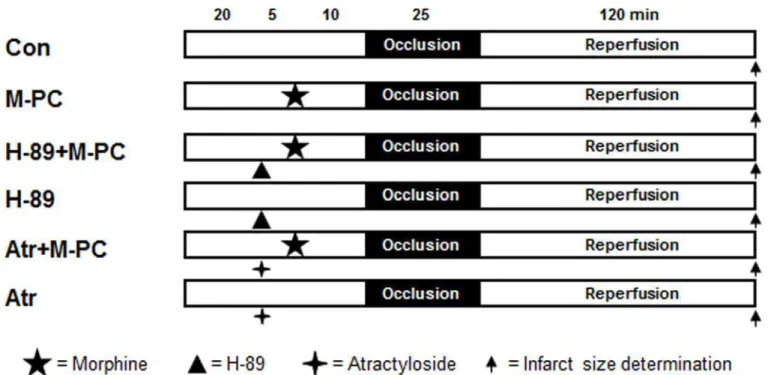
Rats for infarct size experiments were randomly allocated into six groups (seeFig 1). Morphine
(0.3 mg/kg) [16] was administered intravenously 10 minutes before ischemia. The PKA
Fig 1. Experimental protocol forin vivoinfarct size measurement.Con = control, M-PC = morphine preconditioning, H-89 = protein kinase A inhibitor, Atr = atractyloside (mPTP opener).
inhibitor H-89 (10μg/kg) [17] and the mPTP opener atractyloside (5 mg/kg) [18] were given
15 minutes before the onset of ischemia. At the end of the experiments hearts were excised and
infarct sizes were determined using a previously described method [14].
Isolated heart experiments
Surgical preparation was performed as described previously. In brief, rats were anesthetized by intraperitoneal injection of pentobarbital (90 mg/kg). After thoracotomy hearts were excised and mounted on a Langendorff system. Perfusion of the hearts was performed at constant pres-sure (80 mmHg) with a Krebs-Henseleit solution, containing (in mM): 116 NaCl, 4.7 KCl, 1.1 MgSO4, 1.17 KH2PO4, 24.9 NaHCO3, 2.52 CaCl2, 8.3 glucose, and 2.2 pyruvate at 37°C. A
fluid filled balloon was inserted into the left ventricle and end-diastolic pressure was set at 1–4
mmHg. All hearts underwent an equilibration period of 20 minutes. Thereafter, heart rate, the
rate pressure product (RPP, calculated as heart rate x (maximal left ventricular pressure–
mini-mal left ventricular pressure)), left ventricular end-diastolic pressure (LVEDP), and coronary flow were measured continuously and digitized using an analogue to digital converter (Power-Lab/8SP, ADInstruments Pty Ltd, Castle Hill, Australia) at a sampling rate of 500 Hz. The data were continuously recorded on a personal computer using Chart for Windows v5.0 (ADInstru-ments Pty Ltd, Castle Hill, Australia). Maximal contracture and time to maximal contracture were detected by checking the course of contracture development during index ischemia and selecting the time point when contracture reached its highest level in each experiment. Arrhythmic intervals were not used for data analysis.
Experimental protocol
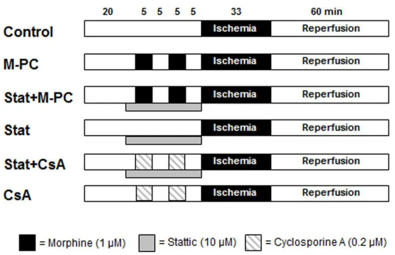
Fig 2summarizes the experimental design. Hearts were randomly assigned to one of six experi-mental groups. In group 1 (Control) hearts were kept under baseline conditions for 35 minutes before they underwent 33 minutes of ischemia followed by 60 minutes of reperfusion.
Mor-phine (1μM) and mPTP inhibitor cyclosporine A (0.2μM) [19] were administered twice for 5
minutes starting 20 and 10 minutes before onset of ischemia. STAT3 inhibitor Stattic (10μM)
[20] was administered over 23 minutes before performing global ischemia.
After 60 minutes of reperfusion, the hearts were cut into transverse slices, which were then stained with 0.75% triphenyltetrazoliumchloride (TTC) solution. The infarcted area was
deter-mined by planimetry using SigmaScan Pro 51computer software (SPSS Science Software,
Chi-cago, IL).
Cardiomyocytes
To investigate whether morphine regulates PKA activity, three additional experimental groups
(Con, Morphine 1μM, Morphine 10μM, each n = 7) were determined. Cardiomyocytes were
isolated from male Wistar rats. Hearts were perfused with oxygen-enriched medium (NaCl 110
mmol/l, KH2PO41.2 mmol/l, KCl 2.5 mmol/l, MgSO41.2 mmol/l, HEPES 25 mmol/l, glucose
10 mmol/l; perfusion flow 10 ml/min). Subsequently, recirculating perfusion was continued for
25 minutes (5 ml/min) with added ~0.06 w/v% collagenase and 25μM CaCl2. Afterwards, the
ventricular mass was minced into small pieces and incubated at 37°C for 5 minutes in oxygen-enriched medium. To isolate intact cardiomyocytes from large cellular detritus, the cell suspen-sion was filtrated through a nylon-mesh. The cell suspensuspen-sion was centrifuged 3 times for 2
minutes at 20 xg whilst gradually increasing CaCl2concentrations (0.2 mmol/l, 0.4 mmol/l and
1.0 mmol/l). The resulting pellet was resuspended in serum free M199 (Biochrom AG, Berlin, Germany; supplemented with pen/strep 2%, HEPES 15 mmol/l, L-carnitine 2 mmol/l, creatine
pre-coated (4 v/v% fetal calf serum) culture dishes. After 1.5–2 hours of incubation, cardiomyocytes
adhered and non-viable cells were washed off leading to cultures of>90% rod shaped cells.
The percentage of vital cardiomyocytes was quantified with trypan blue staining.
Western blot analysis
As a marker for PKA activation, phosphorylation of phospholamban, a protein that is phos-phorylated by PKA, was determined. Western blot analysis was performed as described previ-ously [15].
Statistical analysis
The sample size was calculated using GraphPad StatMateTMVersion 1.01 (GraphPad Software,
San Diego, CA, USA). Sample size analysis revealed a group size of n = 6 was necessary to
detect a difference in infarct size of 14% with a power of 80% andα<0.05. Data from infarct
size analysis are expressed as mean and standard deviation (SD), data from western blot analy-sis as median with 25th and 75th percentiles. Statistical analyanaly-sis (SPSS Science Software, ver-sion 17.0) of the hemodynamic variables was performed by using a two-way analysis of variance followed by Tukey test. Infarct sizes were analysed by a one-way analysis of variance followed by Tukey test. Statistical analysis of the western blot data was performed by the
Fig 2. Experimental protocol forin vitroinfarct size measurement.Con = control, M-PC = morphine preconditioning, Stat = STAT3 inhibitor stattic, CsA = atractyloside (mPTP blocker).
Kruskal–Wallis H-test followed by the Student–Newman–Keuls post hoc test. Changes within
and among groups were considered statistically significant if P<0.05.
Results
Infarct size measurement
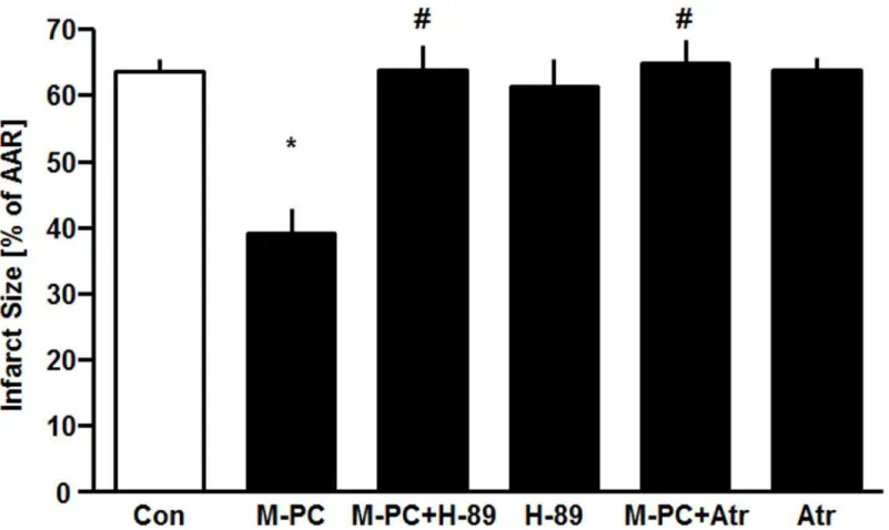
Administration of morphinein vivoreduced infarct size from 64±5% in the control group to
39±9% (P<0.05, seeFig 3). In combination with PKA-Inhibitor H-89 the effect of morphine
was completely abolished (H-89+M-PC: 63±10%, P<0.05 vs. M-PC). Single administration of
H-89 did not show any reduction of the infarct size compared to the control group (H-89: 61±10%, ns vs. Con).
The cardioprotective effect of morphine was completely abolished by atractyloside (Atr
+M-PC: 65±9%, P<0.05 vs. M-PC) suggesting a critical role for prevention of mPTP opening
in M-PC. Atractyloside alone had no effect on infarct size (Atr: 64±5%, ns vs. Con).
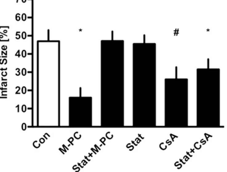
In Langendorff experiments administration of morphine reduced infarct size from 47±6%
in the control group to 16±5% (seeFig 4). In combination with STAT3 inhibitor Stattic the
effect was completely abolished (Stat+M-PC, 47±5% P<0.05 vs. Con). Stattic alone had no
effect on infarct size (Stat 45±5%, P<0.05 vs. M-PC). Administration of Stattic and mPTP
inhibitor cyclosporine A reduced infarct size to 31±6% (Stat+CsA, P<0.05 vs. Con).
Cyclo-sporine A alone reduced infarct size to 26±7% (CsA P<0.05 vs. Con).
Fig 3. Infarct size measurement.Histogram shows the infarct size (percent of area at risk) of controls (Con), morphine preconditioning (M-PC), H-89 combined with morphine preconditioning (H-89+M-PC), H-89 alone (H-89), atractyloside combined with morphine preconditioning (Atr+M-PC) and atractyloside alone (Atr). Data are presented as mean±SD,*P<0.05 vs. Con,#P<0.05 vs. M-PC.
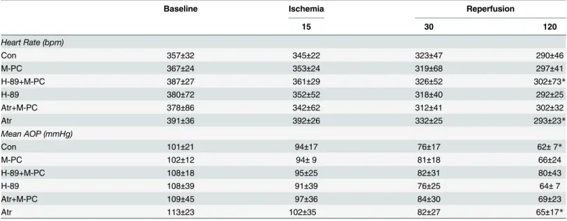
Hemodynamic variables
As shown inTable 1, no significant differences in heart rate and aortic pressure were observed
among thein vivoexperimental groups during baseline, ischemia or reperfusion. After 120
min-utes of reperfusion, heart rate was significantly lower in the H-89+M-PC and in the Atr group
(P<0.05 vs. baseline) compared to baseline. Mean aortic pressure was significantly decreased at
the end of reperfusion in the Con and Atr group (P<0.05 vs. baseline). As summarized inTable 2
at none of the different time points during Langendorff experiments was there a significant differ-ence in the heart rate, RPP, LVEDP, and coronary blood flow among the different experimental groups. Over the duration of the experiments heart rate remained stable in all groups, whereas
LVEDP was increased during the reperfusion period (all P<0.05 versus baseline). In contrast,
coronary flow decreased during the reperfusion period (P<0.05 versus baseline).
Animal characteristics
There were no differences in body weight or heart weight, respectively, between the different
experimental groups (seeTable 3).
Table 1. Hemodynamic variables.
Baseline Ischemia Reperfusion
15 30 120
Heart Rate (bpm)
Con 357±32 345±22 323±47 290±46
M-PC 367±24 353±24 319±68 297±41
H-89+M-PC 387±27 361±29 326±52 302±73*
H-89 380±72 352±52 318±40 292±25
Atr+M-PC 378±86 342±62 312±41 302±32
Atr 391±36 392±26 332±25 293±23*
Mean AOP (mmHg)
Con 101±21 94±17 76±17 62±7*
M-PC 102±12 94±9 81±18 66±24
H-89+M-PC 108±18 95±25 82±31 80±43
H-89 108±39 91±39 76±25 64±7
Atr+M-PC 109±45 97±36 84±30 69±23
Atr 113±23 102±35 82±27 65±17*
Data are mean±SD. Con = control; M-PC = morphine preconditioning; H-89+M-PC = PKA inhibitor H-89 prior to morphine preconditioning; H-89 = PKA inhibitor H-89 alone; Atr+M-PC = mPTP opener atractyloside prior to morphine preconditioning; Atr = atracyloside alone
*p<0.05 vs. Baseline.
doi:10.1371/journal.pone.0151025.t001
Table 2. Hemodynamic variables.
Baseline Reperfusion
30 45 60
Heart Rate (bpm)
Con 325±41 274±53 292±28 267±40
M-PC 334±48 297±33 282±44 267±43
Stat+M-PC 303±12 272±59 271±37 250±55
Stat 297±27 259±51 248±60 247±44
Stat+CsA 330±26 270±25 289±33 292±38
CsA 308±43 241±39 218±52 253±61
LVEDP (mmHg)
Con 3±2 68±10* 62±9* 60±8*
M-PC 4±1 71±7* 65±5* 63±5*
Stat+M-PC 3±2 69±11* 66±9* 63±8*
Stat 4±2 70±11* 66±10* 64±9*
Stat+CsA 3±2 60±10* 55±11* 52±9*
CsA 5±3 72±7* 67±6* 63±5*
CF (ml*min-1)
Con 15±2 10±2* 10±2* 10±2*
M-PC 14±2 10±2* 10±3* 10±3*
Stat+M-PC 13±3 7±2* 7±2* 7±2*
Stat 13±3 7±2* 7±2*§ 6±2*§
Stat+CsA 14±2 8±2* 7±2* 7±2*
CsA 13±2 8±1* 8±1* 7±1*
Data are mean±SD. Con = control; M-PC = morphine preconditioning; Stat = stattic; CsA = cyclosporine A. *P<0.05 vs. Baseline
§P<0.05 vs. Con.
Western blot analysis
Fig 5illustrates that morphine in a concentration of 10μM significantly increased
phospho-lamban phosphorylation (1.21(0.19) vs. Con, P<0.05). In contrast, 1μM morphine had no
effect on phospholamban phosphorylation (1.03(0.21) ns vs. Con).
Discussion
The main findings of our study suggest that morphine-induced preconditioning is mediated by activation of STAT3 which is located upstream of mPTP. Furthermore, there is some evidence that protein kinase A is involved within the signalling pathway.
Because morphine can protect the heart by pre- and postconditioning [7,8] it is suitable
agent for patients undergoing organ ischemia (e.g. heart surgery, vascular surgery, organ trans-plantation) or for patients who were recently exposed to regional ischemia (e.g. stroke, angina pectoris, myocardial infarction). Therefore, it is of particular interest to elucidate the underly-ing mechanisms of morphine-induced cardioprotection.
Previously, we could demonstrate that morphine-induced preconditioning is mediated by
activation of mKCachannels [7]. The mKCachannel inhibitor paxilline completely abrogated
the cardioprotective effect of morphine [7]. Cao et al. could show that the cardioprotective
effect of mKCachannel activation is abolished by opening of the mPTP [9]. The authors
con-cluded that the mKCachannel is an upstream regulator of the mPTP. This conclusion was
con-firmed by the fact that inhibition of the mKCachannel with paxilline and inhibition of the
mPTP with cyclosporine A at the same time was still protective [9]. Together with our own
data showing that morphine preconditioning is mediated by activation of mKCachannels [7] it
is possible that activation of the mKCachannel is an intermediate step in the signal
transduc-tion cascade of morphine preconditransduc-tioning. A possible upstream regulator of mKCachannels is
protein kinase A [10,11]. For desflurane preconditioning protein kinase A is located upstream
of mKCachannels [21]. The activity of protein kinase A depends on the cellular level of cAMP.
Morphine can lead to an increase in cellular cAMP levels [22] but the underlying mechanism is
unknown. However, it is conceivable that the increase in cAMP levels depends either on an ele-vated synthesis of cAMP by stimulation of adenylate cyclase or a less prominent decrease in cAMP by inhibition of phosphodiesterase. The first aim of the present study was to investigate the signal pathway of morphine preconditioning consisting of protein kinase A activation via
mKCachannel and inhibition of mPTP opening. We can exclude that the protective effect of
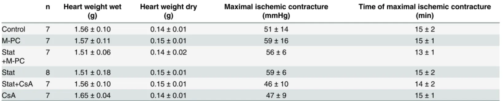
morphine is caused by differences in mechanical heart function as we determined no differ-ences in left ventricular function or coronary flow at baseline or during drug administration among treatment groups. Additionally we do not see significant differences in ischemic con-tracture or heart weights among the groups.
Table 3. Weights and ischemic contracture.
n Heart weight wet (g)
Heart weight dry (g)
Maximal ischemic contracture (mmHg)
Time of maximal ischemic contracture (min)
Control 7 1.56±0.10 0.14±0.01 51±14 15±2
M-PC 7 1.57±0.11 0.15±0.01 59±16 15±1
Stat +M-PC
7 1.51±0.06 0.14±0.02 56±6 13±1
Stat 8 1.51±0.18 0.15±0.01 59±6 15±2
Stat+CsA 7 1.56±0.10 0.15±0.01 46±10 14±2
CsA 7 1.65±0.04 0.14±0.01 47±9 15±1
Data are mean±SD. M-PC = morphine preconditioning; Stat = stattic; CsA = cyclosporine A.
Our finding that the protein kinase A inhibitor H-89 completely abolished the cardioprotec-tive effect of morphine suggest that morphine-induced preconditioning is critically dependent on activation of protein kinase A. However, this conclusion is challenged by investigations
demonstrating non-specific actions of H-89 [23].
Interestingly, the existence of a possible mechanistic link between morphine and PKA might be supported by our finding that morphine increases phospholamban phosphorylation.
It is important to note that this effect was seen using a high morphine concentration of 10μM.
This concentration is usually not reached in blood plasma after morphine bolus injection and therefore has to be considered as "supra-pharmacological" concentration. In conclusion, our findings 1) that morphine-induced cardioprotection is abrogated by PKA inhibitor H-89, and
2) morphine is capable to activate PKA in cardiomyocytes at least at 10μM might indicate that
PKA activation is involved in morphine-induced preconditioning.
In addition, it has been demonstrated that activation of protein kinase A can regulate the
mKCachannels [10,11]. Redel et al demonstrated using a desflurane-induced preconditioning
model that protein kinase A is located upstream of the mKCachannel [21].
Mitochondrial mechanisms are involved in myocardial pre- and postconditioning. Activa-tion of mitochondrial potassium channels might lead to an inhibiActiva-tion of mPTP opening
[24,25]. Hausenloy et al. reported that inhibition of mPTP is involved in cardioprotection by
ischemic preconditioning [26]. Potassium influx into the mitochondrial matrix via activated
potassium channels was suggested to change mitochondrial function. This change leads to the release of oxygen radicals, probably caused by a loss of electrons from the mitochondrial elec-tron transport chain. These oxygen radicals are crucially involved in activating different
kinases, which act as trigger and/or mediator of cardioprotection [27,28]. Mitochondria might
play a role as an endeffector of cardioprotection. A possible candidate is the mPTP [26,29]. As
mentioned above the mPTP is located downstream of mKCachannels [9]. Based on the results
from this study together with data from a recent publication of our group showing that
mor-phine preconditioning is abolished by inhibition of mKCachannels [30], we suggest the
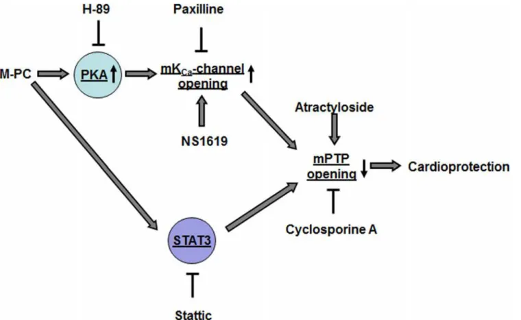
follow-ing signal transduction pathway for morphine preconditionfollow-ing (seeFig 6): Morphine enhances
protein kinase A activity, which in turn leads to activation of mKCachannels with the
conse-quence of prevention of mPTP opening during myocardial ischemia and reperfusion. However, despite the significance of these findings, our results represent only a small pro-portion of mechanisms underlying morphine-induced cardioprotection. Other signalling path-ways have been described for morphine-induced preconditioning including a role for PI3K/ Akt signalling and/or JAK/STAT3 activation. To integrate our results into some aspects of other signalling mechanisms described we conducted supplementary experiments in isolated hearts focusing on the role of STAT3. It has been demonstrated that STAT3 plays an essential
role in morphine-induced cardioprotection [16]. Furthermore there is evidence that STAT3
might be regulated by activation of PKA [31] and is mechanistically linked to prevention of
mPTP-opening [32].Therefore, we tested the protective properties of morphine in the presence
of STAT3 inhibitior Stattic, and whether pharmacological prevention of the mPTP-opening by cyclosporine A (CsA) is cardioprotective during STAT3 inhibition.
Our data show that the protective effect of morphine is abrogated by the STAT3 inhibitor Stattic. This is in line with results from other groups showing a role for STAT3 in morphine-PC. In addition, our finding that CsA offers cardioprotection during STAT3 inhibition suggests that mPTP might be located downstream of STAT3 within the protective signalling pathway.
Fig 5. Western Blot analysis.Summarized data presenting ratio of phosphorylated phospholamban (phospho-PLB) to beta-actine from control and morphine treated hearts are shown. The boxes represent the lower (25th) and upper (75th) quartiles, the horizontal line represents the mean, the whiskers extend to 95% confidence intervals for the mean,*P<0.05 vs. Con.
It remains unclear whether the cAMP/PKA/mKCasignalling pathway and the STAT3 path-way are redundant signalling pathpath-ways that only converge at the mitochondrial level (mPTP) to initiate protection or whether these pathways directly interact, e.g. by PKA-STAT3 interac-tion. These specific questions have to be addressed in future studies.
Fig 6shows a schematic representation of the signalling pathways of morphine-induced pre-conditioning that has been investigated within this study. It has to be mentioned that several other signalling pathways have been described to be involved in morphine-induced cardiopro-tection that are not included in this simplified model. Morphine initiates cardioprocardiopro-tection by
activation of the PKA/mKCa/mPTP pathway. Furthermore, morphine-induced
cardioprotec-tion is dependent on STAT3 activacardioprotec-tion; however it remains unclear whether morphine acti-vates STAT3 via PKA dependent or independent mechanisms. Furthermore, within the signalling pathway, STAT3 is located upstream of mPTP.
Acknowledgments
This work was supported by a Research Fund (SFF 701301765) from the Heinrich-Heine-Uni-versity Duesseldorf, Germany.
Author Contributions
Conceived and designed the experiments: AH RH. Performed the experiments: MR DB HG AH RH. Analyzed the data: MD FB MWH AH RH. Wrote the paper: MD AH RH.
Fig 6. Schematic model of the proposed cardioprotective pathway of morphine preconditioning.M-PC = morphine preconditioning, PKA = protein kinase A, H-89 = protein kinase A inhibitor, Paxilline = mKCa-channel inhibitor, NS1619 = mKCa-channel activator, mPTP = mitochondrial permeability transition pore, Stattic = STAT3 inhibitor
References
1. Murry CE, Jennings RB, Reimer KA. Preconditioning with ischemia: a delay of lethal cell injury in ische-mic myocardium. Circulation. 1986 Nov; 74(5):1124–36. PMID:3769170
2. Zhao Z-Q, Corvera JS, Halkos ME, Kerendi F, Wang N-P, Guyton RA, et al. Inhibition of myocardial injury by ischemic postconditioning during reperfusion: comparison with ischemic preconditioning. American Journal of Physiology—Heart and Circulatory Physiology. 2003 Aug 1; 285(2):H579–88. PMID:12860564
3. Cason BA, Gamperl AK, Slocum RE, Hickey RF. Anesthetic-induced preconditioning: previous admin-istration of isoflurane decreases myocardial infarct size in rabbits. Anesthesiology. 1997 Nov; 87 (5):1182–90. PMID:9366471
4. Müllenheim J, Ebel D, Frässdorf J, Preckel B, Thämer V, Schlack W. Isoflurane preconditions myocar-dium against infarction via release of free radicals. Anesthesiology. 2002 Apr; 96(4):934–40. PMID: 11964602
5. Gross ER, Gross GJ. Ligand triggers of classical preconditioning and postconditioning. Cardiovasc Res. 2006 May 1; 70(2):212–21. PMID:16448635
6. Halestrap AP, Clarke SJ, Khaliulin I. The role of mitochondria in protection of the heart by precondition-ing. Biochimica et Biophysica Acta (BBA)—Bioenergetics. 2007 Aug; 1767(8):1007–31.
7. Frässdorf J, Huhn R, Niersmann C, Weber NC, Schlack W, Preckel B, et al. Morphine induces precon-ditioning via activation of mitochondrial K(Ca) channels. Can J Anaesth. 2010 Aug; 57(8):767–73. doi: 10.1007/s12630-010-9325-1PMID:20461490
8. Huhn R, Heinen A, Weber NC, Schlack W, Preckel B, Hollmann MW. Ischaemic and morphine-induced post-conditioning: impact of mK(Ca) channels. Br J Anaesth. 2010 Nov; 105(5):589–95. doi:10.1093/ bja/aeq213PMID:20693178
9. Cao C-M, Xia Q, Gao Q, Chen M, Wong T-M. Calcium-activated potassium channel triggers cardiopro-tection of ischemic preconditioning. J Pharmacol Exp Ther. 2005 Feb; 312(2):644–50. PMID: 15345753
10. Sanada S, Asanuma H, Tsukamoto O, Minamino T, Node K, Takashima S, et al. Protein kinase A as another mediator of ischemic preconditioning independent of protein kinase C. Circulation. 2004 Jul 6; 110(1):51–7. PMID:15210595
11. Sato T, Saito T, Saegusa N, Nakaya H. Mitochondrial Ca2+-activated K+ channels in cardiac myo-cytes: a mechanism of the cardioprotective effect and modulation by protein kinase A. Circulation. 2005 Jan 18; 111(2):198–203. PMID:15623543
12. Obal D, Dettwiler S, Favoccia C, Scharbatke H, Preckel B, Schlack W. The influence of mitochondrial KATP-channels in the cardioprotection of preconditioning and postconditioning by sevoflurane in the rat in vivo. Anesth Analg. 2005 Nov; 101(5):1252–60. PMID:16243977
13. Toma O, Weber NC, Wolter JI, Obal D, Preckel B, Schlack W. Desflurane preconditioning induces time-dependent activation of protein kinase C epsilon and extracellular signal-regulated kinase 1 and 2 in the rat heart in vivo. Anesthesiology. 2004 Dec; 101(6):1372–80. PMID:15564945
14. Heinen A, Huhn R, Smeele KMA, Zuurbier CJ, Schlack W, Preckel B, et al. Helium-induced precondi-tioning in young and old rat heart: impact of mitochondrial Ca(2+) -sensitive potassium channel activa-tion. Anesthesiology. 2008 Nov; 109(5):830–6. doi:10.1097/ALN.0b013e3181895aa0PMID: 18946295
15. Huhn R, Weber NC, Preckel B, Schlack W, Bauer I, Hollmann MW, et al. Age-related loss of cardiac preconditioning: impact of protein kinase A. Exp Gerontol. 2012 Jan; 47(1):116–21. doi:10.1016/j. exger.2011.11.003PMID:22100641
16. Gross ER, Hsu AK, Gross GJ. The JAK/STAT pathway is essential for opioid-induced cardioprotection: JAK2 as a mediator of STAT3, Akt, and GSK-3 beta. Am J Physiol Heart Circ Physiol. 2006 Aug; 291 (2):H827–34. PMID:16517948
17. Puana R, McAllister RK, Hunter FA, Warden J, Childs EW. Morphine attenuates microvascular hyper-permeability via a protein kinase A-dependent pathway. Anesth Analg. 2008 Feb; 106(2):480–5, table of contents. doi:10.1213/ane.0b013e318160648bPMID:18227303
18. Zhang S, Wang N, Xu J, Gao Q, Lin G, Bruce IC, et al. Kappa-opioid receptors mediate cardioprotection by remote preconditioning. Anesthesiology. 2006 Sep; 105(3):550–6. PMID:16931988
19. Li J, Iorga A, Sharma S, Youn J-Y, Partow-Navid R, Umar S, et al. Intralipid, a clinically safe compound, protects the heart against ischemia-reperfusion injury more efficiently than cyclosporine-A. Anesthesi-ology. 2012 Oct; 117(4):836–46. PMID:22814384
by Reperfusion Injury Salvage Kinase and Survival Activating Factor Enhancement Pathways. Circ Res. 2015 Jul 17; 117(3):279–88. doi:10.1161/CIRCRESAHA.117.306878PMID:26058828 21. Redel A, Lange M, Jazbutyte V, Lotz C, Smul TM, Roewer N, et al. Activation of Mitochondrial
Large-Conductance Calcium-Activated K+ Channels via Protein Kinase A Mediates Desflurane-Induced Pre-conditioning. Anesthesia & Analgesia February 2008. 2008; 106(2):384–91.
22. Muraki T, Uzumaki H, Kato R. Effect of morphine on the tissue cyclic AMP and cyclic GMP content in two strains of mice. J Pharm Pharmacol. 1984 Jul; 36(7):490–2. PMID:6146702
23. Makaula S, Lochner A, Genade S, Sack MN, Awan MM, Opie LH. H-89, a non-specific inhibitor of pro-tein kinase A, promotes post-ischemic cardiac contractile recovery and reduces infarct size. J Cardio-vasc Pharmacol. 2005 Apr; 45(4):341–7. PMID:15772523
24. Cohen MV, Yang XM, Liu GS, Heusch G, Downey JM. Acetylcholine, bradykinin, opioids, and phenyl-ephrine, but not adenosine, trigger preconditioning by generating free radicals and opening mitochon-drial K(ATP) channels. Circ Res. 2001 Aug 3; 89(3):273–8. PMID:11485978
25. Murphy E, Steenbergen C. Preconditioning: the mitochondrial connection. Annu Rev Physiol. 2007; 69:51–67. PMID:17007587
26. Hausenloy DJ, Duchen MR, Yellon DM. Inhibiting mitochondrial permeability transition pore opening at reperfusion protects against ischaemia–reperfusion injury. Cardiovasc Res. 2003 Jan 12; 60(3):617– 25. PMID:14659807
27. Pain T, Yang XM, Critz SD, Yue Y, Nakano A, Liu GS, et al. Opening of mitochondrial K(ATP) channels triggers the preconditioned state by generating free radicals. Circ Res. 2000 Sep 15; 87(6):460–6. PMID:10988237
28. Stowe DF, Aldakkak M, Camara AKS, Riess ML, Heinen A, Varadarajan SG, et al. Cardiac mitochon-drial preconditioning by Big Ca2+-sensitive K+ channel opening requires superoxide radical genera-tion. American Journal of Physiology—Heart and Circulatory Physiology. 2006 Jan 1; 290(1):H434–40. PMID:16126810
29. Halestrap AP, Clarke SJ, Javadov SA. Mitochondrial permeability transition pore opening during myo-cardial reperfusion—a target for cardioprotection. Cardiovasc Res. 2004 Feb 15; 61(3):372–85. PMID: 14962470
30. Heinen A, Ströthoff M, Schmidt A, Stracke N, Behmenburg F, Bauer I, et al. Pharmacological options to protect the aged heart from ischemia and reperfusion injury by targeting the PKA-BK(Ca) signaling pathway. Exp Gerontol. 2014 Aug; 56:99–105. doi:10.1016/j.exger.2014.03.029PMID:24727217 31. Park SY, Kim J-H, Lee SJ, Kim Y. Involvement of PKA and HO-1 signaling in anti-inflammatory effects
of surfactin in BV-2 microglial cells. Toxicol Appl Pharmacol. 2013 Apr 1; 268(1):68–78. doi:10.1016/j. taap.2013.01.017PMID:23360889