online | memorias.ioc.fiocruz.br Malnutrition is a serious public health problem that
has been linked to a substantial increase in the risk of mortality and morbidity (Blössner & Onis 2005), affect-ing mainly women and young children. In Africa and South Asia, for example, 27-51% of women of reproduc-tive age were underweight and it is believed that about 130 million children (21% of all children) suffered of malnutrition in 2005 (Blössner & Onis 2005). Globally, protein-energy malnutrition (PEM) is the most frequent cause of human immunodeficiency (Revillard & Cozon 1991). The development of a normal immune system will be impaired by malnutrition during critical periods of pregnancy, neonatal maturation and weaning (McDade et al. 2001, Keusch 2003). Many studies have shown that the cellular immune system is more directly affected than the humoral immune system by malnutrition (Mc-Murray 1981). Primary malnutrition leads to atrophy of the lymphoid organs, profound T-lymphocyte deficien-cy, increased susceptibility to pathogens and develop-ment of opportunistic infections (Prentice 1999). Zinc and iron deficiencies are usually associated with PEM. Zinc is an essential trace element for the immune system and zinc deficiency compromises primarily the function
Financial support: FAPEMIG, CNPq, Biotério Central/UFOP + Corresponding author: [email protected] Received 11 March 2010
Accepted 26 May 2010
of T cells, but also affects several other immune cells. Zinc deficiency affects about one-third of the world’s population. Zinc is an essential cofactor for the activity of many enzymes and its deficiency leads to a reduc-tion in Th1 cytokines and thymic hormone activity, ulti-mately leading to lymphopaenia. Zinc supplementation improves pathogen-specific cell-mediated immunity in severely malnourished children with shigellosis (Raqib et al. 2004). Iron is also a critical cofactor to immune function and its deficiency is the most common micro-nutrient deficiency in the world, especially in the tropics (Oppenheimer 2001). Iron deficiency has been associ-ated with defects in both adaptive and innate immunity (Cunningham-Rundles et al. 2005). Furthermore, iron deficiency anaemia in children is associated with reduc-tions in both phagocytic activity and immunoglobulin levels (Ekiz et al. 2005).
Epidemiologic and experimental studies have docu-mented the importance of visceral leishmaniasis (VL) in public health. VL is the most devastating form among the clinical forms of leishmaniasis (cutaneous, muco-cutaneous and visceral) and it is caused by intracellular protozoan parasites of the Leishmania donovani complex (Garg & Dube 2006). Human infection with Leishmania (Leishmania) chagasi/infantum (Maurício et al. 2000) is characterized by long-term low-grade fever, enlarged spleen and liver and weight loss. Laboratory findings reveal pancytopaenia, low levels of albumin and hyper-gammaglobulinaemia. Host protection against Leishma-nia infection is dependent on the development of type-1 immunity, which triggers enhanced leishmanicidal activity by infected macrophages (Caldas et al. 2005).
Immune response to Leishmania (Leishmania) chagasi
infection is reduced in malnourished BALB/c mice
Tiago Donatelli Serafim1, Guilherme Malafaia2/+, Marcelo Eustáquio Silva3,
Maria Lúcia Pedrosa4,5, Simone Aparecida Rezende1,6
1Laboratório de Imunoparasitologia 3Laboratório de Nutrição Experimental 4Laboratório de Bioquímica e Biologia Molecular, Núcleo de Pesquisa em Ciências Biológicas 5Departamento de Ciências Biológicas, Instituto de Ciências Exatas e Biológicas
6Departamento de Análises Clínicas, Escola de Farmácia, Universidade Federal de Ouro Preto, Ouro Preto, MG, Brasil 2Departamento de Ciências Biológicas, Instituto Federal de Educação, Ciência e Tecnologia, Urutaí, GO, Brasil
Protein-energy malnutrition and micronutrient deficiencies may down-regulate immune response and increase morbidity and mortality due to infection. In this study, a murine model was used to study the effects of protein, iron and zinc deficiencies on the immune response to Leishmania (Leishmania) chagasi infection. Mice were initially fed a standard diet or with a diet containing 3% casein but deficient in zinc and iron. After malnutrition was established, mice were inoculated with L. chagasi and sacrificed four weeks later in order to evaluate liver and spleen para-site loads and serum biochemical parameters. Significant decreases in liver and spleen weight, an increase in the parasite loads in these organs and decreases in serum protein and glucose concentrations in malnourished animals were observed. Furthermore, the production of interferon-gammaby spleen cells from infected malnourished mice stimulated by Leishmania antigen was significantly lower compared with that in control diet mice. These data sug-gest that malnutrition alters the immune response to L. chagasi infection in the BALB/c model and, in association with the effects on biochemical and anatomical parameters of the host, favored increases in the parasite loads in the spleens and livers of these animals.
Clinical forms may range from sub-clinical infection to progressive fatal disease (Wilson & Streit 1996). If left untreated, the disease may be fatal.
In an innovative prospective study, Badaró et al. (1986) analyzed the epidemiology, clinical patterns and risk factors of VL in an endemic area of Brazil. The authors observed that 45% of VL cases developed among the 11% of the population suffering from second and third-degree malnutrition and that the risk factors included young age (average, 3 years) and PEM before the onset of disease. By using data from the same study, Cerf et al. (1987) concluded that a child with moderate or severe malnutrition had about a 9-fold increased risk of developing VL compared to a well-nourished child. More recently, Maciel et al. (2008) assessedwhether nu-tritional status influenced the outcome of Leishmania infectionby comparing relatives of children with VL with either self-resolvingLeishmania spp infection or apparently uninfected households.The authors observed decreases in the body mass index andmid-upper arm circumference by age z-scores forchildren with VL. Furthermore, levels of vitamin A were lower in chil-dren with active VL as measured by serum retinol and the modified-relative-dose-responsetest. Higher birth weight and albuminconcentrations protected against the disease. Increased breastfeeding was associated with asymptomaticinfection. These results indicate that modifiable nutritionalaspects are associated with the outcome of Leishmania spp infectionin humans.
Although it is accepted that immunity or susceptibil-ity to infect-parasitic diseases is directly related to host nutritional status, the immunologic mechanisms that govern the relationship between PEM and the course of VL are multiple and not well explained. The impact of PEM specifically on immune response against Leishma-nia infection is not totally understood and the nutritional status of the host is often neglected (Malafaia 2009). Anstead et al. (2001) studied the effects of malnutrition on innate immunity and early visceralization following L. donovani infection and observed that malnutrition causes a failure of lymph node barrier function after L. donovani infection, which may be related to excessive production of prostaglandin E2 and decreased levels of interleukin (IL) 10 and nitric oxide (NO). In this study, a murine model of polynutrient deficiency (diets deficient in iron, zinc and protein) was established and demon-strated that malnourished mice have an altered innate immune defence and an increased risk of visceralization following cutaneous L. donovani infection.
Despite the evidence provided by Anstead et al. (2001) indicating that PEM affects the innate immune response in mice infected with L. donovani, little is known about the effects of PEM on the adaptive immune response in mice infected with L. chagasi. Thus, in the present study, a murine model of PEM was created based on that described by Anstead et al. (2001) to evaluate the effects of combined protein, iron and zinc deficien-cies on some aspects of the adaptive immune response against L. chagasi, the causative agent of VL in the New World. Many experimental animal models have been used to study VL, but few studies investigated the evolu-tion of infecevolu-tion in malnourished mice.
SUBJECTS, MATERIALS AnD METhoDS
Leishmania parasite and antigen - L. chagasi strain MHOM/BR/1974/M2682 was used for the preparation of Leishmania antigen. Promastigotes were grown in Grace´s Insect Medium (Gibco BRL, Grand Island, NY) pH 6.5 supplemented with 10% heat-inactivated foe-tal bovine serum, 2 mM l-glutamine, 25 mM HEPES,
50 µM 2-mercaptoethanol and 20 µg/mL gentamicin at 25°C. Infectivity was maintained by serial passage in BALB/c mice. Promastigotes of L. chagasi were har-vested from late-log-phase cultures by centrifugation and washed three times in PBS. For the experimental infection, 1 × 10⁷ promastigotes were suspended in 200 µL RPMI pH 7.2. For antigen preparation, parasites were disrupted by three rounds of freezing and thawing (freeze-thawed antigen), protein content was estimated by the Lowry method (Lowry et al. 1951) and the prepa-ration was frozen at -20°C until use.
Animals, diets and experimental infection - Female BALB/c mice (3 weeks old) were obtained from Central Biotherium, Federal University of Ouro Preto (UFOP). After weaning, all mice received standard mouse chow (Nuvilab CR-II) until the change to experimental diets. These mice were initially divided in two groups (control and malnourished) that received control and malnutri-tion diets, respectively (n = 12 mice per group). The con-trol group received a diet based on Reeves et al. (1993) that contained 14% casein and sufficient concentrations of iron and zinc. The malnourished group was fed a diet containing 3% casein but deficient in iron and zinc (Ta-ble). The protein level of the casein ranged from 70-80%. Mineral and vitamin mixtures were prepared according to the recommendations for adult mice of the American Institute of Nutrition (Reeves et al. 1993). Diets were pre-pared at the Experimental Nutrition Laboratory, School of Nutrition, UFOP.
Mice were initially weight-matched and subsequently assigned a diet. Total body weight was measured weekly. As soon as body weights stabilized in the malnutrition diet group, half of the mice in each group (control and
TABLE
Composition of experimental diets (g/Kg)
Ingredients Control Malnutritionb
Casein 140 30
Corn oil 40 40
Sucrose 100 100
Cellulose 50 50
Vitamin mixturea 10 10
Mineral mixturea 35
-Mineral mixture without iron and zinca - 35
malnutrition) were inoculated with L. chagasi. For the ex-perimental infection, mice were inoculated with 1 × 10⁷ promastigotes of L. chagasi given intravenously in the lateral tail vein. After inoculation, there were four experi-mental groups: non-infected control group (CG), non-in-fected malnourished group (MG), innon-in-fected control group (ICG) and infected malnourished group (IMG).
All animal procedures were approved by the Commit-tee on Ethics in Research of the UFOP, MG, Brazil and followed the guidelines for the use and care of animals for research published by the Canadian Council on Animal Care (1980, 1984).
Biochemical parameters - To evaluate mouse nu-tritional status, haemoglobin, total protein, albumin, globulins and glucose were measured in mice from all experimental groups on the first experimental day, be-fore experimental infection (7th experimental week) and on the 28th day post-infection (11th experimental week). Mice were fasted for 12 h for determination of biochemi-cal parameters and blood samples were collected via the ocular plexus. The haemoglobin concentration was deter-mined immediately after collection using a commercial kit (Labtest Kit, Cat 43, Lagoa Santa, MG, Brazil). Serum was separated by blood centrifugation and total protein, albumin and glucose levels were determined by the stan-dard methods used in medical analysis (Labtest Kit, Cat 18, 19 and 84 respectively). Globulin concentrations were calculated by the difference between the total protein and albumin concentrations.
Determination of tissue parasite burden - To evaluate the effect of PEM on the parasite load, spleens and livers were collected and weighed separately. A fragment from each organ was collected to be used for parasite quanti-fication. Quantitative limiting dilution culture was per-formed as described by Titus et al. (1985) and modified by Marques-da-Silva et al. (2005). One weighed fragment from each organ (liver and spleen) was homogenized in a
tissue grinder and suspended in 500 μL of Grace’s Insect
Medium (Gibco BRL, Grand Island, NY, USA) supple-mented with 10% heat-inactivated foetal calf serum (FCS) (Cripton, Andradina, SP, Brazil), 2 mM L-glutamine (Gibco BRL) and 100 U/mL penicillin G potassium (USB Corporation, Cleveland, OH, USA), pH 6.5 in 96-well
flat-bottom microtitre plates (160 μL per well). Five-fold serial
dilution was done and, after 14 days, plates were scored microscopically for determination of parasite burden. The number of parasites was determined from the reciprocal of the highest dilution at which promastigotes could be detected after 14 days of incubation at 25°C.
Determination of cytokine and NO production - Sin-gle-cell suspensions of spleen were obtained by tissue grinder homogenization and processed as described pre-viously (Marques-da-Silva et al. 2005). These cells were cultured in 48-well flat-bottom microtitre plates for 72 h
in medium (Medium) or stimulated with 50 μg/mL of
freeze-thawed L. chagasi antigen (L. chagasi Ag.). Cell culture supernatant was collected from 3-day cultures
and the production of interferon-gamma (IFN-γ) and IL-4 was determined in these supernatants by enzyme linked
immunosorbent assay (Afonso & Scott 1993). IL-10 pro-duction was evaluated using a PeproTech kit (PeproTech, 900-K53, Rocky Hill, NJ, USA). TNF production was evaluated by biologic assay using WEHI cells (Lattime et al. 1988). The production of NO was determined by the Griess method (Green et al. 1982).
Statistical analyses - All data were analyzed by the D’Agostino & Pearson omnibus normality test. Data with normal distribution were analyzed by Student’s t test. Data with distributions that were not considered normal were submitted to non-parametric Mann-Whitney’s test. Differ-ences were considered statistically significant at p < 0.05.
RESULTS
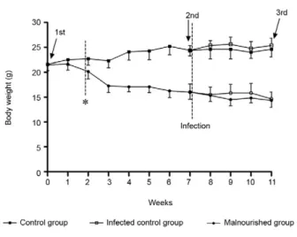
Body weight and biochemical parameters - Total body weight was evaluated weekly after mice were fed with the control or malnutrition diet. Mice fed the malnutrition diet had a progressive decrease in body weight and, in two weeks, it was possible to observe a statistical difference in body weight between the two experimental groups that was maintained until the end of the experiment (Fig. 1). We ob-served no differences in body weight between mice inocu-lated with L. chagasi and mice that were not infected.
The concentrations of biochemical parameters (haemoglobin, total protein, albumin, globulins and glucose) were determined 28 days after infection to evaluate the nutritional status of experimental mice.
A significant reduction in total protein concentration was observed in malnourished mice compared to con-centration in control diet mice starting from the 7th experimental week and this reduction was maintained until the end of the experiment (Fig. 2A). In addition, reductions in albumin (Fig. 2B), globulins (Fig. 2C) and glucose (Fig. 2D) levels were also observed in the mal-nourished mice compared to the levels in control mice. Albumin levels were not reduced in infected mice in comparison to uninfected mice, as is observed in hu-man cases. There were no differences in haemoglobin concentrations between mice fed control or malnutri-tion diets (data not shown). Moreover, no differences were observed in the levels of total protein, albumin or globulins in infected mice when compared to the levels in uninfected mice.
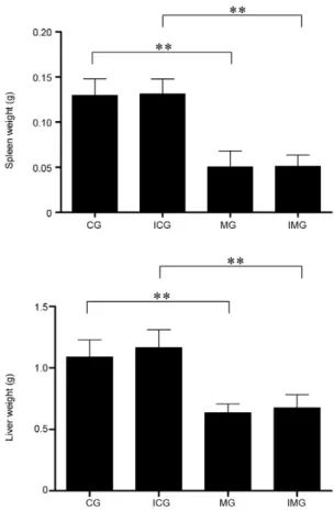
Spleen and liver weight - Four weeks after experi-mental infection, mice were sacrificed and spleens and livers were collected and weighed. Significant reduc-tions in liver and spleen weight in malnourished mice
were observed in comparison to those in control diet mice (Fig. 3). No difference in spleen or liver weight was observed among infected and non-infected mice. We also observed fatty livers in malnourished animals, irrespective of infection (figure not shown).
Parasite loads in spleen and liver -Spleen and liver parasite loads were evaluated in order to study the ef-fects of PEM on the response to the parasite. Although no difference was observed in the parasite load per spleen (Fig. 4B), we observed a significant increase in the parasite load per liver (Fig. 4A) and per milligram of tissue in both livers and spleens from mice fed the malnutrition diet compared to those of the control diet-fed mice (Fig. 4C, D).
Cytokine and NO production by spleen cells - Since PEM caused an increase in parasite load, the produc-tion of cytokines by spleen cells in response to L. cha-gasi antigen was also evaluated. Significant production
of IFN-γ by spleen cells was observed in the infected
in BALB/c mice infected with L. chagasi and the parasite load is increasing in the spleen (Marques-da-Silva et al. 2005). Total body weight was analyzed weekly and two weeks after the change in diet, a significant reduction in body weight was observed in mice fed the low protein diet. Despite the fact that the diets were isocaloric, ex-Fig. 3: spleen and liver weight from mice fed with control or
malnutri-tion diet, infected or not. Mice were sacrificed 28 days after inocula-tion with 1 x 10⁷ promastigote forms of Leishmania chagasi and spleen and liver weight was determined. The markers over the bars represent significant organ weight decrease (p < 0.01) comparing control group (CG) and malnourished group (MG) and infected control (ICG) and malnourished group (IMG). The bars represent the mean + standard deviation of two independent experiments (n = 6 for each experimental group). Statistical differences were determined by Student’s t test.
Fig. 4: parasite load from mice fed with control or malnutrition diet. Parasite load per liver (A), per spleen (B), per milligram of hepatic tis-sue (C) and per milligram of splenic tistis-sue (D) were harvested at 28 days after inoculation with 1 × 10⁷Leishmania chagasi stationary-phase pro-mastigotes and parasite load was quantified by limiting-dilution quan-titative culture. The bars represent the mean + standard deviation from two independent experiments [n = 6 for control infected group (CG) and n = 6 for malnourished infected group (MG)]. Statistical differences were determined by Student’s t test. The markers over the bars indicate a statistical difference between CG and MG (p < 0.05).
Fig. 5: interferon-gamma (IFN-γ) production by spleen cells from
mice fed with control or malnutrition diet. Animals were sacrificed 28 days after inoculation with 1 × 10⁷Leishmania chagasi stationary-phase promastigotes, spleen cells were collected and stimulated with 50 µg/mL of L. chagasi Ag in vitro. IFN-γ production was measured
by enzyme linked immunosorbent assay. Group division was: infected control group (ICG), non-infected control group (CG), infected mal-nourished group (IMG) and non-infected malmal-nourished group (MG). The bars represent the median + interquartile range of two indepen-dent experiments (n = 6 for each experimental group). Statistical dif-ferences were determined by non-parametric Mann-Whitney’s test.
The markers over the bars represent significant difference in IFN-γ
production (*: p < 0.05; **: p < 0.01). pared to uninfected animals (Fig. 5). On the other hand,
spleen cells obtained from infected malnourished mice
were not able to produce IFN-γ when stimulated with
L. chagasi antigen. Since IL-10 is a cytokine with many suppressive effects (Nylén & Sacks 2007), we analyzed the production of this cytokine by spleen cells obtained from the different experimental groups. No differences were observed in the levels of IL-10 between mice fed the control or malnutrition diets or between infected and uninfected mice, indicating that VL in mice is not characterized by high levels of IL-10. Significant levels of TNF, IL-4 and NO were not detected in the superna-tants studied (data not shown).
DISCUSSIon
perimental data obtained in other studies have shown de-creased diet consumption when a hypoproteic diet is used (Chatraw et al. 2008, Fock et al. 2008). Therefore, reduced consumption of calories, macro and micronutrients was observed. Consequently, body weight reduction was ac-companied by a reduction in the levels of total protein, al-bumin and globulins in the mice fed the hypoproteic diet in comparison to mice fed the control diet, as observed in the literature (Fock et al. 2008). These data show typical signs of malnutrition. Interestingly, data from human VL indicate polyclonal hypergammaglobulinaemia and a de-crease in the level of albumin (Bouree et al. 2000), but this was not observed in the BALB/c model of L. chagasi in-fection. These data may appear somewhat strange, but the BALB/c mouse is an experimental model widely used in the study of VL that is comparable to self-controlled oli-gosymptomatic cases in humans. Therefore, it is expected that the signs, symptoms and laboratory data are less se-vere if compared to humans with VL. We could have used the golden hamster, an excellent model to study active human disease (Melby et al. 2001). In this animal, infec-tion is characterized by a relentless increase in visceral parasite burden, progressive cachexia, splenomegaly, pan-cytopaenia, hypergammaglobulinaemia and eventually death. However, since infection with L. donovani complex parasites causes a severe reduction in weight, this model cannot be used to study the effects of malnutrition in the evolution of infection, as it is not possible to have a con-trol well-nourished group (Melby et al. 2001). Concern-ing haemoglobin levels, no difference between the groups was verified. In this case, it is important to emphasize that three stages of iron deficiency have been described in the literature. We suggest that the experimental time used in our study was not sufficient for the occurrence of the de-layed stages of iron deficiency and that this explains the lack of differences between the haemoglobin concentra-tions in the experimental groups.
Body weight was analyzed weekly until mice were killed and no difference was observed in the body weight of infected mice in comparison to that in non-infected mice (control and malnourished mice). This result differs from studies using the hamster model in which an 18% reduction in body weight in L. donovani infected animals was detected by the 56th day of infection (Melby et al. 2001). We suggest that this is due to the time of evaluation (28 days post-infection) and that it might be explained by the virulence of the chosen L. chagasi strain.
Although human VL is characterized by spleen and liver enlargement, we did not observe any increase in the spleen and liver in mice infected with L. chagasi com-pared to those in non-infected control mice on 28 days post-infection. A study performed by Smelt et al. (1997) in which 2 × 10⁷L. donovani amastigotes were inoculated by tail vein injection shows that parasites are not detected in the spleen after two weeks of infection, but a progressive increase is observed in the beginning of the third week of infection followed by an increase in spleen weight. In the study by Mukherjeeet al. (2006), VL produced in BALB/c mice through intracardial administration of L. donovani amastigotes was accompanied by
hepatosple-nomegaly with high organ parasite loads and lymphade-nopathy when animals were followed up to four months. In our model, no increase was detected at 28 days post-infection, but this can be related to the species, number and form of parasites since we used 1 × 10⁷ L. chagasi promastigotes. Moreover, one should consider the route of inoculation of parasites used in the study performed by Mukherjeeet al. (2006). We suggest that splenomegaly can be detected in chronically infected mice in our model of infection, since uncontrolled parasite multiplication is observed in this organ. Otherwise, significant decreases in spleen and liver weight were detected in both infected and non-infected mice that were fed the hypoproteic diet. This reduction might be related to a reduction in cellular mi-gration to these organs. Since cellular mimi-gration depends on the expression of adhesion molecules and chemokine production, we suggest that a protein-deficient diet may alter cellular migration to these organs. Along these lines, it was demonstrated that a short-term dietary restriction impaired neutrophil exudation into local inflammatory sites in murine peritonitis by reducing the cluster of dif-ferentiation molecule 11b/18 expression and macrophage-inflammatory protein-2 production (Ikeda et al. 2001).
In order to evaluate the effects of malnutrition on the parasite load, this parameter was evaluated in the liver and spleen. We observed an increase in parasite load per liver in infected mice but no increase was observed in the spleen. We suggest that this result is due to the strong reduction observed in spleen weight. Otherwise, a significant increase in the number of parasites per mil-ligram of tissue was detected in both spleens and livers in malnourished mice compared to those of control mice. This increase was accompanied by a significant change in cytokine production. We observed significant production
of IFN-γ by spleen cells obtained from control mice in re -sponse to L. chagasi antigen, while no IFN-γ production was observed when spleen cells obtained from
malnour-ished mice were stimulated with this antigen. Since IFN-γ
is involved in the activation of leishmanicidal activity by macrophages (Aguilar-Torrentera & Carlier 2001), we
suggest that the reduced levels of IFN-γ may be one of the
factors responsible for the alterations detected in the para-site load. In fact, in BALB/c mice, the control of visceral infection is associated with the development of parasite-specific cell-mediated immune responses involving both CD4+ and CD8+ T cells (Carrión et al. 2006). Although IL-10 is a cytokine with suppressive effects (Mansueto et al. 2007, Nylén & Sacks 2007), no difference was detected in its level between control and malnutrition groups. No production of IL-4 and NO by spleen cells was detected in the experimental groups through the methods used.
In conclusion, in this work, not many characteristics present in human VL were detected, such as hepatosple-nomegaly, increased IL-10 levels, hypergammaglobuli-naemia and low albumin levels, by 28 days post-infection. Furthermore, we show that PEM associated with iron and zinc deficiencies may alter mouse immune response,
leading to a decrease in the production of IFN-γ that can
ACKnoWLEDGEMEnTS
To Dr Luís Carlos Croco Afonso and Dr Eduardo de Almeida Marques da Silva, for helpful discussions.
REFEREnCES
Afonso LC, Scott P 1993. Immune responses associated with suscep-tibility of C57BL/10 mice to Leishmania amazonensis. Infect Im-mun61: 2952-2959.
Aguilar-Torrentera F, Carlier Y 2001. Immunological factors governing resistance and susceptibility of mice to Leishmaniamajor infec-tion. Rev Latinoam Microbiol43: 135-142.
Anstead GM, Chandrasekar B, Zhao W, Yang J, Perez LE, Melby PC 2001. Malnutrition alters the innate immune response and increas-es early visceralization following Leishmania donovani infection.
Infect Immun69: 4709-4718.
Badaró R, Jones TC, Lorenço R, Cerf BJ, Sampaio D, Carvalho EM, Rocha H, Teixeira R, Johnson WD Jr 1986. A prospective study of visceral leishmaniasis in an endemic area of Brazil. J Infect Dis 154: 639-649.
Blössner M, Onis M 2005. Malnutrition: quantifying the health impact at
national and local levels, World Health Organization, Geneva, p. 51.
Bouree P, Botterel F, Lancon A 2000. Study of protein profile in the visceral leishmaniasis. J Egypt Soc Parasitol30: 885-893.
Caldas A, Favali C, Aquino D, Vinhas V, van Weyenbergh J, Brodskyn C, Costa J, Barral-Netto M, Barral A 2005. Balance of IL-10 and interferon-gamma plasma levels in human visceral leishmaniasis: implications in the pathogenesis. BMC Infect Dis5: 113.
CCAC - Canadian Council on Animal Care 1980. Guide to the care and use of experimental animals. Available from: ccac.ca.
CCAC - Canadian Council on Animal Care 1984. Guide to the care and use of experimental animals. Available from: ccac.ca.
Carrión J, Nieto A, Iborra S, Iniesta V, Soto M, Folgueira C, Abanades DR, Requena JM, Alonso C 2006. Immunohistological features of visceral leishmaniasis in BALB/c mice. Parasite Immunol28: 173-183.
Cerf BJ, Jones TC, Badaró R, Sampaio D, Teixeira R, Johnson WD Jr 1987. Malnutrition as a risk factor for severe visceral leishmania-sis. J Infect Dis156: 1030-1033.
Chatraw JH, Wherry EJ, Ahmed R, Kapasi ZF 2008. Diminished primary CD8 T cell response to viral infection during protein energy malnu-trition in mice is due to changes in microenvironment and low num-bers of viral-specific CD8 T cell precursors. J Nutr138: 806-812.
Cunningham-Rundles S, McNeeley DF, Moon A 2005. Mechanisms of nutrient modulation of the immune response. J Allergy Clin
Im-munol115: 1119-1128.
Ekiz C, Agaoglu L, Karakas Z, Gurel N, Yalcin I 2005. The effect of iron deficiency anemia on the function of the immune system.
He-matol J5: 579-583.
Fock RA, Vinolo MA, Crisma AR, Nakajima K, Rogero MM, Borelli P 2008. Protein-energy malnutrition modifies the production of interleukin-10 in response to lipopolysaccharide (LPS) in a murine model. J Nutr Sci Vitaminol (Tokyo)54: 371-377.
Garg R, Dube A 2006. Animal models for vaccine studies for visceral leishmaniasis. Indian J Med Res123: 439-454.
Green LC, Wagner DA, Glogowski J, Skipper PL, Wishnok JS, Tannenbaum SR 1982. Analysis of nitrate, nitrite, and [15N]ni-trate in biological fluids. Anal Biochem126: 131-138.
Ikeda S, Saito H, Fukatsu K, Inoue T, Han I, Furukawa S, Matsuda T, Hidemura A 2001. Dietary restriction impairs neutrophil exuda-tion by reducing CD11b/CD18 expression and chemokine produc-tion. Arch Surg136: 297-304.
Keusch GT 2003. The history of nutrition: malnutrition, infection and immunity. J Nutr133: 336S-340S.
Lattime EC, Stoppacciaro A, Stutman O 1988. Limiting dilution analy-sis of TNF producing cells in C3H/HeJ mice. J Immunol141: 3422-3428.
Lowry OH, Rosebrough NJ, Farr AL, Randall RJ 1951. Protein mea-surement with the Folin phenol reagent. J Biol Chem193: 265-275.
Maciel BL, Lacerda HG, Queiroz JW, Galvão J, Pontes NN, Dimenstein R, McGowan SE, Pedrosa LF, Jerônimo SM 2008. Association of nutritional status with the response to infection with Leishmania
chagasi. Am J Trop Med Hyg 79: 591-598.
Malafaia G 2009. Protein-energy malnutrition as a risk factor for vis-ceral leishmaniasis: a review. Parasite Immunol31: 587-596.
Malafaia G, Serafim TD, Silva ME, Pedrosa ML, Rezende SA 2009. Pro-tein-energy malnutrition decreases immune response to Leishmania
chagasi vaccine in BALB/c mice. Parasite Immunol31: 41-49.
Mansueto P, Vitale G, Di Lorenzo G, Rini GB, Mansueto S, Cillari E 2007. Immunopathology of leishmaniasis: an update. Int J
Immu-nopathol Pharmacol20: 435-445.
Marques-da-Silva EA, Coelho EA, Gomes DC, Vilela MC, Masioli CZ, Tavares CA, Fernandes AP, Afonso LC, Rezende SA 2005. Intramuscular immunization with p36(LACK) DNA vaccine in-duces IFN-gamma production but does not protect BALB/c mice against Leishmania chagasi intravenous challenge. Parasitol Res 98: 67-74.
Maurício IL, Stothard JR, Miles MA 2000. The strange case of
Leish-maniachagasi. Parasitol Today16: 188-189.
McDade TW, Beck MA, Kuzawa CW, Adair LS 2001. Prenatal under-nutrition and postnatal growth are associated with adolescent thy-mic function. J Nutr131: 1225-1231.
McMurray DN 1981. Cellular immune changes in undernourished chil-dren. Prog Clin Biol Res 67: 305-318.
Melby PC, Chandrasekar B, Zhao W, Coe JE 2001. The hamster as a model of human visceral leishmaniasis: progressive disease and impaired generation of nitric oxide in the face of a prominent Th1-like cytokine response. J Immunol166: 1912-1920.
Mukherjee P, Sen PC, Ghose AC 2006. Lymph node cells from BALB/c mice with chronic visceral leishmaniasis exhibiting cellular aner-gy and apoptosis: involvement of Ser/Thr phosphatase. Apoptosis 11: 2013-2029.
Nylén S, Sacks D 2007. Interleukin-10 and the pathogenesis of human visceral leishmaniasis. Trends Immunol28: 378-384.
Oppenheimer SJ 2001. Iron and its relation to immunity and infectious disease. J Nutr131: 616S-633S.
Prentice AM 1999. The thymus: a barometer of malnutrition. Br J Nutr 81: 345-347.
Raqib R, Roy SK, Rahman MJ, Azim T, Ameer SS, Chisti J, Andersson J 2004. Effect of zinc supplementation on immune and inflamma-tory responses in pediatric patients with shigellosis. Am J Clin Nutr 79: 444-450.
Reeves PG, Nielsen FH, Fahey GC Jr 1993. AIN-93 purified diets for laboratory rodents: final report of the American Institute of Nutri-tion ad hoc writing committee on the reformulation of the AIN-76A rodent diet. J Nutr123: 1939-1951.
Revillard JP, Cozon G 1991. Secondary deficiencies of humoral immu-nity. Rev Prat41: 795-798.
Smelt SC, Engwerda CR, McCrossen M, Kaye PM 1997. Destruction of follicular dendritic cells during chronic visceral leishmaniasis.
J Immunol158: 3813-3821.
Titus RG, Marchand M, Boon T, Louis JA 1985. A limiting dilution as-say for quantifying Leishmania major in tissues of infected mice.
Parasite Immunol7: 545-555.
Wilson ME, Streit JA 1996. Visceral leishmaniasis. Gastroenterol Clin