Energetic Calculations to Decipher
pH-Dependent Oligomerization and Domain
Swapping of Proteins
Prashant Shingate1, Jim Warwicker2, Ramanathan Sowdhamini1*
1National Centre for Biological Sciences, GKVK Campus, Bellary Road, Bangalore, 560065, India, 2Faculty of Life Sciences, John Garside Building (MIB), 131 Princess Street, Manchester, M1 7DN, University of Manchester, Manchester, United Kingdom
Abstract
Domain swapping mechanism is a specialised mode of oligomerization of proteins in which part of a protein is exchanged in a non-covalent manner between constituent subunits. This mechanism is highly affected by several physiological conditions. Here, we present a de-tailed analysis ofthe effect of pH on different regions of the domain swapped oligomer by considering examples which are known to be sensitive to pH in transiting from monomeric to domain-swapped dimeric form. The energetic calculations were performed using a spe-cialized method which considers changes in pH and subsequent changes in the interactions between subunits. This analysis provides definitive hints about the pH-dependence switch from monomer to domain-swapped oligomer and the steps that may be involved in the swapping mechanism.
Introduction
3D-domain swapping is an oligomerization mechanism in which two or more chains exchange their identical or similar structural elements. 3D-domain swapping term was coined by David Eisenberg [1]. Oligomerisation through domain swapping in some proteins depends on envi-ronmental factors like pH, temperature and mutation. pH is one of the crucial physiological factors which decide the fate of protein-protein oligomerization. There are natural pH gradi-ents within the cell wherein different cellular compartmgradi-ents like endosomes differ in pH than that of cytoplasm and affect overall oligomerization process. In this study, we have examined energy considerations to observe pH-dependence of domain-swapping of proteins, whose structures are available in the Protein Data Bank (PDB) in both monomeric and domain-swapped form at widely different pH conditions. Few well-known proteins like RNAses [2–4], human cystatin C [3–6], Cyanovirin-N [7–13], which were experimentally shown to undergo domain-swapped oligomerisation by altering pH, were selected for the analysis.
Structure of RNase enzyme [14], the first domain-swapped molecule was available around five decades before, but still the swapping mechanism is not completely understood. This
a11111
OPEN ACCESS
Citation:Shingate P, Warwicker J, Sowdhamini R (2015) Energetic Calculations to Decipher pH-Dependent Oligomerization and Domain Swapping of Proteins. PLoS ONE 10(6): e0127716. doi:10.1371/ journal.pone.0127716
Academic Editor:Eugene A. Permyakov, Russian Academy of Sciences, Institute for Biological Instrumentation, RUSSIAN FEDERATION
Received:November 26, 2014
Accepted:April 17, 2015
Published:June 4, 2015
Copyright:© 2015 Shingate et al. This is an open access article distributed under the terms of the
Creative Commons Attribution License, which permits unrestricted use, distribution, and reproduction in any medium, provided the original author and source are credited.
Data Availability Statement:All relevant data are within the paper and its Supporting Information files.
Funding:This work was supported by the UK-India Education and Research Initiative (Grant # SA070015). The authors would like to thank UKIERI for financial support for PS’s visit to JW’s laboratory to carry out this work. PS is supported by a Department of Biotechnology fellowship.
oligomerization process is either facilitated by formation of open conformers or by formation of more stable interactions or both. Such domain swapping mechanisms might be dependent on local environment such as pH. Alteration in pH causes changes in type and magnitude of interactions at the interacting regions. There is also a possibility that different interaction ener-gy components contribute in various combinations with diverse magnitudes to facilitate do-main swapping process.
Majority of the pH-dependent domain-swapped proteins undergo domain swapping at acidic pH. For instance, diphtheria toxin [1,14–16], exists as a stable but in active monomer at neutral pH and forms highly stable and active domain-swapped dimer once inside endosome at acidic pH after endocytosis. In the active domain-swapped dimeric form, the receptor-bind-ing domains bind to the receptor to trigger a complex signallreceptor-bind-ing cascade.
On the contrary, in few cases, proteins form stable domain-swapped structure at neutral pH and re-forms monomeric structures at acidic pHe.g. queen bee pheromone binding protein [17]. The pH-dependent domain swapping mechanism is utilized in many different ways in bi-ological systems. In honeybee (Apis mellifera) colonies, the queen bee manages structure and activities of the hive by controlling the activities of the members. Queen bees secrete a major pheromone, 9-keto-2(E)-decenoic acid (9-ODA) which controls the worker bees and male bees, further modulating social or sexual responses. 9-ODA enters in the antennal lymph and binds to pheromone binding proteins (PBPs) whichtransport 9-ODA to the pheromone recep-tor in the sensory neuron membranes. The pheromone, once tightly bound to its PBP, is re-leased to activate the receptor and this binding and release of pheromone is controlled by pH at different locations. At physiological pH,PBP viz. ASP1, exists as domain-swapped dimer and tightly binds to one or two 9-ODA molecules per monomer, while at acidic pH(in sensory neuronal membrane) dissociation of domain swapped dimer is coupled with release of 9-ODA. This pheromone then binds to pheromone receptor to initiate further signalling cascade.
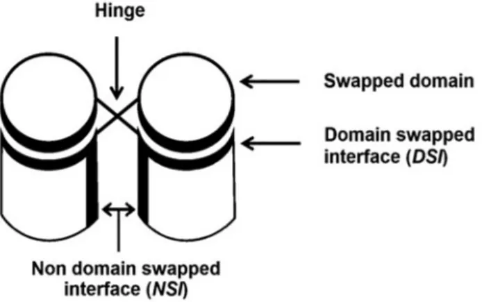
3D-domain-swapped proteins contain two kinds of interfaces (Fig 1). One is domain-swapped interface (DSI) also called as closed interface [1,17]. This interface is present in the mo-nomeric form as intramolecular interface and retains in domaswapped oligomeric form as in-termolecular interface. Another type is non-domain swapped interface (NSI), which is a newly formed interface between two monomeric subunits in domain-swapped molecule due to proxim-ity of monomeric subunits. DSI is present in both domain-swapped and non-domain-swapped form, hence its contribution to interaction energy may not be significant during domain
Fig 1. Domain-swapped oligomers with different regions marked.
swapping process [18,19]. Hence, newly formed interactions in NSI might be the key for the con-version of monomer to domain-swapped dimer.
We have examined the local energetics of domain-swapped dimer and non-domain swapped monomer at different interface regions for protein examples that are observed to ex-hibit pH-dependent domain swapping. pH-dependent energy calculation program [20,21] was employed for this purpose. This program was designed based on continuum electrostatic model. Continuum electrostatics models are highly helpful in analyzing pH-dependent proper-ties: for instance, thermal free energy employed or released during different folding pathways at varying pH. When salt-bridges are considered individually, they are often weak and insignif-icant in folding or oligomerization process. However, their joint contribution is highly crucial in terms of maintaining thermal stabilities of folded protein subunit and protein-proteincom-plex. For instance, several folded proteins lose their thermal stability at acidic or basic pH. This program has wide application to study neurodegenerative diseases associated with misfolding of proteins [5,8,22,23], pH-dependent enzyme activity, viral coat structure and intracellular trafficking.
Warwicker’s group has developed methods to calculate position and environment depen-dent pKa values and pH-dependepen-dent energy profiles for interacting entities. In this method, the pH-dependent pKa shifts were handled according to the extent of burial. This method has been developed by combining Finite Difference Poisson-Boltzmann and Debye-Huckel meth-ods into a FD/DH algorithm. This method was successfully applied on various systems, for in-stance comparison of proteins of thermophilic and mesophilic organisms [24], binding of potassium ion in ion channels [25], and to study proteins according to their location in cellular compartments [26].
This pH-dependence program, which calculates pH-dependent energy profiles based on FD/DH hybrid scheme and also helpful in analyzing misfolded proteins, is highly useful to study pH-dependent domain swapping. As domain swapping process includes partial unfold-ing step prior to oligomerization, this method is highly relevant to this study. Therefore this program was applied on domain-swapped proteins and therespective non-domain-swapped forms available in PDB [27]. While selecting the dataset, we also ensured that both the mono-meric and domain-swapped forms were crystallised under different pH conditions. The effect of pH change over wide range was observed on the thermal stability of DSI and NSI, by mea-suring the energetics at the interface in both domain-swapped and non-domain-swapped forms. Further, types of interactions stabilizing DSI or NSI were identified. The change in ener-gy during formation of NSI and disruption of DSI appears to determine the rate of oligomeriza-tion through domain-swapping process.
Materials and Methods
Dataset
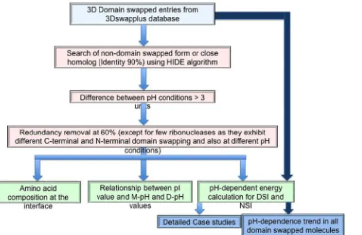
All (2057) domain-swapped examples were selected from 3DSwapplus database [18,28]. Each entry was checked for availability of crystal structure of the same or close homologous protein (identity more than 90%) in non-swapped form, using an in-house HIDE server [18]. Non-domain-swapped forms could be identified for 732 Non-domain-swapped protomers from this data-set. Further, only those pairs (of monomer-domain-swapped forms) which have a pH differ-ence of 3 was considered and a redundancy filter (threshold sequdiffer-ence identity of 60% using Cd-hit program [29]) was applied to give rise to a final dataset comprising of 16 pairs (please seeFig 2for a flow-chart of the steps involved).
unbiased towards the limited protein structural availability. Redundancy check was performed on 2057 total entries from 3DSwapplus database, using Cd-hit tool at 60% threshold, leading to 1432 non-redundant domain-swapped entries. In the final dataset an exception was a RNase molecule which can form two distinct dimers under different physiological conditions involv-ing domain swappinvolv-ing of two structural segments [30].
Datasets of general homodimers and transient oligomers, as employed by Srinivasan’s
group [31], were used to compare pH-dependent energy profiles with that of the domain-swapped molecules.
DSI and NSI
Boundaries of swapped domains were extracted from 3DSwapplus database. The interfaces in which no residue from swapped domain was involved were considered as NSI. New intermo-lecular interactions formed between swapped domains (here in only 1DDT) are considered as NSI. Residues within the two interfaces were identified using 7Å intermolecular distance crite-ria. Equivalence between residues from monomers and domain-swapped form were obtained using structural alignment. The percent composition for each type of amino acid was calculated for both and DSI and compared.
Amino acid propensity calculations at DSI and NSI
Sequence of NSI and DSI interfaces were extracted from the dataset of pH-dependent domain swapped entries and non-redundant domain swapped in 3DSwapplus database. The amino acid propensity for each amino acid type was calculated using following statistical parameters:
AAPPI i ¼
nPI i
ti
NPI i
Ti
AAPSI i ¼
nSI i
ti
NSI i
Ti
AAPPI
i = Amino acid propensity of amino acid type“i”to be in NSI in pH-dependent
do-main swapped proteins with reference to general dodo-main swapped proteins
Fig 2. Strategy used to study pH-dependent domain swapping process.
nPI
i = Number of amino acid type“i”in NSI in dataset of pH-dependent domain swapped
proteins
NPI
i = Number of amino acid type“i”in NSI in non-redundant dataset of general domain
swapped proteins from 3DSwapplus database
AAPSI
i = Amino acid propensity of amino acid type“i”to be in DSI in pH-dependent
do-main swapped proteins with reference to general dodo-main swapped proteins
nSI
i = Number of amino acid type“i”in DSI in dataset of pH-dependent domain
swapped proteins
NSI
i = Number of amino acid type“i”in DSI in non-redundant dataset of general domain
swapped proteins from 3DSwapplus database
ti= Total number of amino acid type“i”in dataset of pH-dependent domain swapped
pro-teinsTi= Total number of amino acid type“i”in non-redundant dataset of general domain
swapped proteins from 3DSwapplus database
Calculations of pH-dependent energy profile
pH—dependence program was applied for calculating and monitoring energies. This program requires interface between two protein structural entities as an input and employs Finite Differ-ence method and Debye Hückel equation to calculate electrostatic interaction energy at varying
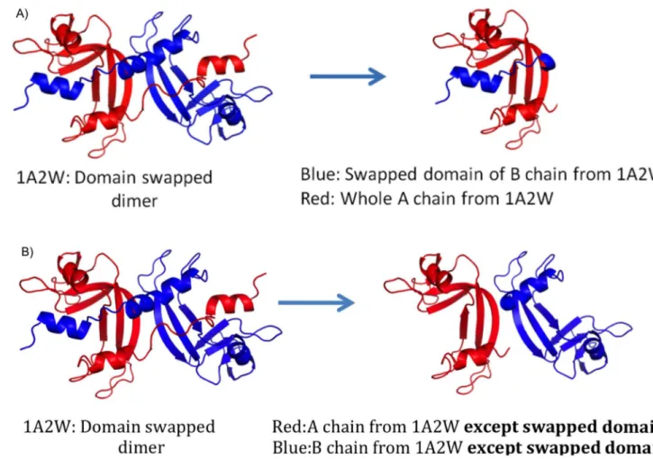
Fig 3. A) Input for pH-dependent energy profile for DSI using (Swapped domain N-terminal alpha-helix in this case). B) Input for pH-dependent energy profile for NSI (Excluding swapped domain N-terminal alpha-helix in this case).
pH [20,21]. This program also considers change in side chain conformation, change in ioniza-tion state of amino acids, varying dielectric constant according to posiioniza-tion of the residue, etc. There is change in magnitude of interaction in response to the change in sidechain conforma-tion. Most suitable rotamer for altered interaction is selected using mean field algorithm which considers solvent accessible surface area of polar and non-polar components of sidechains. Change in sidechain conformation causes changes in other interactions like Van der Waals in-teractions. Hence, all interactions will be calculated separately at different pH. pH-dependent energy profile of DSI for each domain-swapped oligomer in the dataset (swapped domain of one chain and its counterpart from another chain) were used as input (Fig 3A).
To estimate energy contributed by NSI at varying pH, protein complex without swapped domains from both monomeric subunits were used as input (Fig 3B). For diphtheria toxin alone (PDBID: 1DDT), we observed new interface formation (NSI) between swapped domains, hence pH-dependence profile for this NSI was also considered. Final output of the pH-depen-dence program provides energy profile of the DSI and NSI at varying pH.
An in-house server PPCheck [32], which calculates pseudo energy (a combination of elec-trostatic interaction, Van der Waals interaction and hydrogen bond energy) for each interface residue was further employed for detailed analysis.
Results and Discussion
Amino acid composition at interface regions
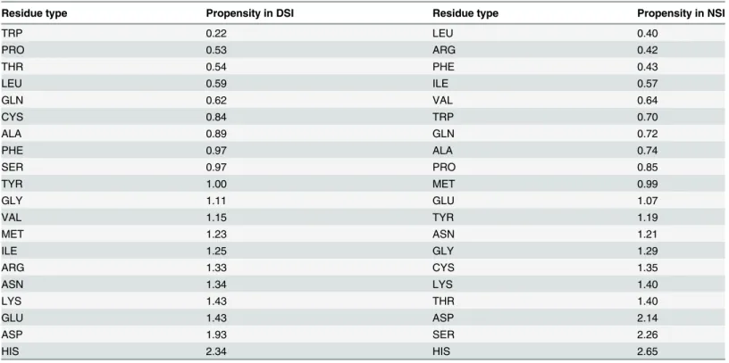
Analysis of amino acid propensities showed that DSI is rich in charged amino acids compared to general 3D-domain swapped proteins within pH-dependent domain swapped proteins (Table 1). This class of amino acids is the most sensitive to pH change. Before domain-swapped
Table 1. Amino acid propensity of DSI and NSI of pH dependent domain-swapped proteins with reference to non-redundant 3Dswapplus entries.
Residue type Propensity in DSI Residue type Propensity in NSI
TRP 0.22 LEU 0.40
PRO 0.53 ARG 0.42
THR 0.54 PHE 0.43
LEU 0.59 ILE 0.57
GLN 0.62 VAL 0.64
CYS 0.84 TRP 0.70
ALA 0.89 GLN 0.72
PHE 0.97 ALA 0.74
SER 0.97 PRO 0.85
TYR 1.00 MET 0.99
GLY 1.11 GLU 1.07
VAL 1.15 TYR 1.19
MET 1.23 ASN 1.21
ILE 1.25 GLY 1.29
ARG 1.33 CYS 1.35
ASN 1.34 LYS 1.40
LYS 1.43 THR 1.40
GLU 1.43 ASP 2.14
ASP 1.93 SER 2.26
HIS 2.34 HIS 2.65
oligomer formation, DSI has to unfold. Hence, the higher presence of charged amino acids at this interface suggests that unfolding is majorly facilitated by pH change in DSI in proteins of the domain swapping kind. Besides, charged amino acids at DSI favoured few hydrophobic amino acids, suggesting weak hydrophobic interactions are preferred as they do not offer much hindrance during unfolding. These trends are not present for NSI.
Both DSI and NSI interfaces favoured histidine. Histidine residue is well-known for its buff-ering action in response to pH change in biological system, as it possesses an imidazole nitro-gen atom which has pK value close to physiological pH. For instance,in Semliki forest virus fusion protein, histidine residue was shown to be involved in regulating low-pH-dependent re-folding[33]. Re-formation of DSI during domain swapping process is an important step and perhaps explains why histidine is observed in pH-dependent domain swapped proteins over general domain swapped proteins. Other polar amino acid residues are not as prevalent as histdine in both interfaces. In NSI, some amino acids like serine and threonine, which are prone to form strong hydrogen bonds in side chains, were favoured. Most of the hydrophobic amino acids were least favoured in both, as this class of amino acids is the least sensitive to pH change.
The order of amino acid types reported inTable 1strongly agrees with the preferred order of amino acids according to point mutations at the interface in domain swapped protomers [7,
34–36]. Besides composition of the interface, the interactions between these interface residues
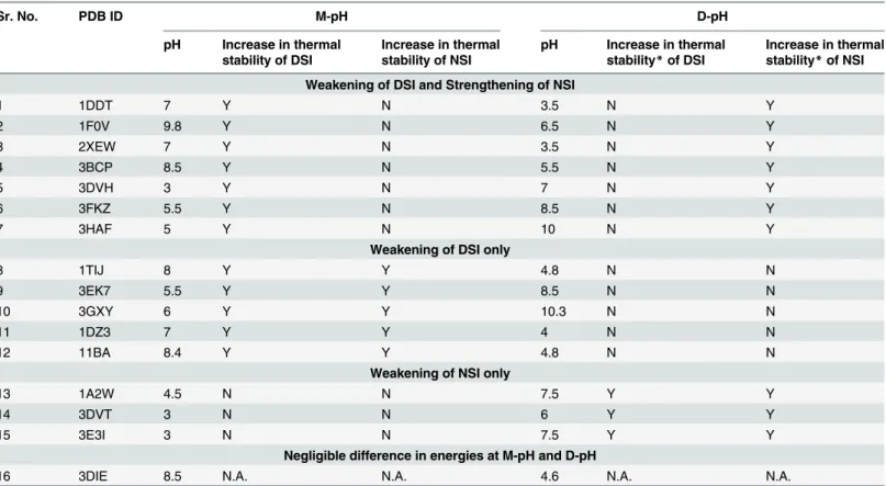
Table 2. Relative thermal stability of DSI and NSI at D-pH and M-pH.
Sr. No. PDB ID M-pH D-pH
pH Increase in thermal stability of DSI
Increase in thermal stability of NSI
pH Increase in thermal stability*of DSI
Increase in thermal stability*of NSI
Weakening of DSI and Strengthening of NSI
1 1DDT 7 Y N 3.5 N Y
2 1F0V 9.8 Y N 6.5 N Y
3 2XEW 7 Y N 3.5 N Y
4 3BCP 8.5 Y N 5.5 N Y
5 3DVH 3 Y N 7 N Y
6 3FKZ 5.5 Y N 8.5 N Y
7 3HAF 5 Y N 10 N Y
Weakening of DSI only
8 1TIJ 8 Y Y 4.8 N N
9 3EK7 5.5 Y Y 8.5 N N
10 3GXY 6 Y Y 10.3 N N
11 1DZ3 7 Y Y 4 N N
12 11BA 8.4 Y Y 4.8 N N
Weakening of NSI only
13 1A2W 4.5 N N 7.5 Y Y
14 3DVT 3 N N 6 Y Y
15 3E3I 3 N N 7.5 Y Y
Negligible difference in energies at M-pH and D-pH
16 3DIE 8.5 N.A. N.A. 4.6 N.A. N.A.
*Thermal stability of interface means energy at that pH is lower than other pH (Thermal stability at D-pH means“energy of interface at D-pH”<“energy of interface at M-pH”).
are crucial as well. Hence, a change in behavior of these interactions according to change in pH were studied.
pH-dependent energy profile of domain-swapped molecules
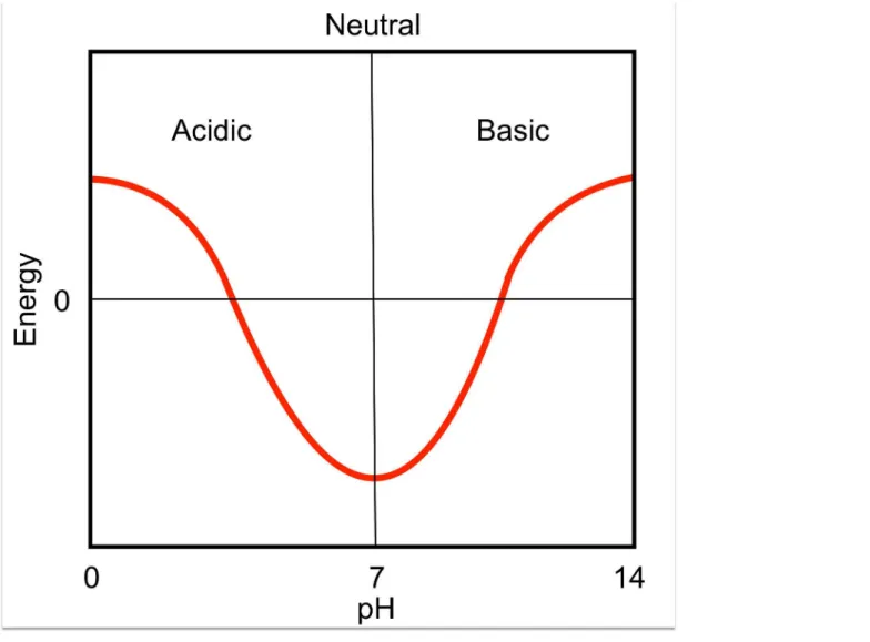
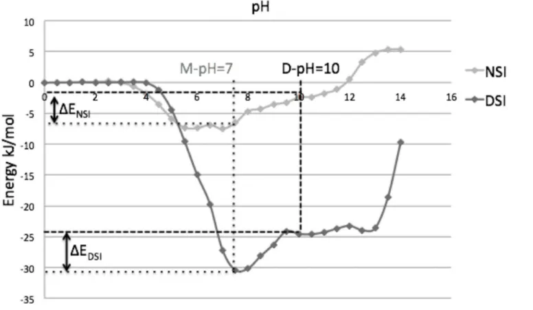
pH—dependence program was used to calculate pH-dependent energy profiles for all 16 do-main-swapped molecules (Table 2) that showed classical pH-dependence (Fig 4). pH—
dependent energy profiles showed following trends at M-pH (pH at which given protein adopts monomeric form) and D-pH (pH at which given protein adopts domain-swapped form).
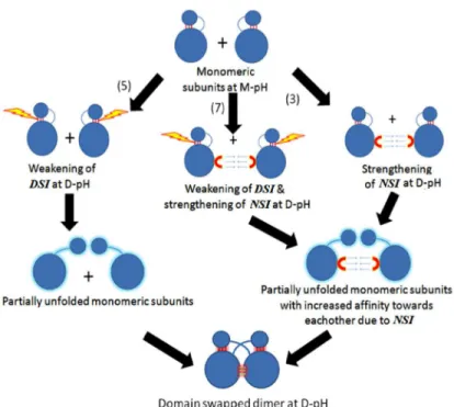
In this study, 10 domain-swapped molecules showed strengthening of NSI, while 12 mole-cules showed weakening of DSI, at a pH where domain-swapped form is observed and struc-turally characterised (S1 File). Hence, it strongly suggests that weakening of interactions at DSI facilitates the formation of partially unfolded open conformers and increase in affinity within NSI at D-pH facilitates oligomer formation.
Fig 4. Classical pH-dependent profile.
Both weakening of DSI and strengthening of NSI were observed together in seven cases. Five cases showed only weakening of DSI, whereas strengthening of NSI alone was present in three cases (Table 2andFig 5). In only one molecule (3DIE), no significant change between en-ergies at M-pH and D-pH for both the interfaces could be observed from our energy
calculations.
Few structures were analysed in greater detail to obtain a structural rationale behind weaken-ing of DSI and strengthenweaken-ing of NSI and how it might drive pH-dependent domain swappweaken-ing.
Fig 5. Proposed model representing possible effects of pH on DSI(SI) and NSI (PI) during pH-dependent domain-swapping.
doi:10.1371/journal.pone.0127716.g005
Fig 6. pH-dependent energy profile of diphtheria toxin (1DDT).
Case studies
1) Diphtheria toxin. Diphtheria toxin (1DDT) is one of the best studied domain-swapped molecules. This protein undergoes domain swapping at acidic pH (3.6). Its pH-dependent en-ergy profile showed both weakening of DSI and strengthening of NSI (Fig 6).
A detailed structural analysis revealed that the major role for domain-swapping is played by negatively charged amino acids, Aspartic and Glutamic acid (Asp and Glu). Both Asp and Glu have isoelectric point near pH 3.6. Hence at D-pH, the net charge on these acidic amino acids is close to zero and all the salt bridges, where one of the interacting amino acids is either Asp or
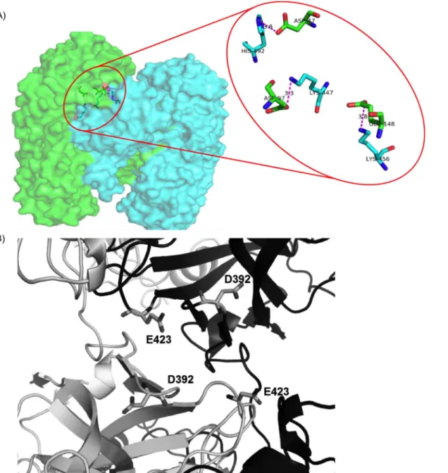
Fig 7. Interactions within domain swapped dimer of diphtheria toxin.A) Three salt bridges within DSI. B) Unfavourable electrostatic interactions between negatively charged residues within NSI.
Glu, get severely weakened. There are three salt bridges present at DSI (Fig 7A). This is the major reason for weakening of DSI at D-pH.
While in pH-dependent energy profile of NSI, four negatively charged residues (Fig 7B) showed unfavourable interactions, when evaluated using in-house PPCheck[32] server. The magnitude of these unfavourable interactions drastically reduces at D-pH (around pH 3). Hence, reduction in electrostatic repulsion at NSI leads to strengthening of the NSI.
2) Prion protein. Prion protein (3HAF) is one of the most important proteins due to its role in amyloid formation and neurodegenerative diseases. This protein forms domain-swapped dimer at basic (pH 10, referred as D-pH). The pH-dependent energy profile of this molecule showed both weakening of DSI and strengthening of NSI in D-pH (Fig 8).
An in-depth structural analysis of the interactions in this protein dimer interface revealed that the crucial amino acids for domain-swapping are positively charged amino acids, predom-inantly arginine, moderately histidine and lysine (Arg, His and Lys) which provide salt bridge interaction at neutral pH. Both Arg and Lys have isoelectric point near pH 10. At D-pH, the net charge on these basic amino acids is close to zero leading to weak salt bridge interactions at D-pH (Fig 9A). pH-dependent energy profile of NSI and detailed analysis using PPCheck serv-er [32] revealed unfavourable interactions between eight positively charged residues (Fig 9B). The magnitude of these unfavourable interactions drastically reduces at D-pH around 10, since net charge on most of these residues changed to zero leading to the strengthening of the NSI region.
In addition, two more pH-dependent domain swapped proteins (viz. ubiquitin and cyano-virin-N) showed similar behaviour as explained in cases above (please seeS1 Textfor details).
Fig 8. pH-dependent energy profile of prion dimer (3HAF).
Relation between pH-dependent change in DSI energy and
oligomerization pH (D-pH)
We observed that energy change from neutral to acidic pH (pH = 3) is a strong indicator of pH-dependence of a protein molecule. The plot (Fig 10A) represents distribution of energy dif-ference of DSI of all domain-swapped molecules available. It revealed an interesting relation-ship between the change in energy and difference between experimentally observed domain-swapped pH and monomer pH,ΔpH. This plot showed very interesting trend after mapping
very well-known literature-curated cases of pH dependence (shown in red circles inFig 10A). This suggests a strong relationship between predicted energy difference at acidic and neutral pH with experimentally known pH of dimerization. DSI is present in monomeric as well as do-main–swapped form. Hence, this pH-dependent energy difference has potential predictive ap-plication for inducing domain-swapping, through modulating pH conditions, if monomeric structure is available.
Fig 9. Interactions within domain swapped dimer of prion protein.A) Several strong and weak favourable ionic interactions between charged residues at DSI(shown in blue dotted lines). B) Several positively charged residues at NSI (shown in red color).
Since the occurrence of charged residues at the interface is observed in transient complexes as well, the distributions of energy difference of DSI at different pH of all domain-swapped molecules and interfaces from transient complexes were compared. In this analysis, we consid-ered transient complexes since they associate to form a complex as well as dissociate into mo-nomeric subunits at the same pH, unlike pH-dependent–domain swapped molecules. The dataset of transient complexes was chosen for this study as mentioned in Swapna and co-work-ers [31]. It was observed that their distributions were considerably different from each other (Fig 10B). Most of the transient dimeric molecules drastically lose their thermal stability, as pH changed to acidic pH compared to that of domain-swapped molecules. The reason might be the size of the interface, in most of the cases, DSI is formed by single secondary structural ele-ments compared to whole interface of transient complexes. This observation suggests that it is very unlikely for monomeric subunits of transient complexes to oligomerize at acidic pH due to comparatively large reduction in thermal stability, while for 3D-domain swapped complexes,
Fig 10. Distribution of difference between binding energies at pH 7 and 3.A) For DSI in 3Dswapplus entries. B) For DSI in 3DSwapplus entries and protein-protein interface in transient complexes
the difference in thermal stability is comparatively small at low pH and hence they are more likely to reform DSI.
pH-dependent change in energy also showed good correlation (correlation coefficient = 0.72) with number of salt bridges present at the DSI. Hence, the number of salt bridges present at DSI is another feature which can provide clues to predict D-pH for monomeric
structure alone.
Relation between pI of protein with D and M-pH
The information of pH dependence for domain swapping might be hidden within protein se-quence. Does pI of the protein indicative of pH-dependence of the protein? To check whether this characteristic is true for pH-dependent domain-swapped proteins or not, we further com-pared pI value of the protein and D-pH. It was observed that in eight proteins, out of 16 cases (50%), D-pH differs from to pI value by 1 (Fig 11,Table 3). While in three cases, it is close to M-pH and in five cases it is in between M-pH and D-pH. We also compared pI and D-pH value for general dataset of homodimers (as in [37]). In this dataset of general homodimers, on the other hand, only 9.5% have a pH difference less than 1 between pI and D-pH (please see
Fig 11).
Fig 11. Distribution of difference between dimerization pH (D-pH) and pI in general homodimers.
Conclusions
We report a novel approach of using the tool of pH-dependence of interactions in our energy calculations to study different types of interfaces observed in few domain-swapped proteins. The amino acid propensity analysis suggests that charged residues and few polar residues at the interface are crucial for pH-dependent domain swapping. Abundance of these residues at DSI or NSI was a driving force for pH-sensitive domain swapping, while hydrophobic amino acids, least sensitive to pH change, are not favoured in the interface regions of pH-dependent domain swapped molecules. This analysis confirms that electrostatic interactions play an im-portant role in pH-dependent domain swapping.
Further observation of these interactions at the interfaces revealed detailed insights of pH-dependent domain swapping process. It was observed that, in domain-swapped proteins, all protomers have to undergo partial unfolding by breaking of DSI in order to form domain-swapped molecule. Analysis of the pH-dependent energy profiles of such domain-domain-swapped molecules showed weakening of DSI is one of the initial and essential steps in domain swap-ping. The energy required to break open or reform this DSI reduced after weakening of DSI at D-pH. Hence, despite negligible energy contribution in the domain-swapped complex, lower-ing the energy barrier required to break open DSI as a result of change in pH might be crucial event in pH-dependent domain-swapping. In majority of the cases under study, pH-dependent energy profile showed increase in thermal stability in NSI, this is another complementary fea-ture to weakening of DSI which further increases in affinity between partially unfolded mono-meric subunits towards each other and provides additional thermal stability to domain-swapped oligomers. In this study, we report the weakening of DSI and strengthening of NSI ob-served in majority of pH-dependent proteins. As these proteins were experimentally verified to be pH-dependent from earlier well-established studies, it further validates all the reported ob-servations in this study. Amino acids at the interface were analysed in detail for few cases.
Table 3. pI, D-pH and M-pH values for pH-dependent domain-swapped proteins.
Sr. No. PDBID pI M-pH D-pH Close to*
1 1DDT 5.93 7.5 3.5
-2 3HAF 7.95 5 10
-3 11BA 9.54 8.4 4.8
-4 1TIJ 8.75 7 4.8
-5 3GXY 4.94 6 10.3
-6 3DVH 6.15 3 7 D
7 3E3I 6.3 3 7.5 D
8 1A2W 8.41 4.5 7.5 D
9 1F0V 7.34 9.8 6.5 D
10 1DZ3 4.98 7 4 D
11 3DIE 4.47 8.5 4.6 D
12 3DVT 6.26 3 6 D
13 3FKZ 8.35 5.5 8.5 D
14 2XEW 6.56 7 3.5 M
15 3BCP 9.04 8.5 5.5 M
16 3EK7 4.97 5.5 8.5 M
*D indicates that the difference between pI and D-pH is less than 1 while M indicates that the difference between pI and M-pH is less than 1
In 13 out of 16 cases in the dataset, domain-swapping was observed at acidic pH. However, it may not be possible to conclude that acidic conditions promote domain swapping in general. Further, pH-dependent energy profiles and pI values both were strong indicators of tendency of a protein to be engaged in pH-dependent domain swapping (D-pH). The analysis of behav-ior of DSI at different pH (especially at pH 3 and 7) showed a pattern in depicting D-pH. How-ever, a detailed study is required for further understanding of this relationship and to develop a predictive tool if monomeric structure is available. Effects of pH on the hinge region can be ex-plored to complement this study on interfaces and for availing structural insights towards the understanding of pH-dependent domain swapping.
Supporting Information
S1 File. pH-dependent energy profiles of all 16 pH dependent 3D domain swapped entries.
(Figure A in S1 File) pH dependent energy profile of 1A2W, (Figure B in S1 File) pH dependent energy profile of 1DDT, (Figure C in S1 File) pH dependent energy profile of 1F0V, (Figure D in S1 File) pH dependent energy profile of 1TIJ, (Figure E in S1 File) pH dependent energy pro-file of 2XEW, (Figure F in S1 File) pH dependent energy propro-file of 3BCP, (Figure G in S1 File) pH dependent energy profile of 2CZ2, (Figure H in S1 File) pH dependent energy profile of 11BA, (Figure I in S1 File) pH dependent energy profile of 3DVH, (Figure J in S1 File) pH de-pendent energy profile of 3DVT, (Figure K in S1 File) pH dede-pendent energy profile of 3E3I, (Figure L in S1 File) pH dependent energy profile of 3EK7, (Figure M in S1 File) pH dependent energy profile of3FKZ, (Figure N in S1 File) pH dependent energy profile of 3GXY, (Figure O in S1 File) pH dependent energy profile of 3HAF
(TIF)
S2 File. Interactions within domain swapped dimer of Ubiquitin.(Figure A in S2 File) Two salt bridges within DSI in Ubiquitin. (Figure B in S2 File) Unfavourable electrostatic interac-tions between negatively charged residues within NSI in Ubiquitin.
(TIF)
S1 Fig. Two salt bridges within DSI in Cyanovirin-N.
(TIF)
S1 Text. Additional case studies.
(DOCX)
Acknowledgments
This work was supported by the UK-India Education and Research Initiative (Grant #
SA070015). The authors would like to thank UKIERI for financial support for PS’s visit to JW’s laboratory to carry out this work. PS is supported by a Department of Biotechnology fellow-ship. RS and PS thank NCBS for infrastructural facilities.
Author Contributions
Conceived and designed the experiments: RS JW. Performed the experiments: PS. Analyzed the data: PS. Contributed reagents/materials/analysis tools: JW PS. Wrote the paper: PS JW RS.
References
2. Liu Y, Hart PJ, Schlunegger MP, Eisenberg D. The crystal structure of a 3D domain-swapped dimer of RNase A at a 2.1-Åresolution.Proc Natl Acad Sci. 1998; 95: 3437–3442. PMID:9520384
3. Zegers I, Deswarte J, Wyns L. Trimeric domain-swapped barnase. Proc Natl Acad Sci. 1999; 96: 818–822. doi:10.1073/pnas.96.3.818PMID:9927651
4. Liu Y, Gotte G, Libonati M, Eisenberg D. Structures of the two 3D domain-swapped RNase A trimers. 2002; 371–380. doi:10.1110/ps.36602.versa
5. Abrahamson M, Jonsdottir S, Olafsson I, Jensson O, Grubb A. Hereditary cystatin C amyloid angiopa-thy: identification of the disease-causing mutation and specific diagnosis by polymerase chain reaction based analysis. Hum Genet. 1992; 89: 377–380. PMID:1352269
6. Janowski R, Abrahamson M, Grubb A, Jaskolski M. Domain swapping in N-truncated human cystatin C. J Mol Biol. 2004; 341: 151–160. doi:10.1016/j.jmb.2004.06.013PMID:15312769
7. Szymańska A, Radulska A, Czaplewska P, Grubb A, Grzonka Z, Rodziewicz-Motowidło S. Governing the monomer-dimer ratio of human cystatin c by single amino acid substitution in the hinge region. Acta-Biochim Pol. 2009; 56: 455–463. PMID:19636441
8. Orlikowska M, Jankowska E, Kołodziejczyk R, Jaskólski M, Szymańska A. Hinge-loop mutation can be used to control 3D domain swapping and amyloidogenesis of human cystatin C. J Struct Biol. 2011; 173: 406–413. doi:10.1016/j.jsb.2010.11.009PMID:21074623
9. Boyd MR, Gustafson KR, McMahon JB, Shoemaker RH, O’Keefe BR, Mori T, et al. Discovery of cyano-virin-N, a novel human immunodeficiency virus-inactivating protein that binds viral surface envelope glycoprotein gp120: potential applications to microbicide development. Antimicrob Agents Chemother. 1997; 41: 1521–1530. PMID:9210678
10. Bewley CA, Gustafson KR, Boyd MR, Covell DG, Bax A, Clore GM, et al. Solution structure of cyano-virin-N, a potent HIV-inactivating protein. Nat Struct Biol. 1998; 5: 571–578. doi:10.1038/828PMID: 9665171
11. Yang F, Bewley C a, Louis JM, Gustafson KR, Boyd MR, Gronenborn a M, et al. Crystal structure of cyanovirin-N, a potent HIV-inactivating protein, shows unexpected domain swapping. J Mol Biol. 1999; 288: 403–12. doi:10.1006/jmbi.1999.2693PMID:10329150
12. Barrientos LG, Louis JM, Botos I, Mori T, Han Z, O’Keefe BR, et al. The domain-swapped dimer of cya-novirin-N is in a metastable folded state: reconciliation of X-ray and NMR structures. StructLondEngl 1993. 2002; 10: 673–686.
13. Barrientos LG, Gronenborn AM. The domain-swapped dimer of cyanovirin-N contains two sets of oligo-saccharide binding sites in solution. Biochem Biophys Res Commun. 2002; 298: 598–602. doi:10. 1016/S0006-291X(02)02489-0PMID:12408994
14. Anfinsen CB, Haber E, Sela M, White FH. The kinetics of formation of native ribonuclease during oxida-tion of the reduced polypeptide chain.Proc Natl AcadSci U S A. 1961; 47: 1309–1314. PMID:13683522 15. Bennett MJ, Schlunegger MP, Eisenberg D. 3D domain Swapping: Mechanism of oligomer Assembly.
Mol Biol. 1995; 2455–2468.
16. Francis JW, Brown RH, Figueiredo D, Remington MP, Castillo O, Schwarzschild MA, et al. Enhance-ment of diphtheria toxin potency by replaceEnhance-ment of the receptor binding domain with tetanus toxin C-fragment: a potential vector for delivering heterologous proteins to neurons. J Neurochem. 2000; 74: 2528–2536. PMID:10820215
17. Pesenti ME, Spinelli S, Bezirard V, Briand L, Pernollet J-C, Campanacci V, et al. Queen bee phero-mone binding protein pH-induced domain swapping favors pherophero-mone release. J Mol Biol. 2009; 390: 981–90. doi:10.1016/j.jmb.2009.05.067PMID:19481550
18. Shingate P, Sowdhamini R. Analysis of domain-swapped oligomers reveals local sequence prefer-ences and structural imprints at the linker regions and swapped interfaces. PloS One. 2012; 7: e39305. doi:10.1371/journal.pone.0039305PMID:22848353
19. Bennett MJ, Eisenberg D.The evolving role of 3D domain swapping in proteins.StructLondEngl 1993. 2004; 12: 1339–41. doi:10.1016/j.str.2004.07.004
20. Warwicker J. Simplified methods for pKa and acid pH-dependent stability estimation in proteins: remov-ing dielectric and counterion boundaries. Protein SciPubl Protein Soc. 1999; 8: 418–25. doi:10.1110/ ps.8.2.418
21. Warwicker JIM. Improved pK a calculations through flexibility based sampling of a water-dominated in-teraction scheme. 2004; 2793–2805. doi:10.1110/ps.04785604.ProteinPMID:15388865
22. Jask M. 3D Domain swapping, protein oligomerization, and amyloid. Rev Lit Arts Am. 2001; 48: 807–827.
24. Greaves RB, Warwicker J. Stability and solubility of proteins from extremophiles. BiochemBiophys Res Commun. 2009; 380: 581–585. doi:10.1016/j.bbrc.2009.01.145PMID:19285004
25. Watson HR, Wunderley L, Andreou T, Warwicker J, High S. Reorientation of the first signal-anchor se-quence during potassium channel biogenesis at the Sec61 complex. Biochem J. 2013; 456: 297–309. doi:10.1042/BJ20130100PMID:24015703
26. Chan P, Warwicker J. Evidence for the adaptation of protein pH-dependence to subcellular pH. BMC Biol. 2009; 7: 69. doi:10.1186/1741-7007-7-69PMID:19849832
27. Berman HM, Westbrook J, Feng Z, Gilliland G, Bhat TN, Weissig H, et al. The Protein Data Bank. Nu-cleic Acids Res. 2000; 28: 235–242. doi:10.1093/nar/28.1.235PMID:10592235
28. Shameer K, Shingate PN, Manjunath SCP, Karthika M, Pugalenthi G, Sowdhamini R. 3DSwap: curated knowledgebase of proteins involved in 3D domain swapping. Database J Biol Databases Curation. 2011; 2011: bar042. doi:10.1093/database/bar042
29. Li W, Godzik A. Cd-hit: a fast program for clustering and comparing large sets of protein or nucleotide sequences. Bioinformatics. 2006; 22: 1658–1659. doi:10.1093/bioinformatics/btl158PMID:16731699 30. Liu Y, Gotte G, Libonati M, Eisenberg D. A domain-swapped RNase A dimer with implications for
amy-loid formation. Nat Struct Biol. 2001; 8: 211–214. doi:10.1038/84941PMID:11224563
31. Swapna LS, Bhaskara RM, Sharma J, Srinivasan N. Roles of residues in the interface of transient pro-tein-protein complexes before complexation. Sci Rep. 2012; 2: 334. doi:10.1038/srep00334PMID: 22451863
32. Sukhwal A, Sowdhamini R. Oligomerisation status and evolutionary conservation of interfaces of pro-tein structural domain superfamilies. MolBiosyst. 2013; 9: 1652. doi:10.1039/c3mb25484dPMID: 23532342
33. Qin Z-L, Zheng Y, Kielian M. Role of conserved histidine residues in the low-pH dependence of the Semliki Forest virus fusion protein. J Virol. 2009; 83: 4670–7. doi:10.1128/JVI.02646-08PMID: 19244325
34. O’Neill JW, Kim DE, Baker D, Zhang KYJ. Structures of the B1 domain of protein L from Peptostrepto-coccusmagnuswith a tyrosine to tryptophan substitution.ActaCrystallogr D BiolCrystallogr. 2001; 57: 480–487. doi:10.1107/S0907444901000373PMID:11264576
35. Bergdoll M, Remy M-H, Cagnon C, Masson J-M, Dumas P. Proline-dependent oligomerization with arm exchange. Structure. 1997; 5: 391–401. doi:10.1016/S0969-2126(97)00196-2PMID:9083108 36. Byeon I-JL, Louis JM, Gronenborn AM. A protein contortionist: core mutations of GB1 that induce
di-merization and domain swapping. J Mol Biol. 2003; 333: 141–152. PMID:14516749