ORIGINAL ARTICLE
Persistent sciatic vein
Persistência da veia ciática
Bárbara Borges Cardoso1, Camila Oliveira Alvarenga1, Maíra de Souza Miyahara1, Marcelo Calil Burihan2, Maria Raphaella
Queiroz Alves de Lima1, Mariana Cardoso Kuwahara1, Rafael Capobianco Maia e Silva1
Abstract
Background: During a period of embryonic life, the sciatic vein is the main lower limb collector. In vascular embryogenesis, there is a diferentiation of the angioblasts in a primitive vascular plexus, with posterior remodeling and expansion. Consequently, anomalies may form during this process. When there is persistent sciatic vein, it may communicate with the small saphenous vein or with the popliteal vein at some point of its path, being anastomosed to the superior perforating vein and to the medial circumlex femoral vein.
Objective: To report a case of bilateral persistent sciatic vein on the lower limbs and review the literature on the subject.
Methods: hirty-two lower limbs from 16 cadavers preserved in formaldehyde were dissected at the Laboratory of Anatomy of the Discipline of Topographic Anatomy of the Medical School of Universidade Santo Amaro (Unisa), during 2006 and 2007, and the sciatic vein was observed in both lower limbs of one cadaver.
Results: On the left lower limb of a cadaver that presented bilateral anomaly, the vein was 37 cm long, originating in the popliteal vein, accompanying the sciatic nerve, perforating the long adductor muscle and draining into the deep femoral vein. On the right lower limb, it was 36 cm long, emerged receiving the veins of the anterior tibial compartment alongside the sciatic nerve, perforated the long adductor muscle and drained into the internal iliac vein.
Conclusion: he anatomical variations of the lower limb venous system are the most common ones. Persistent sciatic vein may cause chronic venous failure in the lower limbs, therefore it must be investigated aiming at a better clinical or surgical management.
Keywords: Anatomy; vein; sciatica.
Resumo
Contexto: Durante um período da vida embrionária, a veia ciática é a principal coletora do membro inferior. Na embriogênese vascular, há diferenciação dos angioblastos em um plexo vascular primitivo, com posterior remodelagem e expansão. Consequentemente, durante esse processo, podem ocorrer anomalias. Quando ocorre persistência da veia ciática, esta pode se comunicar com a veia safena parva ou com a veia poplítea durante seu percurso, anastomosando-se com a veia perfurante superior e com a veia circunlexa medial do fêmur.
Objetivo: Relatar o caso da persistência bilateral de veia ciática nos membros inferiores, comparando à literatura.
Métodos: Foram dissecados 32 membros inferiores de 16 cadáveres formolizados no Laboratório de Anatomia pela Disciplina de Anatomia Topográica da Faculdade de Medicina da Universidade de Santo Amaro (Unisa), durante 2006 e 2007, observando-se em 2 membros inferiores de um único cadáver, a presença de veia ciática.
Resultados: No membro inferior esquerdo de um cadáver que apresentou a anomalia bilateralmente, a veia media 37 cm, tinha origem na região da veia poplítea, acompanhava o nervo ciático, perfurava o músculo adutor magno e desembocava na veia femoral profunda. No membro inferior direito, ela media 36 cm, originava-se recebendo as veias do compartimento tibial anterior, acompanhava o nervo ciático, perfurava o músculo adutor magno e desembocava na veia ilíaca interna.
Conclusão: As variações anatômicas do sistema venoso do membro inferior são as mais prevalentes. A persistência da veia ciática pode causar insuiciência venosa crônica no membro inferior e, dessa forma, deve ser investigada para uma melhor conduta clínica ou cirúrgica.
Palavras-chave: Anatomia; veia; ciática.
Study carried out at Medical School of Universidade de Santo Amaro (Unisa), São Paulo (SP), Brazil.
1 Medical Students (5th year) at Universidade de Santo Amaro (Unisa), São Paulo (SP), Brazil.
2 Adjunct Professor of Anatomy at Unisa; Assistant Physician of the Service of Vascular Surgery at Hospital Santa Marcelina, São Paulo (SP), Brazil.
No conlict of interest was declared concerning the publication of this article. Received on: July 20, 2009. Accepted on: July 26, 2010
Persistent sciatic vein - Cardoso BB et al. J Vasc Bras 2010, Vol. 9, Nº 3
138
Introduction
During a long period of embryonic life, when the sci-atic artery is the main artery of the lower limbs, the scisci-atic vein represents the most important collector vein of these limbs’ venous circulation1.
During embryogenesis, blood islands containing an-gioblasts are derived from the extra-embryonic mesoderm. he vascular origin describes the diferentiation of angio-blasts in a primitive vascular plexus, with posterior remod-eling and expansion, hence vascular anomalies may occur during this process2.
he regulatory factors involved in the embryogenesis include the vascular endothelial growth factor and their re-ceptors, as well as the hematopoietic system2.
A sequence of changes in gene expression, even though the molecular basis of vascular morphology is not well es-tablished, is a possible cause of vascular anomalies. On the other hand, hemodynamic alterations and such anoma-lies may be associated with a generalized defect of the mesoderm2.
he persistent sciatic vein (PSV) is a rare anomaly originated in the embryonic life that derives from posterior muscle aluents, ascends with the sciatic artery, receives posterior gluteal aluents and penetrates the pelvis through the subpyramidal portion. It follows the sciatic nerve medi-ally and drains into the internal iliac vein, inferior gluteal veins or deep femoral vein3.
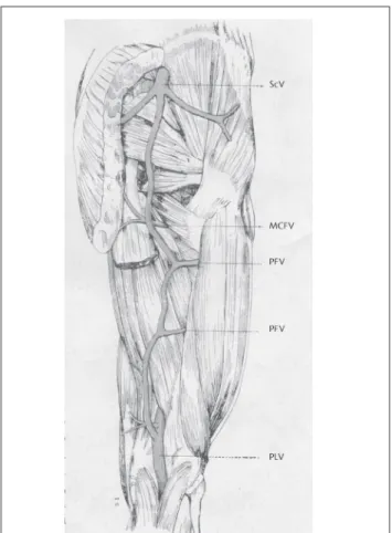
he sciatic vein may communicate with the terminal portion of the small saphenous vein or the popliteal vein, anatomosing with the perforating and medial circumlex femoral veins (Figure 1)1.
his rare anomaly may be classiied in three variations: complete, proximal or superior PSV, or distal or inferior. In complete PSV, the origin is the popliteal vein, ending up on the external iliac vein and crossing the thigh till the buttock. If proximal or superior, it emerges on the higher portion of the thigh and ends on the pelvis, involving the proximal portions of the thigh and buttock. If distal or inferior, it may be found in the inferior and medial portions of the thigh (Figure 2)4.
Objective
To study the frequency of complete bilateral persis-tence of the sciatic vein in the lower limbs, comparing the indings with the literature on the subject.
Figure 2 – (A) Complete persistent sciatic vein; (B) proximal persistence of the sciatic vein; (C) distal persistence of the sciatic vein.
PSV: persistent sciatic vein
Source: Cherry KJ, Gloviczki P, Stanson AW. Persistent sciatic vein: diagnosis and treatment of a rare condition. J Vasc Surg. 1996;23:490-7.
Figure 1 – Anastomosis of the sciatic and popliteal veins
Source: Balli R, Bertelli D. Trattado di anatomia Umana. 2. ed. Milão. Dottor Francisco Veliardi; 1924. v. III.
Persistent sciatic vein - Cardoso BB et al. J Vasc Bras 2010, Vol. 9, Nº 3 139
Method
hirty-two lower limbs from 16 cadavers preserved in formaldehyde were dissected at the Laboratory of Anatomy of the discipline of Topographic Anatomy of the Medical School of Universidade Santo Amaro (Unisa), São Paulo (SP), Brazil, during 2006 and 2007, and the sciatic vein was observed in both lower limbs of one cadaver.
Results
Out of the 32 analyzed lower limbs, 2 presented PSV (6.25%). Among the 28 lower limbs from male cadavers, the PSV was found in 7.14% of them, with one case of complete bilateral persistent vein.
he cadaver that presented complete bilateral persis-tent sciatic vein was that of a 48-year-old mulatto male that was 1.80 m tall.
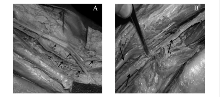
he persistent sciatic vein in the let lower limb was 37 cm long, originating on the popliteal vein region, with contribution of the medial superior genicular and small sa-phenous veins. Its path went along with the sciatic nerve between the heads of the femoral biceps, perforating the lat-eral portion of the long adductor muscle and draining into the deep femoral vein (Figure 3A).
he persistent sciatic vein of the right lower limb was 36 cm long, was originated by receiving the anterior tibial compartment veins, with the contribution of the soleus and the small saphenous veins. It was deeper than the sciatic nerve and presented an ascendant direction, perforating the
lateral portion of the long adductor muscle and draining into the internal iliac vein. here were muscle contributions and it was independent of the popliteal vein that, in this limb, was hypoplastic (Figure 3B).
Discussion
According to Cherry et al.4, out of 41 studied patients,
21 had PSV (51.22%). Among them, 13 were women (61.9%), 8 presented complete PSV (1 bilateral), 6 superior PSV (also 1 bilateral), and 7 inferior PSV. Out of all patients, 19 presented unilateral PSV (90.5%), out of which 10 were found in the let lower limb4. All these data were associated
with the Klippel-Trenaunay syndrome4,5.
According to Pompeo et al.6, in a paper presented at
the XXXV Brazilian Congress of Angiology and Vascular Surgery, a total of 41 lower limbs were dissected at the Service of Death Veriication of the City of São Paulo, and 169 phle-bographies were performed at the Service of Angiology and Vascular Surgery of Hospital Santa Marcelina, São Paulo (SP), Brazil. Among 210 lower limbs analyzed, 7 presented PSV (3.33%). Among these, 5 were male cadavers (71.4%) and 6 presented let lower limb PSV6.
Based on the mentioned papers and on the sample of our study, the anatomical variations of the venous system, espe-cially those of the lower limbs, are the most common ones.
he PSV may lead to cases of chronic venous failure of the lower limb, which must be investigated. Magnetic resonance angiography and CT angiography are useful techniques for diagnostic assessment of the venous system,
Figure 3 – (A) Sciatic vein on the left lower limb: (1) sciatic vein; (2) sciatic nerve; (3) popliteal vein; (4) small saphenous vein; (5) common ibular nerve. (B) Sciatic vein on the right lower limb: (1) sciatic vein; (2) small saphenous vein; (3) common ibular nerve.
Persistent sciatic vein - Cardoso BB et al. J Vasc Bras 2010, Vol. 9, Nº 3
140
for they evaluate characteristics, anatomical origin and re-lations of the varicose veins with venous malformations. Information about precedence, path and anatomical rela-tions of the anomalous vessels helps to plan the treatment adequately, as well as to provide a better clinical and surgi-cal management7.
Acknowledgments
he authors thank the PhD Professor José Carlos Prates for his intellectual contribution, for sharing his ample knowledge about Human Anatomy and for providing the material.
References
1. Balli R, Bertelli D. Trattado di anatomia Umana. 2. ed. Milão: Dottor Francisco Veliardi;1924. v. III.
2. Parry DJ, Aldoon MI, Hammond RJ, Jessil PO, Weston M, Scott DJA. Persistent sciatic vein, varicose veins and lower limb hyper-trophy: an usual case or discrete clinical syndrome? J Vasc Surg. 2002;36:396-400.
3. Latarjet M, Liard AR. Anatomia humana. Paris. Médica Panamericana; 1983.
4. Cherry KJ, Gloviczki P, Stanson AW. Persistent sciatic vein: diagnosis and treatment of a rare condition. J Vasc Surg. 1996;23:490-7.
5. You CK, Rees J, Gillis DA, Steeves J. Klippel-Trenauney syndrome: a review. Can J Surg. 1983;26:399-403.
6. Pompeo ASFL, Grinberg H, Burihan MC, Prates JC. Quão in-frequente é a veia isquiática. Anais do Congresso Brasileiro de Angiologia e Cirurgia Vascular publicado no Jornal Vascular Brasileiro. Jornal Vascular Brasileiro. 2003;2:129.
7. Bastarrika G, Redondo P, Sierra A, et al. New techniques for the evaluation and therapeutic planning of patients with Klippel-Trénaunay syndrome. J Am Acad Dermatol 2007;56:242-9.
Correspondence
Marcelo Calil Burihan Alameda Sarutaiá, n.173 apto 111 - Jardim Paulista CEP: 01403-010 – São Paulo (SP), Brasil E-mail: [email protected]
Authors’ contributions