CASE REPORT
Anomalous formation of the portal vein: a case report
Formação anômala da veia porta: relato de caso
Vasavi Rakesh Gorantla,
1Bhagath Kumar Potu,
1Thejodhar Pulakunta,
1Venkata Ramana Vollala,
2Pavan Kumar Addala,
3Soubhagya Ranjan Nayak
4Abstract
The knowledge about the formation and relations of the portal vein is important for surgeons and radiologists. The variations in the level of formation and the pattern of formation of portal vein might lead to confusions during radiological and surgical procedures. Here we present a rare variation in the formation of the portal vein as found during the cadaveric dissections. The portal vein was formed by the union of splenic vein, superior mesenteric vein and inferior mesenteric veins. The abnormal termination of left gastric vein into superior mesenteric vein before the formation of portal vein was also seen in the same cadaver. Identification of these variations is useful in managing traumatic rupture of the mesentery.
Keywords:Variations, portal vein, obstruction, malrotation.
Resumo
O conhecimento sobre a formação e as relações da veia porta é importante para cirurgiões e radiologistas. As variações no nível de formação e o padrão de formação da veia porta podem causar confusões durante procedimentos cirúrgicos e radiológicos. Neste relato, apresentamos uma variação na formação da veia porta encontrada durante as dissecções cadavéricas. A veia porta foi formada pela união da veia esplênica, veia mesentérica superior e veias mesentéricas inferiores. A terminação anormal da veia gástrica esquerda na veia mesentérica superior antes da formação da veia porta também foi observada no mesmo cadáver. A identificação dessas variações é útil para tratar a ruptura traumática do mesentério.
Palavras-chave:Variações, veia porta, obstrução, má-rotação.
Introduction
The portal vein is normally formed by union of the splenic and superior mesenteric veins, around the level of L2, anterior to the inferior vena cava and posterior to the pancreas. It then passes superiorly and runs pos-terior to the first part of the duodenum. At the porta hepatis it divides into a left and right branch. The portal vein drains blood from all parts of the digestive tract (apart from the lower part of the rectum), the pancreas, spleen and gallbladder. Variant portal architecture has been found in 20-35% of individuals.1,2Two common variations include: (a) trifurcation of the portal vein, where there is absence of the right trunk proper, such that the right anterior and posterior branches stem from the portal trunk at the same point as the left portal vein; and (b) the right posterior branch coming off the main portal vein rather than from the right portal vein.1,2 Indi-vidual segmental branches arising away from their usual
point of origin is another commonly reported varia-tion.1
Knowledge of these variations in portal anatomy is important in the preoperative work-up prior to liver transplantation or resection, as knowledge of the indi-vidual patient’s vascular anatomy helps to minimize the likelihood of postoperative liver ischemia.1There is also a potential benefit for the interventional radiologist in knowing vascular detail prior to catheter-based inter-ventions.1There are several reports on variations on the termination of portal vein in the porta hepatis, but stud-ies on formation of portal vein and its anomalous course are lacking.
Case report
During routine dissections for undergraduate medi-cal students in Department of Anatomy, KMC,
1 . Department of Anatomy, Centre for Basic Sciences, Kasturba Medical College, Manipal, Karnataka, India. 2 . Department of Anatomy, Melaka Manipal Medical College (Manipal Campus), Manipal, Karnataka, India. 3 . Department of Surgery, Kasturba Medical College, Manipal, Karnataka, India.
4 . Department of Anatomy, Centre for Basic Sciences, Kasturba Medical College, Mangalore, Karnataka, India. Manuscript received June 18, 2007, accepted on August 20, 2007.
J Vasc Bras. 2007;6(4):399-401.
Copyright © 2007 by Sociedade Brasileira de Angiologia e de Cirurgia Vascular
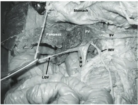
Manipal, a male cadaver about 54 years showed a varia-tion in the formavaria-tion of the portal vein. The formative tributaries were superior mesenteric vein, splenic vein and inferior mesenteric vein. Moreover, the left gastric vein was seen terminating into superior mesenteric vein before the formation of portal vein (Figure 1). All the formative tributaries were carefully dissected and pho-tographs were taken.
Discussion
The portal vein is rarely variable. Identifying devia-tions from normal portal architecture is important in the work-up for surgery such as liver transplantation, and prior to interventional procedures such as stent placement or embolization. There are several reports on variations on the termination of portal vein in the porta hepatis, but studies on formation of portal vein and its anomalous course are lacking. Bergman et al. have reported the absence of portal vein and opening of supe-rior mesenteric and splenic veins into the renal vein.3 Jin Shan et al. have reported doubling of the portal vein
and its clinical implications.4A preduodenal portal vein is a rare congenital anomaly and has been reported a few times.5-7 The main clinical significance of a preduodenal portal vein is its association with intestinal obstruction. This can be due to extrinsic compression of the duodenum, or associated intestinal malforma-tions.8Up to 80% of the obstructions are due to intrin-sic lesions of the duodenum or malrotation.9Two-thirds of children with a preduodenal portal vein present in the first week of life.9Other associations include biliary atresia, annular pancreas, situs inversus, preduodenal common bile duct and cardiovascular malforma-tions.8,9Inoue et al. have reported a prepancreatic post-duodenal portal vein.10
In the embryo, the paired vitelline veins transport blood from the yolk sac to the sinus venosus. During the fourth to fifth weeks of embryonic life, three anasto-moses form between the vitelline veins,11these are the cranial-ventral, dorsal and caudal-ventral anasto-moses, and are named according to their anatomical
IMV = inferior mesenteric vein; LGV = left gastric vein; PV = portal vein; SMV = superior mesenteric vein; SV = splenic vein.
Figure 1- Dissection of the abdomen showing formation of the portal vein by three veins
position and relationship to the primitive foregut that will become the duodenum. From the formation of these anastomoses to the third month of development, there is selective involution of the venous network that even-tually produces the portal vein. It has been proposed that aberrations in this process of involution can result in anatomical variations within the portal venous sys-tem.12
In conclusion, the knowledge of variations in the for-mation and course of the portal vein is very useful for surgeons performing surgeries of pancreas and duode-num. It is also useful in managing the traumatic rupture of the mesentery. Since there are not many studies on variation in the formation of the portal vein, this study might contribute useful data to the literature regarding the same.
References
1. Covey AM, Brody LA, Getrajdman GI, Sofocleous CT, Brown KT.Incidence, patterns, and clinical relevance of vari-ant portal vein anatomy. AJR Am J Roentgenol. 2004;183:1055-64.
2. Atri M, Bret PM, Fraser-Hill MA.Intrahepatic portal venous variations: prevalence with ultrasound. Radiology. 1992;184:157-8.
3. Bergman RA, Thompson SA, Afifi AK, Saadeh FA. Com-pendium of human anatomic variations. Baltimore: Urban & Schwarzenberg; 1988. p. 70.
4. Esscher T.Preduodenal portal vein--a cause of intestinal obstruction?J Pediatr Surg. 1980;15:609-12.
5. Zhang JS, Wang YP, Wang MQ, et al.Diagnosis of an acces-sory portal vein and its clinical implications for portosys-temic shunts. Cardiovasc Intervent Radiol. 1996;19:239-41. 6. Stevens JC, Morton D, McElwee R, Hamit HF.
Preduode-nal portal vein: Two cases with differing presentation. Arch Surg. 1978;113:311-3.
7. Yi SQ, Tanaka S, Tanaka A, Shimokawa T, Ru F, Nakatani T.An extremely rare inversion of the preduodenal portal vein and common bile duct associated with multiple malforma-tions. Report of an adult cadaver case with a brief review of the literature. Anat Embryol (Berl). 2004;208:87-96.
8. Choi SO, Park WH.Preduodenal portal vein: a cause of pre-natally diagnosed duodenal obstruction. J Pediatr Surg. 1995;30:1521-2.
9. Fernandes ET, Burton EM, Hixson SD, Hollabaugh RS.
Preduodenal portal vein: surgery and radiographic appear-ance. J Pediatr Surg. 1990;25:1270-2.
10. Inoue M, Taenaka N, Nishimura S, et al.Prepancreatic post-duodenal portal vein: report of a case. Surg Today. 2003;33:956-9.
11. Sadler T. Cardiovascular system. In: Sadler TW, editor. Lang-man’s medical embryology. 8th ed. Philadelphia: Lippincott Williams & Wilkins; 2000. p. 246-8.
12. Niwa T, Aida N, Tachibana K.Congenital absence of the portal vein: clinical and radiological findings. J Comput Assist Tomogr. 2002;26:681-6.
Correspondence: Bhagath Kumar Potu Department of Anatomy
Centre for Basic Sciences, Kasturba Medical College 576104 – Manipal, Karnataka – India
Tel.: (91) 820-2922327
E-mail: [email protected]