Giant nodular pseudoangiomatous stromal hyperplasia
of the breast with fibroadenomatoid myxoid changes: a
potential pitfall in the differential diagnosis
of phyllodes tumor
Hiperplasia estromal pseudoangiomatosa mamária nodular gigante com padrão
fibroadenomatoide mixoide: diagnóstico diferencial problemático com tumor
phyllodes
Roberto José Medeiros1; Patricia Z. Rebutini2; Thamyres G. V. Vargas1; Fabiola Medeiros3; Ana Paula M. Sebastião2 1. Faculdade Ceres (Faceres), São Paulo, Brazil. 2. Hospital de Clínicas, Universidade Federal do Paraná (UFPR), Paraná, Brazil.
3. Keck Medical Center, University of Southern California (USC), California, USA.
First submission on 08/11/16; last submission on 13/02/17; accepted for publication on 24/04/17; published on 20/06/17
ABSTRACT
A 21-year old woman presented with a 17-cm left breast mass. Physical examination and ultrasound revealed the mass to be well-circumscribed, homogeneous and freely mobile, suggestive of giant ibroadenoma or phyllodes tumor. The mass was surgically excised and initially interpreted as benign phyllodes tumor. Subsequent slide review established the diagnosis of nodular pseudoangiomatous stromal hyperplasia (PASH) associated with ibroadenomatoid areas and myxoid stromal changes. This case illustrates the dificulty encountered in recognizing nodular PASH. A thorough discussion of the histopathologic differential diagnosis of nodular PASH is provided.
Key words: breast; hyperplasia; giant; angiomatous.
INTRODUCTION
Pseudoangiomatous stromal hyperplasia (PASH) is a benign stromal proliferation of the breast characterized by the presence of slit-like spaces within a collagenous stroma. This term is based on the fact that the histomorphologic appearance mimics a vasoformative proliferation, but is in fact composed of myoibroblastic cells. PASH can be found incidentally in breast biopsies or resections performed for other reasons, but more often it forms a mass detectable by physical exam or imaging studies. When the lesion reaches large dimensions, it can be clinically suspicious for malignancy and misinterpreted histologically as such. Herein we report the case of a large nodular PASH that was misinterpreted as phyllodes tumor and discuss the key features that can be useful in its differential diagnoses.
CASE REPORT
A 21-year-old woman presented to a gynecologist with a large nodule in the left breast, which had been rapidly growing over the past six months. It was initially painless, but became painful over the last weeks. There was no nipple discharge. The patient was otherwise healthy, with no history of prior breast disease or any other signiicant past or current medical history. She denied smoking, alcohol consumption, and use of illicit drugs. Menarche was at 12 years, and she had never been pregnant. She was currently taking oral contraceptives and no other prescription or over-the-counter medications. There was no history of breast disease or breast cancer in the family. Physical examination revealed large asymmetrical breasts, with the left being larger than the right. Upon palpation, a 17-cm oval, well-deined, irm,
and freely mobile mass was detected in the lower aspect of the left breast spanning lower outer or lower inner quadrants. No palpable nodules were identiied in the right breast. Axillary lymph nodes were not palpable, and the skin overlying the mass, nipple and areola were unremarkable. Ultrasound examination showed a well-circumscribed, homogenous, hypoechoic solid mass, with areas of cystic degeneration classiied as BI-RADS 4A. Based on the clinical and imaging indings, giant ibroadenoma or phyllodes tumor were provisionally suggested.
A core biopsy of the left breast nodule was performed in a community hospital, and it was diagnosed as PASH. Subsequently, the patient was referred to a tertiary medical center where excision of the nodule with bilateral reduction mammoplasty and immediate plastic reconstruction were performed. Prior to surgery, glass slides and blocks of the left breast core biopsy could not be retrieved and only the pathology report from the core biopsy was
available.
The Anatomic Pathology laboratory received three specimens labeled ‘Right breast, reduction mammoplasty’, ‘Left breast, reduction mammoplasty’ and ‘Left breast, tumor’, respectively. Bilateral reduction mammoplasties weighted 410 grams on the right and 308 grams on the left and showed unremarkable breast parenchyma and skin with no gross abnormalities. The left breast tumor weighted 868 grams and measured 17 × 13 × 10 cm. It
was ovoid, well-circumscribed and surrounded by a ibrous pseudocapsule. On cut sections, it was solid, tan-white, ibroelastic and homogeneous, with no identiiable necrosis or hemorrhage.
Upon examination by light microscopy, the tumor was composed of a densely collagenous stroma containing numerous anastomosing slit-like spaces, which appeared almost empty and intermixed benign breast ducts and lobules (Figure 1). Stromal
spindle cells bordering these spaces resembled endothelial cells. Their nuclei were attenuated, lacked atypia and had no mitotic activity (Figure 2). The spaces were empty. These indings were
diffusely and homogeneously seen throughout the whole tumor, which did not contain adipose tissue. The epithelial component comprised breast ducts and lobules, which in some areas had a normal appearance and in others, ibroadenomatoid changes with compressed ducts. The stroma was abundant and predominated over the epithelial component. Focal usual ductal hyperplasia and ductal dilatation were present. Stromal myxoid change were
seen (Figure 3). Some areas had abundant edematous loose
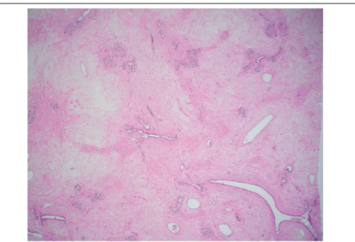
stroma with few intervening epithelial elements (Figure 4). The
presence of these two latter features, along with a predominance of stroma led to an initial diagnosis of benign phyllodes tumor by the general surgical pathologist.
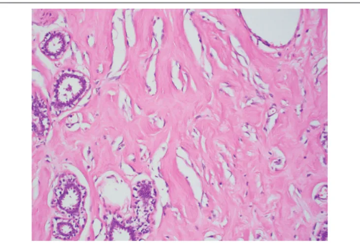
FIGURE 3 − Fibroadenomatoid areas were seen in the left-breast tumor characterized by
compressed ducts with slit-like lumens, surrounded by loose myxoid stroma (HE 40×) HE: hematoxylin and eosin.
FIGURE 2 − Slit-like spaces in left-breast tumor are lined by a discontinuous layer of
bland spindle cells that lack nuclear atypia and mitosis (HE 400×) HE: hematoxylin and eosin.
FIGURE 1 − Left-breast tumor showing numerous anastomosing slit-like spaces in a
densely collagenous stroma, involving interlobular and intralobular stroma (HE 40×)
FIGURE 4 − Areas of abundant edematous stroma in the left-breast tumor (HE 40×)
HE: hematoxylin and eosin.
On the irst post-operative appointment, pathology review of the case was requested by the clinical team. At that time, immunohistochemical staining was performed and showed that the spindle cells were positive for CD34 (Figure 5) and negative
for actin. Both estrogen receptor (ER) and progesterone receptor (PR) showed positivity in breast epithelium with only focal weak staining in breast stroma and spindle cells lining slit-like spaces. Ki-67 showed low proliferation index (< 1%). Based on this immunohistochemical proile, the original diagnosis of the left breast tumor was amended to nodular PASH with ibroadenomatoid hyperplasia and stromal myxoid changes.
FIGURE 5 − CD34 immunostaining in the left-breast tumor showing positivity in
fibroblasts and myofibroblasts (20×)
Bilateral reduction mammoplasties revealed benign breast parenchyma with focal ductal ectasia, usual ductal hyperplasia and ibrosis with areas of ibroadenomatoid hyperplasia, but no
signs of PASH.
DISCUSSION
PASH is a benign stromal proliferation of the breast characterized by the presence of slit-like spaces within a collagenous stroma. PASH can affect a wide age range, from childhood to adolescence, with a mean age ranging from 37 to 51 years(1). Seventy-ive per cent of patients are premenopausal(2).
About 60% of cases form clinically and imaging detectable masses, while 40% are incidental indings(2).
PASH is typically unilateral when it forms a mass with rare documented bilateral tumors. The majority of cases measure between 1 and 15 cm in size, with an average of 5 cm(1). Case reports
in the medical literature characterize rare examples of “giant PASH”. Fine et al. (2015) reported a 21-cm PASH mass in a 31-year-old woman, and Sasaki et al. (2008) details a 23-cm PASH mass in a
48-year-old woman(3, 4). The largest reported case of PASH to date
measured 35-cm and was documented by Abdelrahman et al. (2015) in a 13-year-old girl with small developing breasts(5). Bourke et al.
(2015) reported bilateral PASH tumors measuring 25 and 24 cm in a 46-year-old woman(2). The patient described here was detected with a
17-cm left breast mass associated with bilateral macromastia. Similarly to this case, resected PASH tumors are well-circumscribed, with a smooth external surface and a solid, ibrous and homogeneous cut surface(1). Some contain small cysts. Most
do not show hemorrhage or necrosis grossly.
Microscopically, PASH is composed of benign breast parenchyma with an admixture of stroma and epithelial structures. The lesion is deined by the presence of complex often anastomosing spaces involving both intralobular and interlobular stroma, which is typically expanded and densely collagenous. Myoibroblasts line the spaces in a discontinuous fashion, resembling endothelial cells. These cells often have elongated bland nuclei with no atypia or mitotic activity. The stromal component is often increased with wider separation of ducts and lobular units when compared to the usual arrangement. Collagenization of intralobular stroma with duct attenuation commonly leads to a ibroadenomatoid appearance, which was seen in this case, and likely represented the feature that, along with the large tumor dimension, abundance of stroma and myxoid changes, led to an erroneous initial interpretation of benign phyllodes tumor. Usual ductal hyperplasia and ductal dilatation are common in PASH, and both indings were present in this case.
cells that line the slit-like spaces of PASH show variable expression of myoid and ibroblastic markers. They are usually reactive for CD34 and vimentin, with variable expression of smooth muscle actin and calponin(6). They lack immunoreactivity to keratins and
endothelial markers such as CD31 and factor VIII. The nuclei can be positive for PR but are almost always negative or weak and focal
for ER(6). In this case, myoibroblasts were diffusely positive for
CD34, negative for actin and negative for ER and PR, consistently with what is expected for PASH.
The differential diagnoses of PASH include low-grade angiosarcoma, myoibroblastoma, ibroadenoma, benign phyllodes tumor and mammary hamartoma. Angiosarcoma forms an iniltrative, ill-deined mass unlike PASH, which is mobile and well-circumscribed. On microscopic examination, both PASH and angiosarcoma are characterized by anastomosing spaces lined by spindle cells, but, while the former lacks nuclear atypia and mitosis, the latter shows mild to moderate nuclear atypia and increased mitotic activity. Cells lining the spaces in angiosarcoma are positive for endothelial markers such as CD 31 and factor VIII, whereas these cells in PASH are positive for myoibroblastic markers CD34 and smooth muscle actin (SMA) and negative for endothelial markers.
Myoibroblastoma is characterized by predominant fascicular architecture, being composed of bland spindle cells haphazardly arranged in fascicles with interspersed, thick, hyalinized collagen bundles. Nodular PASH with predominant cellular areas or fascicular foci is more likely to be confused with myoibroblastoma. The immunohistochemical proile is similar between these two lesions that are believed to have a common histogenesis. However, PASH expresses PR, in contrast to the expression of androgen receptors in myoibroblastoma.
Mammary hamartoma is characterized by the presence of mature adipose tissue and nodular aggregates of mammary
parenchyma. Nodular PASH is differentiated from mammary hamartoma by the absence of adipose tissue within the mass, as observed in this case.
Fibroadenoma is the most common differential diagnosis on clinical and imaging studies. Fibroadenomas are biphasic tumors with epithelial and stromal components common in females in the second and third decades of life such as this patient. The epithelial proliferation in ibroadenoma can have intracanalicular or pericanalicular patterns with variable amounts of intervening stroma. The stroma of ibroadenoma characteristically does not contain the slit-like spaces characteristic of PASH, but this has been rarely described(7). Therefore, an important diagnostic consideration
here was a giant ibroadenoma with pseudoangiomatous and myxoid stromal changes. Likewise, ibroadenomatoid areas in PASH, which result from increased intralobular stroma, may be mistaken for ibroadenoma. In this case, there was a predominance of stroma, and ibroadenomatoid areas were admixed with normal appearing ducts and lobules.
Benign phyllodes tumor has close histologic resemblance to ibroadenoma and is always an important consideration in large tumors. Both may show myxoid change in the stroma that is unusual in PASH, but that was seen in this case, contributing to the initial misinterpretation of benign phyllodes tumor. However, this case did not have two key features for the diagnosis of benign phyllodes tumor, namely leaf-like architecture and hypercellular
stroma.
In summary, we present a case of nodular PASH forming a 17-cm unilateral breast mass in association with bilateral macromastia in a 21-year-old woman. It is important to be aware of the differential diagnoses, as nodular PASH may be closely mimicked by several breast tumors, clinically, radiologically and histologically.
RESUMO
Uma mulher de 21 anos desenvolveu uma massa mamária de grandes dimensões (17 cm). Exame físico e ultrassonografia revelaram um nódulo bem delineado, homogêneo e móvel, com características sugestivas de fibroadenoma gigante ou tumor
phyllodes. O nódulo foi excisado e inicialmente interpretado como tumor phyllodes benigno. Subsequente revisão de lâminas estabeleceu o diagnóstico de hiperplasia estromal pseudoangiomatosa nodular associada a áreas de padrão fibroadenomatoide e focos de degeneração mixoide do estroma. Este caso ilustra a dificuldade encontrada no diagnóstico dessa entidade. Segue uma discussão detalhada do diagnóstico diferencial de hiperplasia estromal pseudoangiomatosa nodular.
REFERENCES
1. Rosen PP, Hoda SA, Brogi E, Koerner FC. Rosen’s breast pathology. 4th ed.
Philadelphia: Wolters Kluwer Health/Lippincott Williams Wilkins; 2014. 1379 p.
2. Bourke AG, Tiang S, Harvey N, McClure R. Pseudoangiomatous stromal hyperplasia causing massive breast enlargement. BMJ Case Rep. 2015 Oct 16; 2015.
3. Fine SG, Powell AT, Murray MK. Resection of giant pseudoangiomatous stromal hyperplasia: expectant observation and avoidance of complex breast surgery. Int J Surg Case Rep. 2015; 15: 32-4.
4. Sasaki Y, Kamata S, Saito K, Nishikawa Y, Ogawa J. Pseudoangiomatous stromal hyperplasia (PASH) of the mammary gland: report of a case. Surg Today. 2008; 38(4): 340-3.
5. Abdelrahman T, Young P, Kozyar O, Davies E, Dojcinov S, Mansel RE. Giant pseudoangiomatous stromal hyperplasia presenting in the breast of a prepubertal child. BMJ Case Rep. 2015; 2015.
6. Virk RK, Khan A. Pseudoangiomatous stromal hyperplasia: an overview. Arch Pathol Lab Med. 2010; 134(7): 1070-4.
7. Jaunoo SS, Thrush S, Dunn P. Pseudoangiomatous stromal hyperplasia (PASH): a brief review. Int J Surg. 2011; 9(1): 20-2.
CORRESPONDING AUTHOR
Ana Paula Martins Sebastião