679
Acute Renal Insufficiency after Radiofrequency of Renal Tumor Case Report
International Braz J Urol Vol. 33 (5): 679-682, September - October, 2007
Acute Renal Insufficiency after Radiofrequency of Renal Tumor
Francualdo Barreto, Marcos F. Dall’Oglio, Miguel Srougi
Division of Urology, School of Medicine, University of Sao Paulo, USP, Sao Paulo, SP, Brazil
ABSTRACT
Recent advances in techniques of imaging and ablation have led to the application of several minimally invasive modalities, such as radiofrequency ablation (RFA) with a success rate varying from 79 to 96% and a serious complication rate of 1 to 4% in the treatment of small renal tumors.
The authors report on the case of a 67-year-old patient with a radiofrequency ablation complication, stenosis of the ureteropelvic junction in one kidney, and analyze the results of this modality for the treatment of renal tumors.
Key words: kidney neoplasms; catheter ablation; renal insufficiency
Int Braz J Urol. 2007; 33: 679-82
INTRODUCTION
Traditionally, the treatment of renal tumors included radical or partial nephrectomy. Minimally in-vasive treatment modalities such as cryotherapy and radiofrequency ablation (RFA) by percutaneous ap-proach have been used in the treatment of carcinoma of the renal cells, offering some advantages, such as shorter convalescence, lesser pain, lower costs and better esthetic effect, if compared to conventional surgery (2).
The authors report on the case of a 67-year-old patient with a radiofrequency ablation complica-tion, stenosis of the ureteropelvic junction (UPJ), in one kidney, and analyze the results of this modality for the treatment of renal tumors.
CASE REPORT
680
Acute Renal Insufficiency after Radiofrequency of Renal Tumor
The patient sought out our institution to verify the therapeutic possibilities. Open pyeloplasty was recommended, followed by the enucleation of the re-nal nodule. The pathological examination revealed a necrotic area with the formation of abscesses, fibro-sis and a granulomatose reaction of the foreign-body type, with no evidence of a viable tumor (Figure-3). The patient progressed well (Figure-4), currently hav-ing a creatinine level of 1.9 mg/dL.
Figure 1 – Computed tomography demonstrating renal nodule of 3.5 cm in remaining kidney.
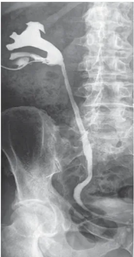
Figure 2 – Stenosis of ureteropelvic junction by magnetic reso-nance.
Figure 3 – Necrotic area, fibrosis and granulomatose reaction of the foreign-body type. Absence of viable residual neoplasia (HE X400).
681
Acute Renal Insufficiency after Radiofrequency of Renal Tumor
COMMENTS
RFA has been used recently as a new treat-ment option for small renal tumors with a success rate of 79 to 96% of the cases (2,3), the incidence of seri-ous complications, such as intestinal lesion, cutaneseri-ous fistula, urethral stenosis and pneumothorax, occurs in 1 to 4% (1,2).
Radiofrequency ablation (RFA) is to be rec-ommended for the treatment of renal tumors of less than 3 cm, which have given signs of growth during the period of one year. Surgical approach may vary either by means of percutaneous puncture (3) or by laparoscopy (2). The principle of RFA involves heat-ing to high temperatures (< 70 degrees C) thus pro-voking necrosis of coagulation and cell death (2,3). The criteria of inclusion for RFA are solid lesions < 3 cm, which have been growing over the previous year, creatinine below 2.0 mg/dL and 24-hour creatinine clearance greater than 60 mL/min (2,3). The position of the tumor (posterior, lateral or medial) has not been considered among the exclusion criteria, although the proximity of the colon, duodenum or of important ves-sels is a limiting factor for this technique (1-3). The most frequent complications arising from RFA are hematuria (4 to 8%), proteinuria (16%), low back pain (16%) and perirenal hematoma (4%), and these are treated conservatively (2,3).
The criterion of cure is confirmed by the ab-sence of the visualization of contrast (< 10UH) on tomography, with a success rate of 79 to 96% (2,3).
Stenosis of the UPJ may occur in 4% of the cases, being presented after two months (2). Accord-ing to some authors, the position of the tumor does not
constitute a criterion of exclusion, but rather a limita-tion of the applicability of the technique (2,3). Accord-ing to Hwang et al., open pyeloplasty is the best way to deal with this complication (2), particularly in the reported case, as we are dealing with a sole kidney in a rather delicate situation. An alternative technique in the case of an extensive lesion would be the interpo-sition of the loop ileal.
The RFA of small renal tumors, whether by percutaneous approach or laparoscopy, still requires further study for the assessment of the method’s effi-ciency and safety. If the long-term results are favor-able, then RFA could be an attractive treatment op-tion for solid renal lesions.
CONFLICT OF INTEREST
None declared.
REFERENCES
1. Rhim H, Dodd GD 3rd, Chintapalli KN, Wood BJ, Dupuy DE, Hvizda JL, Sewell PE, Goldberg SN: Radiofrequency thermal ablation of abdominal tumors: lessons learned from complications. Radiographics. 2004; 24: 41-52. 2. Hwang JJ, Walther MM, Pautler SE, Coleman JA,
Hvizda J, Peterson J, et al.: Radio frequency ablation of small renal tumors:: intermediate results. J Urol. 2004; 171: 1814-8.
3. Pavlovich CP, Walther MM, Choyke PL, Pautler SE, Chang R, Linehan WM, et al.: Percutaneous radio fre-quency ablation of small renal tumors: initial results. J Urol. 2002; 167: 10-5.
Accepted after revision: April 4, 2007
Correspondence address:
Dr. Françualdo Barreto
Rua Vitoriano Palhares, 218, Apto 1201 Recife, PE, 50710-190, Brazil
682
Acute Renal Insufficiency after Radiofrequency of Renal Tumor
EDITORIAL COMMENT
The management of small renal tumors is changing over the years to a nephron-sparing surgery. Of the various ablation techniques, radiofrequency ablation and cryotherapy are being increasingly ap-plied clinically (1). They can be performed both laparoscopically or percutaneously using a combina-tion of probes and imaging techniques for focusing and monitoring the therapy. Noninvasive tumor abla-tion by high-intensity focused ultrasound, and other techniques, are still on experimental stage.
Although the initial outcomes of cryoablation and radiofrequency ablation are encouraging, long-term data are necessary to confirm their efficacy. Early reports of the technique’s effectiveness are promis-ing (2). Dr Inderbir Gill from the Cleveland Clinic pub-lished 51 patients undergoing cryotherapy for a uni-lateral, sporadic renal tumor with a 3-year cancer
spe-cific survival of 98%. There was no open conversion, kidney loss, urinary fistula, dialysis requirement, or perirenal or port site recurrence in any patient.
These ablative techniques should be reserved for carefully selected patients, the data should be pro-spectively studied and the results should be compared to the standard treatment, open or laparoscopic par-tial nephrectomy.
REFERENCES
1. Aron M, Gill IS: Renal tumor ablation. Curr Opin Urol. 2005; 15: 298-305.
2. Gill IS, Remer EM, Hasan WA, Strzempkowski B, Spaliviero M, Steinberg AP, et al.: Renal cryoablation: outcome at 3 years. J Urol. 2005; 173: 1903-7.