Heart rate changes during the
Valsalva maneuver in patients
with isolated aortic insufficiency
1Centro de Investigaciones Cardiovasculares and
2Departamento de Fisiopatologia, Universidad de Los Andes,
Merida, Venezuela A.E. Navarro1, D.F. Dávila1,
A. Torres2, G. Bellabarba2,
J.H. Donis2 and J. Casado1
Abstract
To determine the possible relationship between left ventricular dilata-tion and heart rate changes provoked by the Valsalva maneuver (Valsalva ratio), we studied 9 patients with isolated chronic aortic insufficiency. Left ventricular systolic function was assessed by two-dimensional echocardiography and cardiac catheterization. All pa-tients were asymptomatic (functional class I of the New York Heart Association). The left ventricular internal diameters and volumes were significantly increased in all patients. The asymptomatic patients had either normal or slightly depressed ejection fraction (EF>0.40). The Valsalva ratio of these asymptomatic patients showed no significant correlation with the left ventricular volumes or with the left ventricular ejection fraction. In other words, parasympathetic heart rate control, as expressed by the Valsalva ratio, was normal in the asymptomatic patients with left ventricular dilatation and preserved left ventricular ejection fraction. Therefore, left ventricular dilatation may not be the major mechanism responsible for the abnormal parasympathetic heart rate control of patients with acquired heart disease.
Correspondence
D.F. Dávila Apartado Postal 590 Merida, 5101 Venezuela
Research supported in part by CDCHT-ULA (Nos. M-483-94 and M-484-94).
Received May 16, 1996 Accepted June 25, 1997
Key words
•Valsalva maneuver •Parasympathetic
nervous system •Left ventricular systolic
function
•Chronic aortic insufficiency •Ejection fraction
Introduction
Patients with congestive heart failure have cardiac parasympathetic abnormalities and neurohormonal activation (1-3). Cardiac para-sympathetic heart rate control, as assessed by the heart rate changes during the Valsalva maneuver, is impaired in these symptomatic patients (4-6). The mechanism(s) responsible for these abnormalities is (are) unknown (7-9). It has been postulated that left ventricular dila-tation may be one of the responsible mecha-nisms (10).
In patients who have suffered a myocar-dial infarction (11) and in most patients with
dilated and chagasic cardiomyopathy (12,13), progressive ventricular dilatation takes place on a left ventricle with segmental wall mo-tion abnormalities. Most of these patients, although still asymptomatic, have abnormal heart rate responses during the Valsalva ma-neuver (14-16).
offers a unique opportunity to study the ef-fects of varying degrees of ventricular dilata-tion on the heart rate changes occurring dur-ing the Valsalva maneuver.
In the present investigation, we postulate that if cardiac parasympathetic heart rate control, as assessed by the heart rate changes during the Valsalva maneuver, is normal in asymptomatic patients with chronic aortic insufficiency, left ventricular dilatation may not be the major mechanism responsible for the cardiovascular parasympathetic abnor-malities.
Patients and Methods
Nine patients with a clinical diagnosis of isolated chronic aortic regurgitation, no past medical history of rheumatic fever and no serologic evidence of syphilis were re-ferred to our Institute because of the pres-ence of a heart murmur. The clinical diagno-sis of isolated aortic regurgitation was based on the presence of a high-pitched diastolic murmur along the left sternal border (21). This diagnosis was confirmed by cardiac catheterization and ventricular cineangiog-raphy. The severity of the aortic valve in-competence was established according to Grossman and Dexter (22). Patients with other cardiac, metabolic or systemic dis-eases were excluded. Eleven age-matched healthy sedentary subjects were used as nor-mal controls.
Clinical and echocardiographic protocol
After informed consent was obtained, all patients had their clinical history taken and were submitted to a routine laboratory work-up, chest X-ray and M-mode and two-di-mensional echocardiographic evaluation. The size of the left cardiac chambers was deter-mined as follows: patients were positioned in a shallow left lateral decubitus. We used a Hewlett-Packard 77020-Ac two-dimensional echocardiographic instrument to measure
areas and lengths and to estimate volumes. A 2.25 or 3.25 MHz transducer was used. The left ventricular and atrial internal diameters were measured by M-mode and two-dimen-sional echocardiography. A cursor was placed on the diameter of the chamber of interest in the parasternal short-axis or long-axis two-dimensional display of the heart (23).
Cardiac autonomic function tests
On the same day of the echocardiographic study, the patients were taken to a quiet semidarkened room and allowed to rest for 30 min in the supine position. The following sequence of cardiac autonomic function tests was performed.
Valsalva maneuver. The patients and the control subjects were instructed to maintain an expiratory pressure of 40 mmHg for 10 s by means of forced expiration into a mouth-piece connected to a sphygmomanometer. The EKG was recorded during the maneuver and 15 s thereafter. The Valsalva ratio was calculated by dividing the maximum heart rate observed during the maneuver by the minimum heart rate after the maneuver (24).
Standing up. The patients stood up after a verbal command. The maneuver was per-formed over a period of 2-3 s and the patients remained upright for 30 s. The EKG was recorded before, during and after the maneu-ver. The 30/15 ratio was calculated by divid-ing the minimum heart rate after the 30th beat by the maximum heart rate at the 15th beat (25).
Facial immersion. The patients were in-structed to immerse their faces in a water basin at 18-20oC, after a normal inspiration.
The maneuver was performed over a period of 20-30 s, while the subjects were sitting in front of a table. The magnitude of the brady-cardic response was estimated by subtract-ing the minimum heart rate observed from the baseline heart rate (26,27).
were performed on all patients within 6 months of cardiac catheterization.
Statistical analysis
The left ventricular function parameters and the results of the cardiac autonomic function tests obtained for patients and nor-mal controls were compared by the t-test for unpaired data. The left ventricular diameters and the heart rate changes induced by the cardiac autonomic function tests were corre-lated by standard regression and correlation analysis.
Results
Clinical, cineangiographic and echocardiographic characteristics
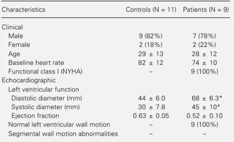
We studied 9 patients (7 men and 2 women) with isolated chronic aortic regurgi-tation. All patients were in functional class I (New York Heart Association, NYHA). The baseline heart rate was similar to that of the controls (Table 1).
The diagnosis of isolated chronic aortic regurgitation was confirmed by cardiac cath-eterization. All patients had severe aortic regurgitation (complete and dense left ven-tricular opacification in 1 beat with an even-tual contrast density greater than that of the aorta). Left ventricular wall motion was nor-mal in all patients. There were no segmental wall motion abnormalities upon a quantita-tive analysis of the left ventricular cinean-giograms (21).
The echocardiographic evaluation per-formed on the same day of the assessment of cardiac autonomic function revealed no dis-crete areas of abnormal wall motion. How-ever, there was marked left ventricular en-largement (Table 2).
The left ventricular ejection fraction was preserved and did not significantly differ from that of the normal controls (Table 1).
Table 1 - Clinical and echocardiographic characteristics.
Data are reported as means ± SD. *P≤0.01 compared to control (t-test).
Characteristics Controls (N = 11) Patients (N = 9)
Clinical
Male 9 (82%) 7 (78%)
Female 2 (18%) 2 (22%)
Age 29 ± 13 28 ± 12
Baseline heart rate 82 ± 12 74 ± 10
Functional class I (NYHA) - 9 (100%)
Echocardiographic Left ventricular function
Diastolic diameter (mm) 44 ± 6.0 68 ± 6.3*
Systolic diameter (mm) 30 ± 7.8 45 ± 10*
Ejection fraction 0.63 ± 0.05 0.52 ± 0.10
Normal left ventricular wall motion - 9 (100%)
Segmental wall motion abnormalities -
-Table 2 - Cardiac autonomic function tests.
Data are reported as means ± SD with the 95% confidence interval given below. There were no significant differences between patients and controls (ANOVA).
Autonomic test Controls (N = 11) Patients (N = 9)
Valsalva ratio 1.90 ± 0.49 1.65 ± 0.33
1.63 ± 2.10 1.39 ± 1.92
Standing up (30/15 ratio) 1.16 ± 0.16 1.14 ± 0.11
1.07 ± 1.25 1.04 ± 1.25
Facial immersion (beats/min) -31 ± 15 -25 ± 22
-19 ± 43 -12 ± 38
Cardiac autonomic function tests
Valsalva maneuver. When compared to the normal sedentary controls the Valsalva ratio was slightly lower in the asymptomatic patients with aortic insufficiency, although not significantly different (Table 2). There was no correlation between the Valsalva ratio and the left ventricular function param-eters.
(brady-cardic response) were similar in both groups of subjects. There were no significant corre-lations between the echocardiographic pa-rameters of left ventricular function and the heart rate changes induced by these two cardiac autonomic function tests (Table 2).
Discussion
The Valsalva maneuver is a well-known and widely accepted test of cardiac parasym-pathetic function. This test, which has been used in multicenter trials to evaluate auto-nomic function, is reliable, consistent and operator-independent (24-28). The heart rate changes provoked during the maneuver and expressed as the Valsalva ratio are mostly dependent on cardiovagal integrity (29).
The Valsalva maneuver is abnormal in most asymptomatic patients with coronary heart disease and cardiomyopathies (14-16). The patients studied in these earlier clinical investigations had a distinct and common pathologic feature, i.e., left ventricular wall motion abnormalities (30-32). Most of these patients also had mild to moderate left ven-tricular dilatation. On the other hand, the ischemic and cardiomyopathic patients with a normal Valsalva ratio had either normal segmental wall motion or highly localized myocardial dyskinesis (i.e., apical aneu-rysms). Thus, asymptomatic patients with acquired heart disease of different etiologies and with an abnormal Valsalva ratio had segmental wall motion abnormalities and ventricular dilatation (33-37).
The mechanism(s) responsible for the abnormal heart rate changes during the Val-salva maneuver is (are) still unknown (1,4,5). We (36) and other investigators (10) have postulated that progressive ventricular dila-tation may diminish the normal inhibitory action of cardiac vagal afferents on the brain-stem vasomotor center which in turn may decrease the efferent parasympathetic traffic to the sinus node and partially explain the abnormal heart rate changes observed
dur-ing the Valsalva maneuver (38,39).
In the present investigation, we studied patients with pure aortic insufficiency. We chose this clinical entity because, in asymp-tomatic chronic aortic insufficiency, myo-cardial fibrosis is discrete, does not affect systolic function and is of rather late appear-ance (18,20). Therefore, this clinical model may allow us to study the effects of left ventricular dilatation on parasympathetic heart rate control. Our results suggest that ventricular dilatation by itself does not sig-nificantly affect the Valsalva ratio in asymp-tomatic aortic insufficiency.
Since the two additional tests of cardiac parasympathetic function were also normal in our asymptomatic patients with ventricu-lar dilatation and no segmental wall motion abnormalities, we are tempted to hypoth-esize that, in order to impair parasympa-thetic heart rate control, ventricular dilata-tion should be associated with segmental wall motion abnormalities and/or congestive heart failure (40).
mechanisms may interfere with the tonic activity of the parasympathetic fibers di-rected to the sinus node (46,47).
In summary, our results indicate that, in asymptomatic patients with left ventricular dilatation and no segmental wall motion ab-normalities, parasympathetic heart rate
con-trol as indicated by the Valsalva maneuver is not impaired.
Acknowledgment
We are indebted to Miss Irlanda Márquez for valuable secretarial assistance.
References
1. Eckberg DL, Drabinsky M & Braunwald E (1971). Defective cardiac parasympathetic control in patients with heart failure. New England Journal of Medicine, 285: 877-883.
2. Mancia G (1990). Neurohormonal activa-tion in congestive heart failure. American Heart Journal, 120: 1532-1537.
3. The SOLVD Investigators (1994). Relation of neurohormonal activation to clinical variables and degree of left ventricular dysfunction. Journal of the American Col-lege of Cardiology, 23: 1410-1420. 4. Nishimura RA & Tajik AJ (1986). The
Val-salva maneuver and response revisited. Mayo Clinic Proceedings, 61: 211-217. 5. Eckberg DL (1980). Parasympathetic
car-diovascular control in human disease: a critical review of methods and results. American Journal of Physiology, 239: H581-H593.
6. Levin AB (1986). A simple test of cardiac function based upon the heart rate changes induced by the Valsalva maneu-ver. American Journal of Cardiology, 18: 90-99.
7. Tsay L & Chen JH (1990). Abnormal he-modynamic response to Valsalva maneu-ver in patients with atrial septal defect evaluated by Doppler echocardiography. Chest, 98: 1175-1178.
8. Bernardi L, Saviolo R & Spodick DH (1989). Do hemodynamic responses to the Valsalva maneuver reflect myocardial dysfunction? Chest, 95: 986-991. 9. Zema MJ (1990). Heart failure and the
Valsalva maneuver. Chest, 97: 772-773. 10. Floras JS (1993). Clinical aspects of
sym-pathetic activation and parasymsym-pathetic withdrawal in heart failure. Journal of the American College of Cardiology, 22 (Suppl A): 72A-84A.
11. Gaudron P, Eilles G, Kugler I & Ertl G (1993). Progressive left ventricular dys-function and remodeling after myocardial infarction. Potential mechanisms and early predictors. Circulation, 87: 755-763.
12. Bestetti RB, Dalbo CMR, Freitas OC, Teno LAC, Castilho OT & Oliveira JSM (1994). Non-invasive predictors of mortality for patients with Chagas’ disease: a multi-variate stepwise logistic regression study. Cardiology, 84: 261-267.
13. Wallis D, O’Connell JB, Hemkin RE, Constanzo-Morin MR & Scanlon PJ (1984). Segmental wall motion abnormali-ties in dilated cardiomyopathy: a common finding and good prognostic sign. Journal of the American College of Cardiology, 4: 674-679.
14. Junqueira LF (1990). Ambulatory assess-ment of cardiac autonomic function in Chagas’ heart disease of patients based on indexes of R-R interval variation in the Valsalva maneuver. Brazilian Journal of Medical and Biological Research, 23: 1091-1098.
15. Marin-Neto JA, Maciel BC, Gallo Jr L, Junqueira Jr LF & Amorin DS (1986). Ef-fect of parasympathetic impairment on the hemodynamic response to handgrip in Chagas’ disease. British Heart Journal, 55: 204-210.
16. Fuenmayor AJ, Dávila DF, Rodríguez L, Donis JH, Torres A, Fuenmayor AM & Navas M (1987). The Valsalva maneuver in chagasic patients with left ventricular segmental wall motion abnormalities and normal ejection fraction. Arquivos Brasi-leirosde Cardiologia, 49: 221-224. 17. Bonow RO, Lakatos E, Maron BJ &
Epstein SE (1991). Serial long term as-sessment of the natural history of asymp-tomatic patients with chronic aortic regur-gitation and normal left ventricular myo-cardium. Circulation, 84: 1625-1635. 18. Krayenbuenhl HP, Hess OM, Monrad S,
Schneider J, Mall G & Turina M (1989). Left ventricular myocardial structure in aortic valve disease before, intermediate and late after aortic valve replacement. Circulation, 79: 744-755.
19. Villari B, Campbell SE, Hess OM, Mall G, Vassalli G & Weber KT (1993). Influence of collagen network on left ventricular sys-tolic function in aortic valve disease. Jour-nal of the American College of Cardiol-ogy, 22: 1477-1484.
20. Buja LM (1985). The heart. In: Robbins SL & Kumar V (Editors), Human Pathology. McGraw Hill, New York, 317-354. 21. Braunwald E (1980). Valvular heart
dis-ease. In: Braunwald E (Editor), Heart Dis-ease. A Textbook of Cardiovascular Medi-cine. WB Saunders, Philadelphia. 22. Grossman W & Dexter L (1980). Profile in
valvular heart disease. In: Grossman W (Editor), Cardiac Catheterization and An-giography. Lea & Febiger, Philadelphia. 23. Schnittger I, Gordon EP, Fitzgerald PJ &
Popp RI (1983). Standardized intracardiac measurements of two-dimensional echo-cardiography. Journal of the American College of Cardiology, 2: 934-938. 24. Schumer M, Miller-Crain G & Pfeiter MA
(1988). The study group. Diabetic auto-nomic neuropathy - Part II: Coefficient of variation of RR-variation and Valsalva ma-neuver tests. American Journal of Medi-cine, 85: 144-146.
25. Ewing DJ, Campbell IW, Murray A, Neilson JM & Clarke M (1978). Immedi-ate heart-rImmedi-ate response to standing: a simple test of autonomic neuropathy in diabetes. BritishMedical Journal, I: 145-147.
26. Blix AS (1988). Cardiovascular responses to diving. Acta Physiologica Scandinavica, 133 (Suppl 571): 61.
28. Moriarty KT, Ryder REJ & Hardisty CA (1994). Cardiovascular autonomic function tests - are three Valsalvas‘ and six deep breaths necessary or will singles do? Jour-nal ofNeurology, Neurosurgery and Psy-chiatry, 54: 938-939.
29. Sandroni P, Benarroch EE & Low PA (1991). Pharmacological dissections of components of the Valsalva maneuver in adrenergic failure. Journal of Applied Physiology, 71: 1563-1567.
30. Ideker RE, Behar VS, Wagner GS, Starr JW, Starmer CF, Lee KL & Hackel DB (1978). Evaluation of asynergy as an indi-cator of myocardial fibrosis. Circulation, 57: 715-725.
31. Zema MJ (1985). Prognosis after myocar-dial infarction: prediction in ambulatory patients by use of the Valsalva maneuver. Angiology, 34: 94-104.
32. Parisi AF, Moylliham PF, Folland ED & Feldman CL (1981). Quantitative detec-tion of regional left ventricular contracdetec-tion abnormalities by two-dimensional echo-cardiography. II. Accuracy in coronary ar-tery disease. Circulation, 63: 761-767. 33. Dávila DF, Donis JH & Rossell R (1989).
Cardiac parasympathetic abnormalities. Cause or consequence of Chagas‘ heart disease? Parasitology Today, 5: 327-329. 34. Dávila DF, Donis JH, Navas M, Fuenmayor
AJ, Torres A & Gottberg CF (1988). Heart rate responses to atropine and left ven-tricular function in Chagas‘ heart disease. International Journal of Cardiology, 21: 143-152.
35. Dávila DF, Donis JH & Torres A (1992). Chagas’ heart disease and neuropathy. American Heart Journal, 124: 1665-1666.
36. Dávila DF, Donis JH, Torres A, Gottberg CF & Rossell O (1991). Cardiac parasym-pathetic innervation in Chagas’ heart dis-ease. Medical Hypothesis, 35: 80-84. 37. Fuenmayor AJ, Rodríguez L, Torres A,
Donis JH, Navas M, Fuenmayor AM & Dávila DF (1988). Valsalva maneuver: a test of the functional state of cardiac in-nervation in chagasic myocarditis. Inter-national Journal of Cardiology, 18: 351-356.
38. Porter TR, Eckberg DL & Fritsch JM (1990). Autonomic pathophysiology in heart failure: sympathetic-cholinergic in-terrelations. Journal of Clinical Investiga-tion, 85: 1362-1369.
39. Sopher SM, Smith ML, Eckberg DL, Tritsch JM & Dibner-Dunlap ME (1990). Autonomic pathophysiology in heart fail-ure: carotid baroreceptor-cardiac reflexes. American Journal of Physiology, 259: H689-H696.
40. Binkley PF, Nunziata E, Haas GJ, Nelson SD & Cody RJ (1991). Parasympathetic withdrawal is an integral component of autonomic imbalance in congestive heart failure: demonstration in human subjects and verification in a paced canine model of ventricular failure. Journal of the Ameri-can College of Cardiology, 18: 464-472. 41. Barber MJ, Mueller TM, Davies BG, Gill
RM & Zipes DP (1985). Interruption of sympathetic and vagal-mediated afferent responses by transmural myocardial in-farction. Circulation, 71: 623-631.
42. Minisi AJ & Thames MD (1992). Distribu-tion of left ventricular sympathetic affer-ents demonstrated by reflex responses to transmural myocardial ischemia and to intracoronary and epicardial bradykinin. Circulation, 87: 240-246.
43. Abboud FM, Thames MD & Mark AL (1981). Role of cardiac afferent nerves in regulation of circulation during coronary occlusion and heart failure. In: Abboud FM, Fozzard HA, Gilmore JP & Reis DJ (Editors), Disturbances in Neurogenic Control of the Circulation. American Physi-ological Society, Bethesda, MD, 317-340. 44. Bonaduce D, Petretta M, Piscione F, Indolfi C, Migaux ML, Bianchi V, Esposito N, Marciano F & Chiarello M (1994). Influ-ence of reversible segmental left ventric-ular dysfunction on heart period variabil-ity. Journal of the American College of Cardiology, 24: 399-405.
45. Airaksinen KEJ, Ylitalo KV, Peuhkurinen KJ, Ikakeimo MJ & Huikuri HV (1995). Heart rate variability during repeated arte-rial occlusion in coronary angioplasty. American Journal of Cardiology, 75: 877-881.
46. Zipes DP (1990). Influence of myocardial ischemia on autonomic innervation of the heart. Circulation, 82: 1095-1105. 47. Tseng CD, Wang TL, Lin JL, Hsu KL,