The Use of High Resolution Optical Coherence Tomography to
Evaluate Robotic Radical Prostatectomy Specimens
Pankaj P. Dangle, Ketul K. Shah, Benjamin Kaffenberger, Vipul R. Patel
Robotic and Minimally Invasive Urologic Surgery, Ohio State University Medical Center, Columbus, Ohio, USA
ABSTRACT
Objective: Optical coherence tomography (OCT) is a unique technology, developed to provide high resolution, cross sectional images of human tissue. The objective of this study was to explore the feasibility of OCT for the evaluation of positive surgical margins and extra capsular extension in robotic prostatectomy specimens and compare it to histopathology.
Materials and Methods: Radical prostatectomy was performed in 100 patients. Twenty OCT images of each specimen were taken from the base of the seminal vesicles (SV), apical and vesicle margins, peripheral and posterolateral area and any palpable nodule. Predictions were made regarding positive surgical margin, SV involvement, capsular invasion and compared with the final histopathology.
Results: A total of 2000 OCT images were taken and analyzed. Out of 100 specimens, 85 had T2 disease, 15 had T3 dis-ease with a median Gleason’s score of 7 (range 6 to 9) and 10 had positive surgical margins. We predicted 21 specimens to have positive margins based on OCT images out of which 7 were truly positive and 14 were falsely positive. Based on OCT images, 79 specimens were predicted to have negative margins out of which 76 were truly negative and 3 were falsely negative. We found the sensitivity, specificity, positive predictive value and negative predictive value to be 70%, 84%, 33% and 96% respectively.
Conclusion: Our initial feasibility study established the template for the visual OCT characteristics of the prostate, SV and cancerous tissue. The negative predictive value of evaluating surgical margins was high.
Key words: prostate cancer; prostatectomy; laparoscopy; robotic assisted; tomography, optical coherence
Int Braz J Urol. 2009; 35: 344-53
INTRODUCTION
Prostate cancer is the second most common solid cancer among men in the United States. It was estimated that in the year 2007 a total of 270,050 new cases of prostate cancer were diagnosed (1).
Anatomic radical prostatectomy as a treat-ment option for localized prostate cancer has been well practiced since its first description by Walsh et al. Over the last two decades more prostatectomies have
been performed by minimally invasive approaches such as laparoscopy or robot assisted.
Solo-way et al. studied 495 patients who underwent open radical prostatectomy and found 30.5% had one or more positive margins. In this subset of patients the detected recurrence rate for various locations was 29% apex/urethra, 30% posterior, 33% anterior, 36% lateral, 48% posterolateral, and 57% bladder neck. In this study the author concluded that recognizing posi-tive margin pre or intraoperaposi-tively could be useful to avoid disease recurrence (3).
Over and above the oncological outcomes, functional outcomes like continence and potency are also crucial. The preservation of neurovascular bundle (NVB) to aid early recovery in urinary continence and potency is of vital importance (4). Thus, the intraop-erative identification of the NVB is impintraop-erative in order to be preserved during dissection. Due to the close proximity (2-3 mm) of the NVB to the prostatic fascia balancing the oncological and functional outcome is critical.
A technique which can precisely identify the positive margin intraoperatively is required. Optical coherence tomography (OCT) is a technique available for imaging the microstructure of tissues. We describe the OCT technique used ex vivo on postoperative prostate specimen to assess its feasibility in detecting the positive margin and extra capsular extension.
MATERIALS AND METHODS
OCT imaging was obtained in 100 consecu-tive patients who underwent robotic assisted radical prostatectomy performed in a standard manner as previously described by our group (5). After the prostate was removed, it was thoroughly cleaned and subjected to a gentle saline wash in an adjacent room, taking care not to alter the gross anatomy. The specimen was washed so as to remove any attached blood clots and non prostatic tissue, which could po-tentially alter the imaging with OCT with increased tissue interface between the prostatic surface and the OCT probe. The specimen was then transferred to the OCT technician within the operating room. The time required to transfer the specimen was variable with average time being 35 min. (25-55 min.) from the time of devascularization to completion of procedure at which point the specimen was removed from the
body. The OCT technician was blinded to any of the pre or intra-operative information.
A systematic and thorough analysis of the specimen was performed in the following manner. The specimen was scanned in circumferential man-ner on all four surfaces (anterior, posterior and two laterals) and two ends (base and apex). The images were obtained from each specimen starting at the base of the seminal vesicles (SV), apical and vesical margins, peripheral and posterolateral area, and any palpable nodule if present (Figure-1). The operator scanned both the right and left sides of the preceding areas. Structures identified based on OCT imaging were tumor location, capsular penetration, and mar-gin status. The scanning was conducted in real-time and an image was saved when the operator identified one of the previously mentioned landmarks. At the same time predictions were made regarding surgical margin. The OCT image predictions were compared with the final histopathology results. The images thus obtained were assessed based on the appearance of the image in correlation to the site on the specimen. The image was assessed for homogenous appearance of the capsule, prostatic surface epithelium and prostatic stroma. Any variation from normal appearance was noted and assessed for the interface between capsule and the surface epithelium to identify positive margin and extra capsular extension.
The outer surface of the specimen was inked; all the scanned areas were color coded. The seminal vesicles were separated from the site of their insertion at the prostate base and subsequently bivalved. The
base and apex were axially separated from the prostate in 1 cm thick sections, which were further cut at 0.3 cm intervals in the sagittal plane. The remaining mid prostate was axially sectioned at 0.3 cm intervals, carefully including a complete inked margin around each slice. All tissue was entirely submitted in whole mount cassettes. The tumor contours were mapped on the glass slides, while additionally noting foci of extraprostatic extension, margin positivity, and/or seminal vesicle involvement.
After analysis by the pathologist, OCT im-ages were correlated with their corresponding histo-pathologic micrographs on the same areas of the same specimens.
OCT is an imaging technique which we em-ployed using a Niris OCT Imaging System made by Imalux (Cleveland, OH) and cleared by the Federal Drug Administration in 2004. OCT is currently be-ing used in ophthalmology departments throughout the country. Similar to ultrasound, it consists of two components. The first is the computer console that projects a user driven application. It can show images, save, and print these images after they are obtained. The second component is the actual scanning probe. In the Niris the probe has a diameter of 2.7 mm, and can be made in lengths up to 4 m while still being flexible. Probes with even smaller diameters have been described (6). The Niris produces one image in 1.5 seconds. Theoretically an image in OCT can be obtained in about 250 msec without decreasing image resolution (7).
Optical Coherence Tomography is comprised of low-coherence near infrared light with a fiber optic interferometer to measure the characteristic scatter in a real-time scan. Despite using infrared light as opposed to acoustic, OCT is analogous to ultrasound as it relies on beaming the infrared wave through the tissue, then measuring the backscatter (Figure-2). The characteris-tic wavelength of near infrared light is between 750 nm and 1µm. The Niris OCT system uses near infrared light centered at 1310 nm allowing for an axial resolution of 20 micrometers (8). OCT produces an image 10-100 times higher in resolution than any other clinically available diagnostic imaging (9).
OCT technology is non invasive and that the energy used does not cause physical injuries. The greatest potential harm from infrared light would
oc-cur by a heating effect but these have shown to be less than 0.5 degrees Celsius when used in the eye (10). The American National Standards Institute classifies near infrared light as being a class 1. Class 1 lasers pose virtually no risk to skin or eyes (11).
RESULTS
Prostate specimens from 100 patients who un-derwent robotic radical prostatectomy were scanned systematically by OCT technology. The small portable machine allowed the analysis to be performed in the operating room in less than 5 minutes.
Figure 2 – A) Imalux Niris Optical Coherence Tomogra-phy Imaging System. B) Illustration of 2D image creation based on scatter.
A
A total of 2000 images were obtained from the 100 specimens that were obtained following robot assisted laparoscopic prostatectomy. The pathological stages of the specimens were T2 in 85% of cases and T3 in 15% of cases. The median Gleason score was 7 with a range from 6 to 9 (Table-1).
Of the 100 samples scanned 10 had positive surgical margin. Of these 10 patients 5 had pT2c disease whereas 3 had pT3a and 2 had pT2a disease. We predicted 21 specimens to have positive margins based on OCT images out of which 7 were truly positive and 14 were falsely positive. Based on these results the sensitivity was 70% where as specificity was 84%. The positive predictive value (PPV) based on above results was 33% and the negative predictive value (NPV) was 96% (Table-2). Of the 7 predicted true positives, correlation with the postoperative pa-thology, revealed that 2 patients had pT3a disease, 1 had pT2a and 4 had pT2c disease.
We predicted extra capsular extension in 12 patients of which 6 were truly positive and 6 were falsely positive. Of these 6 true positives, 3 patients had pT2c and 3 had pT3a disease on final pathology. The sensitivity and specificity was 46% and 84% re-spectively, whereas the PPV and NPV was 50% and 92% respectively (Table-2).
Based on the imaging 3 patients were pre-dicted to have seminal vesicle invasion with 1 being truly positive and 2 being falsely positive. The sensi-tivity and PPV was 33% each whereas the specificity and NPV was 97% respectively (Table-2).
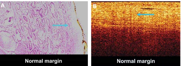
The OCT images were compared to the fi-nal pathology slides. Following Figure-3 shows the normal tissue architecture of prostatic epithelium. The OCT image shows a well defined homogenous appearance of capsular surface, epithelium and subepithelium with no evidence of heterogenous columns or areas of cells suggesting tumor cells. Figure-4 shows positive surgical margin. The characteristics are the heterogenous, low scat-tering columns (arrows) extending continuously from subepithelium to surface serosa suggesting invasion by tumor cells.
In comparing Figures-3 and 4, we can see that Figure-4B shows low scattering areas (arrows) that are not in a columnar form radiating towards the edge of the specimen. These low scattering areas are surrounded by high scattering areas yielding a char-acteristic image of positive capsular extension. The same areas are also covered by normal epithelium. Figure-5 shows capsular invasion by tumor. The figure shows low scattering areas that are not in columnar form radiating towards the edge of the specimen. The low scattering areas are surrounded by high scatter-ing areas yieldscatter-ing a characteristic image of positive capsular extension. There is small normal rim of epithelial layer.
Table 1 – Patient demographics.
Variable Mean
Mean age- 60.3 yrs. ± 5.2 Mean PSA- 6.60 ng/ml ± 3.8 Median Gleason-score 7 (6-9) Pathologic stage
T2 85%
T3 15%
T4 0%
Nerve spare
BNS 55%
UNS 14%
NNS 31%
BNS = bilateral nerve spare; UNS = unilateral nerve spare; NNS = non nerve spare.
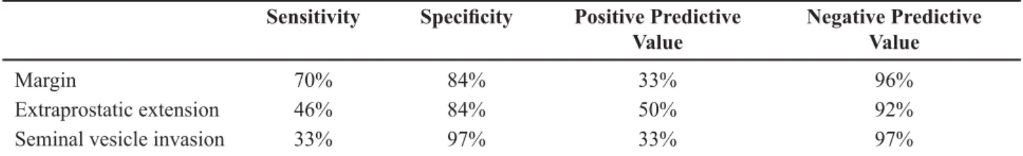
Table 2 – Statistics for positive margin, extraprostatic extension and seminal vesicle invasion.
Sensitivity Specificity Positive Predictive Value
Negative Predictive Value
Margin 70% 84% 33% 96%
Extraprostatic extension 46% 84% 50% 92%
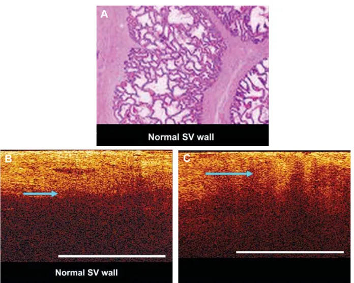
Figure-6 shows normal seminal vesicle (histology, OCT image) and invasion by tumor re-spectively. The image shows areas of high scattering columns of tumor cells seen extending into the wall of seminal vesicle suggesting tumor invasion.
Figure-7 shows OCT view of adipose tissue, blood vessel, neurovascular bundle (NVB) (both the transverse, longitudinal view).
COMMENTS
Presently it is difficult to assess the margin and location of NVB pre and intraoperatively by available modes of investigation. With the tactile and haptic feed back the assessment of microscopic mar-gin and the NVB is unfeasible and restricted. The use of laparoscopic and robotic approach for treatment of
Figure 3 – A) Hematoxylin-eosin staining of normal prostatic epithelium (arrow). B) Corresponding optical coherence tomography image showing a well defined homogenous appearance of capsular surface, epithelium and subepithelium with no evidence of heterogenous columns or areas of cells suggesting tumor extension (arrow).
A
B
Figure 4 – A) Hematoxylin-eosin staining of positive surgical margin of prostate gland (arrow). B) Corresponding optical coherence tomography image characteristics are the heterogeneous, low scattering columns (arrow) extending continu-ously from subepithelium to surface serosa suggesting presence of tumor.
localized prostate cancer has an inherent drawback in the lack of tactile feedback which makes assessment of margin more difficult. Use of frozen section is only for evaluation of focal positivity and not for assess-ing the entire gland. Since the introduction of OCT technology it has been investigated for use in multiple specialties of medicine with variable results.
After being first described in 1991, OCT has been accepted for in vivo use in ophthalmology for over 10 years. While it has quickly found use in ophthalmology departments, it has had less momen-tum with imaging non-transparent tissues. Even in non-transparent tissues OCT was found to have the ability to demonstrate boundaries of the mucosa and differentiate between epithelial types in the esophagus (8).
Feldchtein et al. applied the technology to endoscopy while performing biopsies, guiding surgi-cal procedures, monitoring functional state of organs and monitoring post-operative recovery progress in their in vivo studies of over 100 patients (12).
Zysk et al. in their study involving breast cancer detection and analysis using OCT reported a specificity of 97% and a sensitivity of 56-67% which was higher than both mammograms and ultrasounds (13).
Another study by Zeluaga et al. justified the use of OCT to detect cervical cancer in vivo. Using 16 patients, the study compared image interpretation of OCT against blinded pathologists. A significant difference P < 0.024 was found between normal and cancerous cervical specimens (14).
In the year 1997, Amling et al. used the technol-ogy to study the different structures of the male reproduc-tive system. Using OCT they were able to differentiate the fibrous prostatic capsule, the NVB prostatic urethra, and normal prostatic epithelium (15).
Boppart et al. used OCT to guide radio fre-quency ablation of the prostate. In their study they were able to demonstrate extra control over ablation parameters using OCT in real time (16).
Tearney et al. investigated the capability of OCT to differentiate the architectural morphology of urologic tissue with the long term aim of using OCT as an adjunct to endoscopic imaging and to improve the efficiency of interventional procedures such as transurethral prostatectomy. These authors studied urologic tissues from postmortem subjects, dissected and imaged using OCT. The microstructure thus obtained was delineated in different urologic tissues including the prostatic urethra, prostate, bladder, and ureter. They also studied the ability to achieve
opti-Figure 5 – A) Hematoxylin-eosin staining of prostatic capsular invasion by tumor (arrows). B) Corresponding optical coherence tomography image shows low scatter areas (not in columnar form) radiating towards the edge of the specimen (arrow). The low scatter areas are surrounded by high scatter areas yielding a characteristic image of positive capsular extension. These formations are not consistent with blood vessels as blood typically exhibits high scattering with little if any penetration of light though the vessel. Notice the small normal rim of epithelium at the surface of the image.
B
C
A
cal biopsy with OCT suggesting potential to obtain information on tissue microstructure. The group concluded that high resolution, cross-sectional OCT images acquired in this study provide information on tissue microstructure that could only previously be obtained with conventional biopsy (8).
D’Amico et al. used the OCT technology in ex vivo human benign and malignant prostatic tissues. Images obtained using OCT were directly compared with images acquired after standard his-topathologic processing. These results suggested that microarchitecture on the order of 10-µm can be distinguished in prostatic tissue. The major technical limitation at this point, however, was that the 10-µm
resolution achieved in this study was limited to a depth of approximately 0.5 mm. Further studies us-ing this technique to improve detection and stagus-ing are ongoing. In addition, future trials will investigate whether neurovascular bundle invasion can be ac-curately identified intraoperatively. In these men, the neurovascular bundle would be scanned with OCT intraoperatively, and then the images would be com-pared with the final histopathologic sections from the radical prostatectomy specimen (17).
Our experience based on OCT imaging and comparison with the final pathology is satisfactory for detection of positive margin with 70% and 84% sensitivity and specificity respectively.
Figure 7 – A) Optical coherence tomography image of adipose tissue (arrow). B) Blood vessel with coagulated blood (ar-row). Note poor penetration of light below vessel lumen. C) Typical image of neurovascular bundle with nerve fascicles (arrow), and surrounded by adipose tissue in D (arrow).
The positive predictive value is less (33%) but the high negative predictive value may help detect the true negative margins and avoid overzealous dissec-tion to compromise NVB laterally. The low positive predictive value for detecting positive margin could be due to the heterogenous appearance of the tumor and the low depth of penetration (2-3 mm) with OCT (17). We experienced a potential difficulty in initial 50 cases to better understand the normal and anatomical vari-ants associated with the prostatic anatomy. With each set of 10 cases our diagnostic capabilities improved to define the normal anatomical architecture based on
OCT imaging. Though we experienced a difficulty in understanding the normal and anatomical variants, we did not separately evaluate the results for first and later 50 cases. In our experience one needs to learn, understand the technical issues of OCT machine, the most important being the image interpretation which could contribute to the learning curve.
In our experience, OCT imaging helped to rule out extra capsular extension in prostatectomy specimen in 92% of cases. Of the 6 patients with positive extra capsular extension, three were detected based on OCT technology. All these three patients had
A
B
pathological T3a disease, implying OCT is restricted in identifying those with higher stage disease.
From our initial ex vivo study the OCT im-aging had a detection rate of 33% for assessment of seminal vesicle invasion. Its usefulness in ruling out seminal vesicle invasion is high, approximately 97%. Our initial feasibility study established the template for the visual OCT characteristics of the prostate, SV and cancerous tissue.
Thus, the current OCT imaging has a selected role in identifying the positive margin and ruling out the extraprostatic extension and seminal vesicle inva-sion.
One drawback of our study was that we lim-ited our study to ex-vivo use of OCT. The specimen was not removed from the patient until the surgery was nearly complete. The average time required to transfer the specimen was 35 min. (25-55 min.) from the time of devascularization to completion of procedure at which point the specimen was removed from the body. The tissue characteristics as measured by OCT can be affected by long periods without perfusion, and we did not control the length of time our specimen was without perfusion. We are currently working towards repeating the study in vivo, during robotic-assisted radical prostatectomies. Because there are currently no standards defined for imaging the prostate for mar-gins of resection, or the seminal vesicles for invasion there was a learning curve involved, especially for our early cases when there was a higher false positive rate and low positive predictive value. As we gained experience with the OCT image appearance of normal tissue, and were able to correlate our images with pathological results, our statistics improved.
A further randomized in vivo study is planned to understand the exact role of the OCT technology to predict prostate margin status and seminal vesicle invasion during resection and prior to specimen re-moval as well as using OCT to identify the location of the neurovascular bundle.
CONCLUSION
This initial feasibility study established the template for the visual OCT characteristics of the prostate, SV and cancerous tissue. After a careful
analysis of the corresponding images from OCT with their histopathologic micrographs, there appears to be an opportunity to use OCT in order to assess the mi-crostructure of the prostate. The high negative predic-tive value could be useful to rule out posipredic-tive surgical margin, extraprostatic extension and SV invasion. Our current study was conducted on ex vivo specimens and we are planning in vivo studies to quantify our findings and to test in vivo OCT effectiveness for margins, SV invasion and NVB identification.
ACKNOWLEDGEMENT
Imalux Cleveland Ohio.
CONFLICT OF INTEREST
None declared.
REFERENCES
1. National Cancer Institute: Prostate cancer statistics for 2007. http://www.cancer.gov/cancertopics/types/pros-tate
2. Amling CL: Prostate-specific antigen and detection of prostate cancer: What have we learned and what should we recommend for screening? Curr Treat Op-tions Oncol. 2006; 7: 337-45.
3. Obek C, Sadek S, Lai S, Civantos F, Rubinowicz D, Soloway MS: Positive surgical margins with radi-cal retropubic prostatectomy: anatomic site-specific pathologic analysis and impact on prognosis. Urology. 1999; 54: 682-8.
4. Kaiho Y, Nakagawa H, Ikeda Y, Namiki S, Numahata K, Satoh M, et al.: Intraoperative electrophysiologi-cal confirmation of urinary continence after radielectrophysiologi-cal prostatectomy. J Urol. 2005; 173: 1139-42.
5. Patel VR, Shah KK, Thaly RK, Lavery H: Robotic-assisted laparoscopic radical prostatectomy: The Ohio State University technique. Journal of Robotic Surgery. 2007; 1: 51-59.
7. Brezinski M, Tearney G, Boppart S, Swanson E, Southern J, Fujimoto J: Optical Biopsy with Opti-cal Coherence Tomography: Feasibility for SurgiOpti-cal Diagnosis. J Surgical Research. 1997; 71: 32-40. 8. Tearney GJ, Brezinski ME, Southern JF, Bouma BE,
Boppart SA, Fujimoto JG: Optical biopsy in human urologic tissue using optical coherence tomography. J Urol. 1997; 157: 1915-9.
9. Toth CA, Birngruber R, Boppart SA, Hee MR, Fuji-moto JG, DiCarlo CD, et al.: Argon laser retinal lesions evaluated in vivo by optical coherence tomography. Am J Ophthalmol. 1997; 123: 188-98.
10. Bozkurt A, Onaral B: Safety assessment of near infra-red light emitting diodes for diffuse optical measure-ments. Biomed Eng Online. 2004; 3: 9.
11. American National Standards Institute. Z136.1. http:// www.ansi.org.
12. Feldchtein F, Geliknov G, Gelkonov V, Kuranov R, Sergeev A: Endoscopic applications of Optical Coher-ence Tomography. Optics Express. 1998; 3: 257-70. 13. Zysk AM, Boppart SA: Computational methods for
analysis of human breast tumor tissue in optical
coher-ence tomography images. J Biomed Opt. 2006; 11: 054015.
14. Zuluaga AF, Follen M, Boiko I, Malpica A, Richards-Kortum R: Optical coherence tomography: a pilot study of a new imaging technique for noninvasive examination of cervical tissue. Am J Obstet Gynecol. 2005; 193: 83-8.
15. Amling CL, Blute ML, Bergstralh EJ, Seay TM, Slezak J, Zincke H: Long-term hazard of progression after radical prostatectomy for clinically localized prostate cancer: continued risk of biochemical failure after 5 years. J Urol. 2000; 164: 101-5.
16. Boppart SA, Herrmann JM, Pitris C, Stamper DL, Brezinski ME, Fujimoto JG: Real-time optical coher-ence tomography for minimally invasive imaging of prostate ablation. Comput Aided Surg. 2001; 6: 94-103.
17. D’Amico AV, Weinstein M, Li X, Richie JP, Fujimoto J: Optical coherence tomography as a method for iden-tifying benign and malignant microscopic structures in the prostate gland. Urology. 2000; 55: 783-7.
Accepted after revision: January 13, 2009
Correspondence address:
Dr. Pankaj P. Dangle Robotic Surgery &
Minimally Invasive Urologic Surgery 410 West 10th Avenue, 538 Doan Hall Columbus, Ohio, 43235, USA Fax: + 1 614 293-0982