Arquivos Brasileiros de Cardiologia - Volume 82, Nº 4, Abril 2004
381
Case report
The patient is a 34-year-old female being followed up at the Rheumatology Service of the Hospital das Clínicas of the University of Minas Gerais since May 1994 due to the diagnosis of systemic lupus erythematosus, according to the criteria of the American College of Rheumatology 5. The patient had malar erythema,
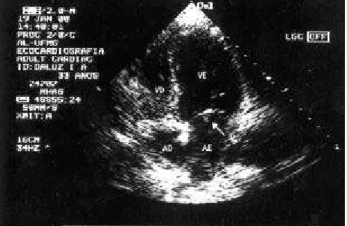
photo-sensitivity, discoid cutaneous lesions, symmetric polyarthritis, oral ulcers, and positive antinuclear factor titers (1:1024). She also had renal impairment, confirmed on biopsy, which revealed diffuse proliferative glomerulonephritis (class IV of the World Health Organi-zation). Prednisone (1 mg/kg/day) and monthly pulse therapy with cyclophosphamide (1 g/month) were initiated with progressive im-provement in clinical findings and later a reduction in medications. The patient evolved stably until March 1998, when she began to experience generalized tonicoclonic convulsive crises. Compu-terized tomography of the brain and examination of the cerebros-pinal fluid were normal, and the investigation of anticardiolipin antibodies revealed an IgG of 12.2 GPL (< 10) and an IgM of 0.5 MPL (< 10). However, the search for lupus anticoagulant with the techniques of the kaolin coagulation test and the tissue thromboplastin inhibition test was positive. The patient had a previous history of 3 abortions in the second gestational trimester, therefore confirming the diagnosis of secondary antiphospholipid antibody syndrome. An anticonvulsant (diphenylhydantoin) at a dosage of 300 mg per day and acetylsalicylic acid at a dosage of 200 mg per day were initiated, and partial control of the convulsive crises was obtained. In January 2000, the physical examination showed a regurgitation cardiac murmur in the inferior left sternal border, and the transthoracic echocardiography showed vegetation in the mitral valve. Blood cultures were negative (fig. 1). The presence of Libman-Sacks endocarditis was hypothesized, an oral anticoagulant (10 mg of warfarin per day) was initiated, and an INR above 2.0 was maintained. In July 2000, the patient was asymptomatic, with no convulsive crises, and a new control echo-cardiography (transesophageal and transthoracic) showed no vege-tation in the mitral valve (fig. 2). Currently, the patient is using prednisone, azathioprine, and warfarin, in maintenance doses, and her disease is inactive.
Discussion
Valvular heart diseases are the most frequent and important cardiac manifestations of systemic lupus erythematosus 4,8.The
Case Report
Libm an-Sacks Endocardit is and
Oral Ant icoagulat ion
Fabiano Almeida Brit o, M agali LM C Tóf ani, Fábio Ávila Tóf ani, Adriana M aria Kakehasi,
Crist ina Cost a Duart e Lanna, M arco Ant onio Parreiras Carvalho
Belo Horizont e, M G - Brazil
Hospital das Clínicas da Universidade Federal de Minas Gerais Mailing address: Fabiano Almeida Brito
Serviço de Reumatologia do Hospital das Clínicas da UFMG Rua Timbiras, 832/701 - Cep 30140-060
Belo Horizonte, MG, Brazil - E-mail: [email protected] Received: 1/24/03
Accepted: 3/10/03
English version by Stela Maris C. e Gandour
The patient is a 34-year-old female with systemic lupus erythe-matosus and secondary antiphospholipid antibody syndrome, who evolved with convulsive crises, partially controlled with an anticonvulsant, and auscultation of a cardiac murmur, whose investigation showed the presence of a mitral valve vegetation. Once the diagnosis of Libman-Sacks endocarditis was established, therapy with warfarin sodium was initiated, and, after 6 months of oral anticoagulation, the patient had total control of the con-vulsive crises and the valvular vegetation disappeared on echo-cardiography. This study discusses the occurrence of Libman-Sacks endocarditis in systemic lupus erythematosus, its asso-ciation with antiphospholipid antibody syndrome, and the anti-coagulant therapy. A literature review is also provided.
Libman-Sacks endocarditis was initially reported in 1924 as the presence of bacterium-free valvular vegetations1, and later as
a manifestation of systemic lupus erythematosus 2. Its incidence
varies and may even reach 60% in postmortemstudies 3. It is
usually asymptomatic, but fragmentation of the vegetations may occur with systemic embolization and a predisposition to infective endocarditis 4. The simultaneous presence of antiphospholipid
anti-bodies has been reported in a small number of studies, and this association is still controversial 5,6. Corticosteroids and
immuno-suppressants are known not to have an effect on the valvular lesions of Libman-Sacks endocarditis. On the other hand, anti-coagulation may be used in the treatment of antiphospholipid antibody syndrome, and some authors have suggested the use of this therapeutic modality when the association of antiphospholipid syndrome and Libman-Sacks endocarditis occurs.
Arquivos Brasileiros de Cardiologia - Volume 82, Nº 4, Abril 2004
382
Libman-Sacks Endocarditis and Oral Anticoagulation
valvular alterations may manifest as Libman-Sacks masses or vege-tations, valvular thickening, regurgitation, and, rarely, stenosis. The mitral valve is the most frequently affected, followed by the aortic valve. Impairment of the tricuspid and pulmonary valves is rarely reported 5-8.
More than half of the patients with systemic lupus erythema-tosus, when assessed through transesophageal echocardiography, have clinically silent valvular alterations with few anatomic and functional repercussions 9. Despite this, these patients have a
greater incidence of stroke, peripheral embolism, heart failure, infective endocarditis, and death as compared with patients with no valvular heart disease 9. No temporal relation seems to exist
between valvular impairment and activity, duration, and therapy for systemic lupus erythematosus, although one study related the presence of valvular alterations to the duration of the disease 9.
The Libman-Sacks vegetations are a sterile build-up of immune complexes, mononuclear cells, hematoxylin bodies, and fibrin and platelet thrombi. They may develop at any site on the endocardial surface, but are more commonly found on the left heart valves, particularly on the atrial surface of the mitral valve. Their healing leads to fibrosis, and, in some cases, to calcification. If the vege-tations are large, the scarring process may cause valvular deformity, possibly leading to mitral or aortic regurgitation 6-10.
On echocardiography, these masses are usually less than 1 square centimeter in size and have irregular margins, he-terogeneous echodensity, and do not move. Most valves with masses have associated thickening or regurgitation 6,9.
The pathogenesis of Libman-Sacks endocarditis has not yet
been completely elucidated. The major mechanisms proposed are as follows: 1) formation of fibrin and platelet thrombi on the impaired valves, whose organization leads to fibrosis, distortion, and subsequent valvular dysfunction. The thrombotic phenomena may result from the following biological effects of antiphospholipid antibodies: an increase in platelet activity, a reduction in antithrom-bin III levels, inhibition of prostacyclin release by endothelial cells, inhibition of thrombomodulin protein C-protein S system, and de-creased activity of the tissue plasminogen activator released by endothelial cells; 2) immunologic injury as an initial insult to the valvular apparatus, triggering the sequence of pathogenetic events. Deposits of immunoglobulins and complement were shown in the subendothelial layer of the valves in patients with antiphospholipid antibodies 6,11.
Some studies have suggested an association between valvular heart disease and the presence of antiphospholipid antibodies, although other studies have not confirmed this relation 5,6. These
divergencies partially result from the different methods used for detecting antiphospholipid antibodies, as well as from variations in the echocardiographic technique used and in the interpretation of the results 6. A correlation between the type and the titer of
anticardiolipin antibodies and the probability of developing valvular heart disease seems to occur: patients with moderate to high IgG anticardiolipin antibody titers have a higher incidence of val-vular alterations when compared with patients whose IgG and IgM anticardiolipin antibody titers are low. However, in some pa-tients with valvular disease, the lupus anticoagulant may be the only antiphospholipid antibody detected 6.
The ideal treatment for patients with antiphospholipid syndro-me has not yet been defined, partially due to the scarcity of infor-mation on the natural history of the disease in untreated patients. Most authors recommend high-intensity (INR>3) anticoagulation as secondary prevention for thromboembolic phenomena. Due to the high risk of recurrence of thrombotic episodes, especially in the first 6 months after the interruption of anticoagulant therapy, indefinite anticoagulation is indicated in patients with persistently high titers of antiphospholipid antibodies 12. Primary prevention of
thrombotic episodes in patients with moderate to high titers of antiphospholipid or anticardiolipin antibodies is controversial. These patients usually receive low doses of acetylsalicylic acid, although no evidence exists about the efficacy of this approach. Corticosteroids and immunosuppressants are not used in patients with antiphos-pholipid syndrome because they do not influence the hypercoagu-lable state 11-16. Our patient had antiphospholipid syndrome secondary
to systemic lupus erythematosus, the use of the immunosuppressant being justified for controlling the clinical manifestations of the primary disease not related to the syndrome.
Five cases have been reported about patients with primary anti-phospholipid antibody syndrome manifested as stroke or acute myo-cardial infarction, who underwent oral anticoagulation. These patients had vegetations in the mitral valve and no evidence of infection, as in our case. After approximately 6 weeks to 4 months of treatment, all patients evolved with resolution of the vegetations 17-19.
A recent study described the echocardiographic characteristics of 29 patients with primary antiphospholipidantibody syndrome 20.
Transesophageal echocardiography was performed in all patients in the beginning of the study, and 22 had valvular lesions consisting of the presence of irregular nodules on the atrial face of the mitral
Fig. 1 - Initial transthoracic echocardiogram, showing a vegetation in the mitral valve.
Arquivos Brasileiros de Cardiologia - Volume 82, Nº 4, Abril 2004
383
Libman-Sacks Endocarditis and Oral Anticoagulationvalve and on the vascular face of the aortic valve. In addition to valvular abnormalities, 2 patients had evidence of myocardial in-farction and a defect in the atrial septum. All patients used an oral anticoagulant or antiplatelet agent for 1 year, and 13 patients ended up undergoing a new transesophageal echocardiogram. The second examination showed unchanged lesions in 6 patients and new lesions in the other 7. The authors concluded that treatment with an oral anticoagulant or antiplatelet agent does not contribute to the di-sappearance of noninfectious valvular vegetations, despite the spora-dic reports on the resolution of vegetations with the use of high-intensity oral anticoagulation for less than 1 year. Our patient evolved with disappearance of the vegetations with oral anticoagulation for 6 months, and, in addition, control of the convulsive crises, which was partial up to then.
One complication of systemic lupus erythematosus is the throm-boembolic phenomenon, the brain being the most affected site 8,21.
In most cases, the embolic episodes are known to be subclinical, but sometimes they may manifest as signs and symptoms of ischemia of the affected organ8,21. Convulsive crises may be a sign of cerebral
ischemia 22. In our patient, the control of convulsive crises coincided
with the beginning of oral anticoagulation, and one may infer that
1. Libman E, Sacks B. A hitherto undescribed form of valvular and mural endocardi-tis. Arch Intern Med 1924; 33: 701-37.
2. Galvi E, Candell-Rivera J, Pigaro C, Permanyer-Miralda G, Garcia-Del-Castillo H, Soler-Soler J. Prevalence morphologic types end evolution of cardiac valvular disea-se in systemic lupus erythematosus. N Engl J Med 1988; 1988: 817-23. 3. Bulkcley BH, Roberts WC. The heart in systemic lupus erythematosus and the
chan-ges induced in it by corticosteroid therapy. Am J Med, 1975; 58: 243-64. 4. Mandell BF. Cardiovascular involvement in systemic lupus erythematosus. Semin
Arthritis Rheum 1987; 17: 126-41.
5. Hojnik M, George J, Ziporen L, Shoenfeld Y. Heart valve involvment (Libman-Sacks endocarditis) in the antiphospholipid antibody syndrome. Circulation 1996; 93: 1579-87.
6. Gabrielli F, Alcini E, Di Prima MA et al. Cardiac valve involvement em SLE and pri-mary antiphospholipid antibody syndrome: lack of correlation with antiphos-pholipid antibodies. Int J Cardiol, 1995; 51: 117-26.
7. Hochberg MC. Updating the American College of Rheumatology revised criteria for the classification of systemic lupus eruthematosus. Arthritis Rheum, 1997; 40: 1725.
8. Roldan CA. Valvular disease associated with systemic illness. Cardiol Clin 1998; 16: 531-50.
9. Roldan CA, Shively BK, Crawford MH. An echocardiografic study of valvular heart disease associated with systemic lupus erythematosus. N Engl J Med 1996; 335: 1424-30.
10. Longo JL, Remetz MS. Cardiovascular manifestations of systemic autoimmune di-seases. Clin Chest Med 1998; 19: 793-808.
11. Levine JS, Branch DW, Rauch J. The antiphospholipid syndrome. N Engl J Med 2002; 346: 752-63.
12. Khamashta MA, Cuadrado MJ, Mujic F, Taub NA, Hunt BJ, Hughes GRV. The
References
management of thrombosis in the antiphospholipid antibody syndrome. N Engl J Med 1995; 332: 993-7.
13. Lockshin MD. Answers to the antiphospholipid antibody syndrome? N Engl J Med 1995; 332: 1025-27.
14. Petri M. Pathogenesis and treatment of the antiphospholipid antibody syndrome. Med Clin North Am 1997; 81: 151-77.
15. Petri M. Management of thrombosis in antiphospholipid antibody syndrome. Rheum Dis Clin North Am 2001; 27: 633-42.
16. Levy RA, Vilela VS. Síndrome do Anticorpo Antifosfolípide. In: Moreira C, Carvalho MAP. Reumatologia- Diagnóstico e Tratamento. 2ed. MEDSI, 2001; 511-22. 17. ONeill D, Magaldi J, Dobkins D, Greco T. Dissolution of intracardiac mass lesions
in the primary antiphospholipid syndrome. Arch Intern Med 1995; 155: 325-7. 18. Skyrme-Jones ARP, Wardrof CAJ, Wiles CM, Fraser AG. Transesophageal
echocardio-graphic demonstration of resolution of mitral vegetations after warfarin in a patient with primary antiphospholipid syndrome. J Am Soc Echocardiogr 1995; 8: 251-6. 19. Agirbasli MA, Hansen DE, Byrd III BF. Resolution of vegetations with anticoagu-lation after myocardial infarction in primary antiphospholipid syndrome. J Am Soc Echocardiogr 1997; 10: 877-80.
20. Espinola-Zavaleta N, Vargas-Barron J, Colmenares-Galvis T, et al. Echocardiogra-phic evaluation of patients with primary antiphospholipid syndrome. Am Heart J 1999; 137: 973-9.
21. Amaral G, Santos Junior EH, de Azevedo LC, de Azevedo LA, Pimenta J. Nonbac-terial thrombotic endocarditis. Arq Bras Cardiol 1997; 68(5): 373-5. 22. Cocito L, Favale E, Reni L. Epileptic seizures in cerebral arterial occlusive disease.
Stroke 1982; 13: 189-95.
23. Herranz MT, Rivier G, Khamashta MA. Association between antiphospholipid antibodies and epilepsy in patients with systemic lupus erythematosus. Arthritis Rheum 1994; 37: 568-71.
the medication may have acted on one of the pathophysiological processes (hypercoagulable state) involved in the formation and release of thrombi from the vegetation 21. However, epilepsy is one
of the most common neuropsychiatric manifestations of lupus and is associated with a high prevalence of antiphospholipid antibodies, in whose pathogenesis the occlusion of small vessels of the cere-bral circulation is implicated, as a result of the hypercoagulable state 23. Therefore, in our case, more than 1 etiology for the
convul-sive crises may have existed, all of which related to the presence of antiphospholipid antibodies and to a hypercoagulable state, upon which oral anticoagulation may have therapeutically acted, con-trolling the convulsive crises.