ISSN 1806-3713 © 2017 Sociedade Brasileira de Pneumologia e Tisiologia
http://dx.doi.org/10.1590/S1806-37562017000000162
Liquid silicone injection in the chest wall
simulating cysticercosis
Luiz Felipe Nobre1, Gláucia Zanetti2, Edson Marchiori2
1. Departamento de Clínica Médica, Universidade Federal de Santa Catarina, Florianópolis (SC) Brasil. 2. Departamento de Radiologia, Universidade Federal do Rio de Janeiro, Rio de Janeiro (RJ) Brasil.
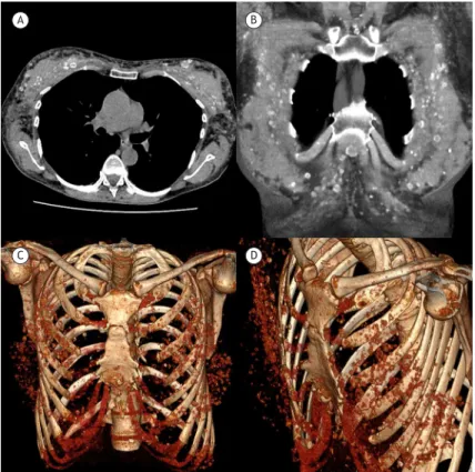
A 37-year-old homosexual Brazilian man presented with fever, chest pain, and several small, palpable lumps in the anterior chest wall. Physical examination revealed numerous mobile subcutaneous and intramuscular nodules measuring 1-2 cm in the anterior chest wall, with signs
of a local inlammatory process. Chest CT scanning
showed numerous round and oval nodules, most of
which had rim calciications, in the anterior chest wall. The nodules were apparently related to the muscles of
the region (Figure 1).
Since the patient was from a cysticercosis endemic region, this infection was initially suspected. However,
the CT indings were not suggestive of cysticercosis. The
characteristic morphology of the calciications observed
in cysticercosis is “rice like” (or “cigar shaped”), and
the calciications are orientated along the long axes of
the muscles. In addition, nodules in cysticercosis are distributed diffusely throughout the muscles.(1) In our case, the calciications presented with a rimmed aspect
and occurred only in the anterior chest wall. Upon further discussion, the patient reported that he had undergone liquid silicone injection into the breasts, performed by an unskilled individual for soft-tissue augmentation, 12 years previously. In conclusion, sequelae of liquid silicone injection should be included in the differential diagnosis
of chest wall calciications.
Figure 1. Axial (in A) and coronal (in B) CT reconstruction scans showing various bilateral round and oval nodules with rim calciications in the anterior chest wall. Three-dimensional coronal (in C) and oblique (in D) CT reconstruction images demonstrated that the nodules were apparently related to the muscles of the region.
A B
C D
REFERENCE
1. Liu H, Juan YH, Wang W, Liang C, Zhou H, Ghonge NP, et al. Intramuscular cysticercosis: starry sky appearance. QJM. 2014;107(6):459-61. https://doi. org/10.1093/qjmed/hct243
J Bras Pneumol. 2017;43(5):399-399
399