Case 9551
Primary ovarian Burkitt lymphoma
Monteiro V, Cunha TM, Saldanha T
Genital (Female) Imaging
Section:
2011, Nov. 20
Published:
23 year(s), female
Patient:
Authors' Institution
V Monteiro , TM Cunha , T Saldanha1 2 3
Unidade Local de Saude do Baixo Alentejo, EPE - Beja (Portugal) 1
Instituto Português de Oncologia de Lisboa Francisco Gentil (Portugal) 2
Centro Hospitalar de Lisboa Ocidental, EPE - Hospital de Egas Moniz (Portugal) 3
Clinical History
A 23-year-old woman was admitted for progressive pelvic pain, weight loss, fever and enlargement of abdomen that had started one month before admission.
On physical examination a large pelvic painful mass was palpated. Laboratory studies revealed anaemia, thrombocytopenia and moderate elevation of levels of CA-125 and LDH.
Imaging Findings
Ultrasonography revealed bilateral solid adnexial masses and a MRI was performed to better characterize these lesions.
MRI revealed bilateral lobulated, heterogeneous, solid ovarian masses which were hypointense on T1-weighted images (WI) (Fig. 1) and of intermediate signal intensity on T2-WI with small, round, high signal intensity lesions in the periphery, consistent with follicles (Fig. 2).The lesions extended up to the level of the liver (Fig. 3) and small amount of ascites and moderate right hydronephrosis were also seen (Fig. 4). After intravenous gadolinium administration the lesions enhanced
The main differential diagnoses were lymphoma, metastases, immature germ cell tumor and granulocytic sarcoma.
Bone marrow biopsy was performed and there was no evidence of involvement by non-Hodgkin lymphoma. Preoperatively CT did not revealed generalized lymphadenopathy.
Total abdominal hysterectomy and bilateral adnexectomy were performed and pathological diagnosis was bilateral ovarian Burkitt lymphoma. Presently the patient is receiving combination chemotherapy treatment.
Discussion
Involvement of the ovary by malignant lymphoma is a well-known late manifestation of
disseminated nodal disease. However, primary ovarian lymphoma is very rare and accounts for only 1.5% of ovarian neoplasms and 0.5% of non-Hodgkin's lymphomas [1]. This low incidence of ovarian involvement by lymphoma is thought to be due to the fact that there is no lymphoid tissue in the ovary and it has been suggested that the tumour originates from rare lymphocytes that are dispersed throughout the ovarian stroma and within ovarian follicles and corpora lutea [2].
The most common type of lymphoma involving the ovary is diffuse large B-cell lymphoma [3]. Burkitt's lymphoma is a very rare highly undifferentiated type of non-Hodgkin's lymphoma derived from B-lymphocyte. It is a rapidly growing lymphoma that occurs mostly in children, and has been described in HIV/AIDS patients.
The majority of primary abdominal lymphoma present with pelvic complaints and common symptoms are abdominal masses and menstrual abnormality.
The diagnosis of primary ovarian lymphoma should be considered in the presence of large bilateral solid ovarian mass, although no specific imaging features can differentiate Burkitt's lymphoma from other neoplasms and only histopathological finding after surgery treatment confirms the diagnosis.
Some criteria for the diagnosis of primary ovarian lymphoma have been suggested [4], and the diagnosis can only be made if at the time of diagnosis, the tumor is confined to the ovary without any evidence of lymphoma elsewhere, except if spread has occurred to immediately adjacent lymph nodes or infiltrates immediately adjacent structures. Also, the peripheral blood and the bone marrow should not contain any abnormal cells and several months should have elapsed between the
appearances of the ovarian and the extra-ovarian lesions.
MRI provides better characterization of ovarian masses than ultrasound [5]. The MRI findings usually include solid bilateral masses, with low signal intensity on T1-weighted images and mildly high signal intensity on T2-weighted images. Lesions tend to show mild to moderate contrast enhancement after intravenous gadolinium administration.
CT is the preferred imaging modality for lymphoma staging in the chest, abdomen, and pelvis as well as in other nodal lymphomas. Bone marrow biopsy is also mandatory for staging.
Final Diagnosis
Bilateral ovarian Burkitt lymphoma
Differential Diagnosis List
Ovarian metastases, Immature germ cell tumour , Granulocytic sarcoma
Figures
Figure 1 Axial T1-WI (A) and axial fat-suppressed T1-WI (B)
:The lesions were hypointense on
T1-WI (A) and on fat-suppressed images
T1-WI (B).:
© Instituto Português de Oncologia de Lisboa Francisco Gentil, Portugal
Area of Interest: Genital /
Reproductive system female;
Imaging Technique: MR;
Procedure: Imaging sequences;
Special Focus: Neoplasia;
Figure 2 Axial T2-WI
:On T2-WI, the lesions were of
intermediate signal intensity,
heterogeneous and with small, round,
high-signal intensity lesions in the
periphery, consistent with follicles
(arrow).:
© Instituto Português de Oncologia de Lisboa Francisco Gentil, Portugal
Area of Interest: Genital /
Reproductive system female;
Imaging Technique: MR;
Procedure: Imaging sequences;
Special Focus: Neoplasia;
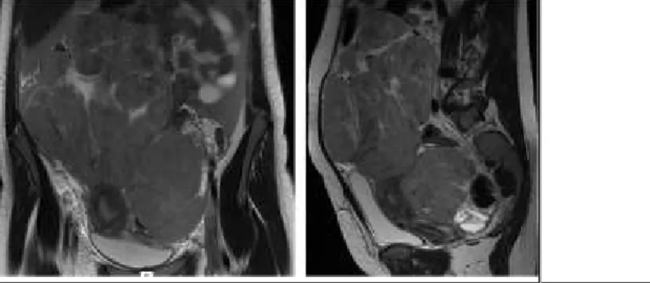
Figure 3 Coronal and sagittal T2 - WI
:The lesions extended from the pelvis
to the upper abdomen.:
© Instituto Português de Oncologia de Lisboa Francisco Gentil, Portugal
Figure 4 Coronal T2-WI and axial fat-suppressed T2-WI
:Small amount of ascites (*) and
moderate right hydronephrosis
(arrow) was also seen.:
© Instituto Português de Oncologia Francisco Gentil de Lisboa, Portugal
Area of Interest: Genital /
Reproductive system female;
Imaging Technique: MR;
Procedure: Imaging sequences;
Special Focus: Neoplasia;
Figure 5 Axial fat-suppressed T1- WI after intravenous gadolinium administration
:After intravenous gadolinium
administration, the lesions enhanced
moderately with some septal
enhancement.:
© Instituto Portugês de Oncologia de Lisboa Francisco Gentil, Portugal
Area of Interest: Genital /
Reproductive system female;
Imaging Technique: MR;
Procedure: Imaging sequences;
Special Focus: Neoplasia;
MeSH
[C15.604.515.569]
Lymphoma
A general term for various neoplastic diseases of the lymphoid tissue.
[A05.360.319.114.630]
Ovary
The reproductive organ (GONADS) in female animals. In vertebrates, the ovary contains two functional parts: the OVARIAN FOLLICLE for the production of female germ cells
References
[1] Dimopoulos MA, Daliani D, Pugh W, Gershenson D,Cabanillas F, Sarris AH (1997) Primary ovarian non-Hodgkin'slymphoma: outcome after treatment with combination chemotherapy Gynecol Oncol 64: 446-450
[2] Vang R, Medeiros LJ, Warnke RA, Higgins JP, Deavers MT (2001) Ovarian non-Hodgkin's Mod Pathol 14:1093-1099
lymphoma: a clinicopathologic study of eight primary cases
[3] Vang R, Medeiros LJ, Fuller GN, Sarris AH, Deavers MT (2001) Non-Hodgkin's lymphoma Adv Anat Pathol 8:200-217
involving the gynecologic tract: a review of 88 cases
[4] Fox H, Langley FA, Govan ADT, Hill AS, Bennett MH (1998) Malignant lymphoma presenting Br J Obstet Gynaecol 95:386-90 as an ovarian tumour: a clinicopathological analysis of 34 cases
[5] Spencer JA et al. (2010) ESUR guidelines for MR imaging of the sonographically indeterminate Eur Radiol Jan;20(1):25-35
adnexal mass: an algorithmic approach
[6] Elharroudi T, Ismaili N, Errihani H, Jalil A (2008) Primary lymphoma of the ovary J Cancer Res Ther 4:195-6
Citation
Monteiro V, Cunha TM, Saldanha T (2011, Nov. 20)
Primary ovarian Burkitt lymphoma {Online}