BRIEF REPORT
Laser treatment for verrucous hemangioma
Juan Manuel Segura Palacios&Pablo Boixeda& Joana Rocha&Javier Alcántara González&
Leticia Alonso Castro&Cristina de Daniel Rodríguez
Received: 22 March 2011 / Accepted: 15 September 2011 / Published online: 6 October 2011
#Springer-Verlag London Ltd 2011
Introduction
Verrucous hemangiomas (VH) are hyperkeratotic vascular malformations characterized by a combination of dilated vasculature in the entire dermis, or even much deeper, and prominent hyperkeratosis, which may result in papules, patches, or violet-colored erythematous tumors. Bleeding after trauma or scratching is frequent. Surgery is nowadays the gold-standard treatment but laser therapy may nevertheless be useful in the superficial component of the lesions. Treat-ments with argon laser and copper vapor laser have been used [1], but both are limited by lack of specificity, poor penetration, and adverse sequelae. Recently, the Nd:YAG 1,064-nm laser has been used in a few cases, either alone or combined with CO2or the pulsed dye laser (PDL) [2].
Materials and methods
We report our experience as a tertiary referral hospital laser unit in the treatment of VH with different combinations of
laser therapy over the last 10 years: CO2 laser (10–30 W
continuous-wave mode), PDL 595 nm (7 mm, 2 ms, 10–
12 J/cm2) and combined dual PDL-Nd:YAG (Cynergy with Multiplex™, Cynosure, Westford, MA) (PDL: 7 mm, 10 ms, 10 J/cm 2with 1-s delay and Nd:YAG: 15 ms, 50–100 J/ cm2). Continuous airflow cooling (Cryo5s, Zimmer Medi-zinsysteme GmbH, Neu-Ulm, Germany) was always applied at its maximum level (6) during laser treatment.
Post-laser care consisted of the daily application of topical antibiotic ointment (Fucidine©) until spontaneous detachment of the scab. Strict avoidance of sun exposure of the treated areas was recommended for at least 2 months to avoid postinflammatory hyperpigmentation.
The study period included eight patients (seven female and one male) evaluated for laser treatment of VH. The mean age of the patients was 6.5 years (range: 3–
16 years). All the patients presented multiple hyperkera-totic erythematous plaques that had developed in early childhood, located on the lower limbs, except for one on the left forearm (patient number 7). The lesions were heterogeneous with hyperkeratotic areas interspersed with flat incipient vascular lesions with no hyperkeratosis, all representing a challenge in terms of surgical treatment. Biopsies confirmed the diagnosis of VH.
None of the patients had any relevant personal or family history and no muscle or bone involvement was noted with magnetic resonance imaging. All the patients underwent laser treatment under local anesthesia, except for one girl who required general anesthesia. Two dermatologists evaluated the effectiveness of the treatment by means of photographs of the patients before and after the therapy. Ulceration, infection, scarring, atrophy, hypopigmentation, hyperpigmentation, or other side-effects were recorded. Patient satisfaction, or parent satisfaction if the patient was a minor, was rated on a scale from 0 to 10.
J. M. Segura Palacios (*)
Department of Dermatology, Hospital Carlos Haya, Plaza Hospital Civil s/n,
29009 Málaga, Spain
e-mail: [email protected]
P. Boixeda
:
J. Alcántara González:
L. Alonso Castro:
C. de Daniel Rodríguez
Department of Dermatology, Hospital Ramón y Cajal, Carretera Colmenar km 9.1,
28034 Madrid, Spain
J. Rocha
Department of Dermatology, Hospital de Braga, Braga, Portugal
Results
The best response in the hyperkeratotic superficial portion was obtained with CO2laser, but the superficial and deep
vascular components responded better to dual PDL-Nd: YAG laser (Figs. 1–4). PDL was only useful in very superficial lesions to prevent the development of the early hyperkeratotic component of some of these lesions [3]. Ulceration, scarring, and hypopigmentation can appear but most of the lesions improved markedly, resulting in a better quality of life. No infectious problems were seen in our patients (Table1).
Discussion
In 1967, Imperial and Helwig introduced the term“ verru-cous hemangioma”, and defined it as a congenital vascular malformation comprising a capillary or cavernous heman-gioma in the dermis and subcutaneous tissue associated with reactive epidermal acanthosis, papillomatosis, and hyperkeratosis, distinguishing it from angiokeratoma [4]. Currently, most authors consider VH to be a malformation of dermal capillaries and encourage use of the term hyperkeratotic vascular malformation to describe this type of vascular change [5]. Nonetheless, the histopathological features and the reactivity against specific immunohisto-chemical markers (focal GLUT-1 endothelial positivity) have led other authors to be cautious about encompassing VH in the group of vascular malformations or tumors [6].
Given the clinical course of these lesions, we prefer the term hyperkeratotic vascular malformation.
The lesions are generally noted at birth or in early childhood [7], although they may appear later or even in adult life. Most VH are multiple lesions and are located on the lower extremities, with unilateral involvement [8]. The
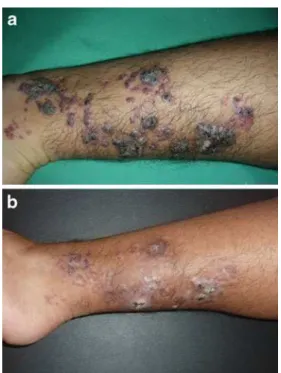
Fig. 1 aVerrucous hemangioma on the lower limb of a 4-year-old girl.bImprovement after sessions with PDL and combined dual PDL-Nd:YAG laser
Fig. 2 aVerrucous hemangioma on the lower limb of a 16-year-old male. b Improvement after sessions with CO2, PDL and combined dual PDL-Nd:YAG laser
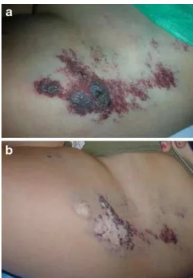
Fig. 3 a Verrucous hemangioma on the lower limb of a 4-year-old girl.bImprovement after sessions with CO2, PDL and combined dual PDL-Nd:YAG laser
early clinical lesions are nonkeratotic, soft, and bluish-red in color, but in time they become increasingly hyperkera-totic and verrucous, usually following trauma or infection [6,8–10].
The differential diagnosis of VH includes angiokera-toma, lymphangioma circumscriptum, angioma serpigino-sum, verrucous epidermal naevus, verrucous cancer, or even malignant melanoma [6–8,10,11]. Occasional venous malformations may also mimic VH, though there is usually minimal hyperkeratosis [9].
Diagnostic histopathological findings are ectatic vessels underlying an acanthotic and hyperkeratotic epidermis in combination with deeply localized vascular formations interspersed with fatty tissue [12]. Inflammatory cells, hemosiderin, and fibrosis may be present in the upper dermis [7]. VH resemble angiokeratoma, although VH involve small, thick-walled, blood-filled vessels with a multilamellated basement membrane involving the dermis as well as fatty tissue [6,9].
VH do not resolve spontaneously, and have a tendency to relapse. Early diagnosis and treatment is important to achieve a better cosmetic result [8]. Management of VH has included excision, cautery, cryosurgery, and laser therapy [11,13]. VH require a large, deep excision because they extend into the subcutis, occasionally involving the fascia. Incomplete extirpation can lead to recurrence [8, 9, 11, 14]. Yang and Ohara performed surgery in combination with PDL, CO2 laser, or argon laser in 23
patients with VH and reported that a combination approach using surgical reconstruction and laser is necessary for large and extensive lesions [15]. Surgery is even more complicated due to the multiplicity of lesions in the same patient. Although laser treatment is only palliative, it cannot treat the deep component, and requires Fig. 4 aVerrucous hemangioma on the lower limb of a 7-year-old
girl.bImprovement after sessions with PDL and combined dual PDL-Nd:YAG laser
Table 1 Clinical characteristics, outcome, and laser used in the treatment sessions
Patient no.
Age (years)
Gender Extension Hyperkeratotic component– Treatment with CO2laser*
Superficial vascular component– Treatment with PDL
Deep vascular component– Treatment with Dual PDL-Nd: YAG
Number of sessions
Response** Degree of satisfaction***
1 7 Female 30 cm2 +++ + + >10 +++ 9
2 16 Male 40 cm2 +++ + + >10 ++++ 10
3 6 Female 30 cm2 + + + 1-5 ++ 10
4 7 Female 15 cm2 + + + 5-10 +++ 8
5 11 Female 40 cm2 ++ + + >10 +++ 8
6 4 Female 25 cm2 +++ + + >10 +++ 8
7 5 Female 20 cm2 + + + >10 +++ 10
8 3 Female 25 cm2 + + + 1-5 ++++ 9
* Hyperkeratotic component–(treated with CO2laser for patients with 2–3 passes): + Hyperkeratosis <2 mm
++ Hyperkeratosis 2–4 mm +++ Hyperkeratosis >4 mm
** Response: 0–25% (+), 25–50% (++), 50–75% (+++), 75–100% (++++)
*** Degree of satisfaction: from 1 (poor) to 10 (excellent), valued by the patient or a parent if a minor
multiple treatment sessions, as in our patients. It is nevertheless worth using in many patients in an attempt to avoid surgery.
Finally, a recent study reported that hypertrophic and PDL-resistant port-wine stains showed improvement with a 755-nm alexandrite laser, which should also be considered in the future as a new therapeutic option for verrucous hemangioma [16,17].
Conclusions
Treatment of VH can be difficult and recurrence is common. Surgery should always be considered for these patients, though laser treatment should be tried beforehand, even though it is only palliative. We have observed a very satisfactory response to laser treatment in some circum-scribed lesions and partial response of the superficial component of deeper lesions, with an improvement in quality of life and patient satisfaction. Thus, combined laser therapy (CO2and dual PDL-Nd:YAG laser) may be useful
in most cases of verrucous hemangiomas.
Conflicts of interests None.
References
1. del Pozo J, Fonseca E (2005) Angiokeratoma circumscriptum naeviforme: successful treatment with carbon-dioxide laser va-porization. Dermatol Surg 31(2):232–236
2. Sommer S, Merchant WJ, Sheehan-Dare R (2001) Severe predominantly acral variant of angiokeratoma of Mibelli: response to long-pulse Nd:YAG (1064nm) laser treatment. J Am Acad Dermatol 45(5):764–766
3. Boixeda P, Pérez-Carmona C, Vanó-Galván S, Jaén P, Lanigan SW (2008) Advances in treatment of cutaneous and subcutaneous vascular anomalies by pulsed dual wavelength 595- and 1,064-nm application. Med Laser Appl 23(3):121–126
4. Imperial R, Helwig EB (1967) Verrucous hemangioma: a clinicopathologic study of 21 cases. Arch Dermatol 96:247–253 5. Requena L, Sangueza OP (1997) Cutaneous vascular
anoma-lies. Part I. Hamartomas, malformations, and dilation of preexisting vessels. J Am Acad Dermatol 37(4):523–549, quiz 549–52
6. Garrido-Ríos AA, Sánchez-Velicia L, Marino-Harrison JM, Torrero-Antón MV, Miranda-Romero A (2008) A histopatho-logic and imaging study of verrucous hemangioma. Actas Dermosifiliogr 99(9):723–726
7. Yasar A, Ermertcan AT, Bilac C, Bilac DB, Temiz P, Ozturkcan S (2009) Verrucous hemangioma. Indian J Dermatol Venereol Leprol 75:528–530
8. Koc M, Kavala M, Kocatürk E, Zemheri E, Zindanci I, Sudogan S, Kural E (2009) An unusual vascular tumor: verrucous hemangioma. Dermatol Online J 15(11):7
9. Tennant LB, Mulliken JB, Pérez-Atayde AR, Kozakewich HP (2006) Verrucous hemangioma revisited. Pediatr Dermatol 23:208–215
10. Calduch L, Ortega C, Navarro V, Martínez E, Molina I, Jordá E (2000) Verrucous hemangioma: report of two cases and review of the literature. Pediatric Dermatol 17:213–216
11. Mankani MH, Dufresne CR (2000) Verrucous malformations: their presentation and management. Ann Plast Surg 45(1):31–36 12. Wentscher U, Happle R (2000) Linear verrucous hemangioma. J
Am Acad Dermatol 42(3):516–518
13. Wang G, Li C, Gao T (2004) Verrucous hemangioma. Int J Dermatol 43(10):745–746
14. Piqué E, Pérez-Cejudo JA, Palacios S, Martínez MS (2005) Malformaciones vasculares hiperqueratósicas. Aportación de 3 casos. Actas Dermosifiliogr 96(10):685–689
15. Yang CH, Ohara K (2002) Successful surgical treatment of verrucous hemangioma: a combined approach. Dermatol Surg 28 (10):913–919, discussion 920
16. Izikson L, Nelson JS, Anderson RR (2009) Treatment of hypertrophic and resistant port wine stains with a 755-nm laser: a case series of 20 patients. Lasers Surg Med 41(6):427–432 17. Izikson L, Anderson RR (2009) Treatment endpoints for resistant
port-wine stains with a 755-nm laser. J Cosmet Laser Ther 11 (1):52–55