63
Radiol Bras. 2015 Jan/Fev;48(1):59–64Letters to the Editor
ernous(1). A purely epidural hemangioma is a rare lesion, repre-senting only 4% of epidural lesions, and the cavernous subtype is the most commonly found in this region(1). The lesion is located in the posterior region of the spine in up to 93% of cases and the dorsal spine is affected in 80% of cases(2). Epidural cavernous hemangioma is most commonly found in men (at a 2:1 ratio) aged over 40(2). Vertebral intraosseous involvement is frequent, with a prevalence of 11%(3).
The clinical condition includes dorsal or lumbar pain, with signs of radiculopathy and myelopathy, and the patient is referred to undergo imaging study for suspicion of disk herniation. The clinical presentation is normally insidious, but acute clinical dete-rioration due to sudden increase in the lesion volume resulting from hemorrhage or venous occlusion(4). As the lesion is highly vascularized, the diagnostic suspicion is very important for the surgical planning, reducing the chances of bleeding during the procedure. Incomplete resection due to bleeding might lead to persistence of clinical symptoms and reoperation would be diffi-cult because of local adhesions(1,4).
Epidural hemangiomas are described as elongated and lobu-lated lesions, possibly with distinctive imaging findings depend-ing on the subtype. Venous and arteriovenous hemangiomas present as cystic masses, generally with hypo- or intermediate sig-nal on T1-weighted and marked hypersigsig-nal on T2-weighted images with peripheral contrast enhancement. Capillary and cav-ernous hemangiomas are seen as solid masses, with hypo- or in-termediate signal on T1-weighted, marked hypersignal on T2-weighted images, and intense contrast-enhancement(1,4–6). The main differential diagnoses of epidural hemangiomas include nerve sheath tumor, meningioma, lymphoma, abscess and extradural hematoma(1,6–8).
Finally, cavernous hemangioma should be considered in the differential diagnosis of epidural lesion with hypersignal on T2-weighted images and prominent contrast enhancement, particu-larly in case where the posterior region of the dorsal spine is af-fected.
REFERENCES
1. Lee JW, Cho EY, Hong SH, et al. Spinal epidural hemangiomas: various types of MR imaging features with histopathologic correlation. AJNR Am J Neuroradiol. 2007;28:1242–8.
2. Aoyagi N, Kojima K, Kasai H. Review of spinal epidural cavernous he-mangioma. Neurol Med Chir (Tokyo). 2003;43:471–5.
3. Castro DG, Lima, RP, Maia MAC, et al. Hemangioma vertebral sinto-mático tratado exclusivamente com radioterapia exclusiva: relato de caso e revisão da literatura. Radiol Bras. 2002;35:179–81.
4. Sanghvi D, Munshi M, Kulkarni B, et al. Dorsal spinal epidural cavern-ous hemangioma. J Craniovertebr Junction Spine. 2010;1:122–5. 5. Rovira A, Rovira A, Capellades J, et al. Lumbar extradural hemangiomas:
report of three cases. AJNR Am J Neuroradiol. 1999;20:27–31. 6. Shin JH, Lee HK, Rhim SC, et al. Spinal epidural cavernous
heman-gioma: MR findings. J Comput Assist Tomogr. 2001;25:257–61. 7. Atlas SW. Magnetic resonance imaging of the brain and spine. 4th ed.
Philadelphia: Lippincott Williams & Wilkins; 2008.
8. El Khamary SM, Alorainy IA. Case 100: spinal epidural meningioma. Radiology. 2006;241:614–7.
Marcelo Mantiolhe Martins1, Flavia Angelica Ferreira Francisco1, Rafael Alfenas de Paula1, Daniella Braz Parente2
1. Universidade Federal do Rio de Janeiro (UFRJ), Rio de Janeiro, RJ, Brasil. 2. Instituto D’Or de Pesquisa e Ensino, Rio de Janeiro, RJ, Brasil. Mailing Address: Dr. Marcelo Mantiolhe Martins. Rua Francisco Otaviano, 23/802, Copacabana. Rio de Janeiro, RJ, Brazil, 22080-040. E-mail: [email protected].
http://dx.doi.org/10.1590/0100-3984.2013.0018
Giant pilomatrixoma: conventional and diffusion-weighted magnetic resonance imaging findings
Dear Editor,
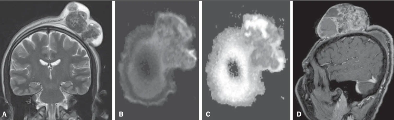
Over the last two years, a 32-year-old man presented growth of a little painful firm nodule located in the high parietal region. Due to the cosmetic deformity, the patient sought medical assis-tance and underwent laboratory tests whose results were normal, and magnetic resonance imaging (MRI) (Figure 1) that demon-strated the presence of a heterogeneous lesion with predominance
of iso/hyposignal on T1-weighted, low signal intensity on T2-weighted, foci of signal drop on magnetic susceptibility sequences and absence of diffusion restriction. After gadolinium injection, exuberant contrast enhancement was observed. Histopathological analysis revealed the presence of basaloid cells associated with phan-tom cells, with areas of foreign-body-type granulomatous reaction compatible with pilomatrixoma. Surgical resection was performed and no recurrence has been observed up to the present moment. Most of times, tumor-like processes in the skull are associ-ated with bone or central nervous system lesions, as reported by
Figure 1.A: Coronal, T2-weighted sequence showing a tumor in the left parietal region with predominance of hyposignal, intermingled with areas of cystic/necrotic degeneration. B: Axial functional MRI, diffusion-weighted sequence does not demonstrate areas of hypersignal. C: Axial image, apparent diffusion coefficient mapping corroborating the absence of areas of diffusion restriction. D: Contrast-enhanced sagittal T1-weighted sequence showing exuberant and heterogeneous contrast enhancement.
64
Radiol Bras. 2015 Jan/Fev;48(1):59–64 Letters to the Editorrecent studies developed by Brazilian authors(1–7). However, skin tumors are rarely similar to each other.
Pilomatrixoma is a rare benign skin tumor originating from hair follicle matrix, most frequently located in the head or neck(8– 10). It is the most common solid skin tumor in patients under the
age of 20(9). Giant pilomatrixomas (> 5 cm) are not frequently found and malignant transformation rarely occurs. Clinically, it manifests as a slow-growing, painless or little painful lesion, some-times in association with bluish coloration of the skin(11). Histo-pathological analysis gives the definitive diagnosis, and the treat-ment is surgical resection with margins of 1 to 2 cm to avoid re-currence.
At MRI, most lesions are well delimited, measuring up to 3 cm, with homogeneous iso-signal on T1-weighted and low signal intensity on T2-weighted sequences. However, reports about stri-ated lesions with hypersignal from the center to the periphery are found in the literature(8,10,12). Calcifications are commonly found and may not present expressive contrast enhancement or even enhance only in the already described areas of hypersignal on T2-weighted sequences(8,10,12). Besides the uncommon lesion size (10.2 cm), heterogeneous signal was observed on T1- and weighted images, with predominance of iso/hyposignal on T2-weighted images, intermingled with areas of cystic/necrotic de-generation and foci of signal drop on magnetic susceptibility se-quences. After gadolinium injection, exuberant contrast enhance-ment of the solid portions of the tumor was observed. Recent stud-ies highlight the utilization of diffusion-weighted sequences in the evaluation of head and neck lesions, demonstrating that ap-parent diffusion coefficient values < 1.22 × 10⁻³ mm2/s are sug-gestive of malignancy(13). In the present case, such a value was 1.35 × 10⁻³ mm2/s, corroborating the previously described find-ings. Other advanced MRI sequences might add further data, particularly in the prediction of benignity and malignancy(14,15). Finally, the diagnosis of pilomatrixoma should be considered in patients under the age of 20 presenting with skin tumors, par-ticularly those located in the head and neck, and typical imaging findings should not be expected in cases of giant pilomatrixomas.
REFERENCES
1. Werner Jr H. Evaluation of the central nervous system of fetuses and neonates. Radiol Bras. 2012;45(6):v–vi.
2. Sanches P, Yamashita S, Freitas CCM, et al. Chordoid glioma of the third ventricle: a new case report. Radiol Bras. 2012;45:288–90. 3. Coeli GNM, Tiengo RR, Silva AC, et al. Nodular calcified
neurocysti-cercosis with signs of reactivation. Radiol Bras. 2012;45:291–3. 4. Reis F, Schwingel R, Nascimento FBP. Central nervous system
lym-phoma: iconographic essay. Radiol Bras. 2013;46:110–6.
5. Brandão LA. Primary and secondary lymphoma of the central nervous system. Conventional and functional magnetic resonance imaging find-ings. Radiol Bras. 2013;46(2):ix–x.
6. Curioni OA, Souza RP, Amar A, et al. Value of PET/CT in the approach to head and neck cancer. Radiol Bras. 2012;45:315–8.
7. Matushita JP, Matushita JS, Simões LAM, et al. Giant cell tumor of the frontal sinus: case report. Radiol Bras. 2013;46:255–8.
8. Hsieh TJ, Wang CK, Tsai KB, et al. Pilomatricoma: magnetic reso-nance imaging and pathological evaluation. J Comput Assist Tomogr. 2008;32:320–3.
9. Beaman FD, Kransdorf MF, Andrews TR, et al. Superficial soft-tissue masses: analysis, diagnosis, and differential considerations. Radiographics. 2007;27:509–23.
10. Laffan EE, Ngan BY, Navarro OM. Pediatric soft-tissue tumors and pseudotumors: MR imaging features with pathologic correlation: part 2. Tumors of fibroblastic/myofibroblastic, so-called fibrohistiocytic, muscular, lymphomatous, neurogenic, hair matrix, and uncertain ori-gin. Radiographics. 2009;29:e36.
11. Whittemore KR, Cohen M. Imaging and review of a large pre-auricu-lar pilomatrixoma in a child. World J Radiol. 2012;4:228–30. 12. De Beuckeleer LH, De Schepper AM, Neetens I. Magnetic resonance
imaging of pilomatricoma. Eur Radiol. 1996;6:72–5.
13. Gonçalves FG, Ovalle JP, Grieb DFJ, et al. Diffusion in the head and neck: an assessment beyond the anatomy. Radiol Bras. 2011;44:308– 14.
14. Wang CK, Li CW, Hsieh TJ, et al. Characterization of bone and soft-tissue tumors with in vivo 1H MR spectroscopy: initial results. Radiol-ogy. 2004;232:599–605.
15. Costa FM, Vianna EM, Domingues RC, et al. Espectroscopia de pró-tons e perfusão por ressonância magnética na avaliação dos tumores do sistema musculoesquelético. Radiol Bras. 2009;42:215–23.
http://dx.doi.org/10.1590/0100-3984.2014.0071
Bruno Niemeyer de Freitas Ribeiro1, Edson Marchiori2