ESCOLA UNIVERSITÁRIA VASCO DA GAMA
MESTRADO INTEGRADO EM MEDICINA VETERINÁRIA
CARDIORENAL SYNDROME: THERAPEUTICAL CHALLENGE
Sara Lopes
ii
ESCOLA UNIVERSITÁRIA VASCO DA GAMAMESTRADO INTEGRADO EM MEDICINA VETERINÁRIA
CARDIORENAL SYNDROME: THERAPEUTICAL CHALLENGE
Coimbra, Junho 2016
Autor
Sara Cristina Cerqueira Lopes
Aluna de Mestrado Integrado em Medicina Veterinária
Orientador Interno
Professora Doutora Maria João Nobre de Matos Pereira Vieira
Co-orientador
Professora Doutora Ana Luísa Nobre de Matos Pereira Vieira
Orientador externo
iv
RESUMOA síndrome cardiorenal caracteriza-se pela coexistência de doença cardíaca e renal num mesmo indivíduo, podendo afetar cães e gatos. A definição mais consensual descreve-o como um “distúrbio fisiopatológico do coração e dos rins, em que a afeção aguda ou crónica de um dos órgãos, origina dano agudo ou crónico no outro”.
O interesse e reconhecimento da importância e prevalência desta síndrome em medicina veterinária tem sido crescente e, recentemente, um comité formado por médicos veterinários especialistas em cardiologia e nefrologia propuseram que a síndrome cardiorenal fosse designada como um distúrbio do eixo cardiovascular-renal, classificando-a em três tipos, consoante a origem do distúrbio: CvRDH para o coração como órgão de falência primário, CvRDK para rins como órgãos de
falência primário e CvRDO para quando é originada por outras causas.
A terapêutica atual para a síndrome cardiorenal é muito variada, e depende em muito da preferência do médico veterinário quanto aos fármacos utilizados. No entanto, há certas premissas que devem ser consideradas no que diz respeito a este desafio terapêutico e que não devem ser subvalorizadas, pois refletem-se seriamente no sucesso da terapêutica e consequentemente na esperança de vida do animal. As dosagens dos fármacos utilizados devem sofrer uma revisão constante, acompanhada de uma cuidada monitorização das funções cardíaca e renal do paciente, dado que o principal desafio reside na tentativa de melhorar ao máximo a função de um dos órgãos sem causar dano, ou deterioração, à função do outro.
Esta dissertação tem como objetivo uma revisão do estado atual do conhecimento sobre a síndrome cardiorenal em animais de companhia, incidindo no seu diagnóstico, classificação e desafios terapêuticos.
v
ABSTRACTCardiorenal syndrome is described by the coexistence of cardiac and renal disease on the same individual, and it can affect both dogs and cats. The most consensual definition describes it as a “pathophysiologic disorder of the heart and kidneys whereby acute or chronic dysfunction of one of the organs causes acute or chronic dysfunction of the other”.
The interest, recognition of the importance and prevailing of this syndrome in veterinary medicine has grown and, recently, a committee of veterinarians specialized in cardiology and nephrology proposed that cardiorenal syndrome would be further designated as cardiovascular-renal axis disorders, classifying it in three types according to the disorders primary cause: CvRDH to heart as
the primary failing organ, CvRDK to kidneys as the primary failing organ and CvRDO to when it has other
causes.
The current therapeutic for cardiorenal syndrome presents many variations, depending on the veterinarian preference as for what drugs should be used. However, there are certain assumptions that must be considered regarding this therapeutic challenge, and shouldn’t be undervalued since they take serious effect on the therapeutics success and, therefore, on the animal life expectancy. The dosages for the drugs used must suffer constant review, together with careful monitoring to the patients cardiac and renal functions, since the main challenge relies on the attempt to improve at the most one the organs function, without causing damage, or deterioration, to the other.
This dissertation objective is to revise the current state of knowledge of cardiorenal syndrome in small animals, focusing on its diagnosis, classification and therapeutic challenge.
vi
No culminar desta etapa tão importante da minha vida não posso deixar de dedicar todo o meu trabalho aos que tanto contribuíram para a sua realização. Por isso, dedico a minha dissertação final aos meus pais e à minha irmã, que sempre me apoiaram, e também à minha avó.vii
AgradecimentosAo chegar ao final desta grande “viagem”, não posso deixar de agradecer a todos aqueles que a tornaram possível, contribuindo para a minha formação profissional e pessoal. Assim agradeço:
Aos meus pais, que sempre lutaram e fizeram tudo ao seu alcance para que eu pudesse seguir os meus sonhos.
À minha irmã, que esteve sempre presente, para me guiar nos momentos piores e para festejar comigo nos melhores.
À minha orientadora interna, Prof. Dra. Mª João Vieira, por toda a amizade, incentivo e pelo grande apoio prestado ao longo do meu estágio final e na elaboração da dissertação.
Ao meu orientador externo, Dr. Carlos Sousa, e a toda a sua equipa, pelo conhecimento transmitido, pela ajuda prestada e por me terem feito sentir em casa.
À Escola Universitária Vasco da Gama, pelo apoio para a realização do meu estudo.
Aos docentes da Escola Universitária Vasco da Gama, pela contribuição preciosa na minha formação como futura Veterinária.
Aos meus amigos, que fizeram valer a pena todos os momentos.
E a todos os outros que, de alguma forma contribuíram para a minha formação, não só académica mas também pessoal e profissional,
viii
LIST OF CONTENTSResumo ... iv
Abstract ... v
List of Figures ... ix
List of Tables ... x
List Of Abbreviations ... xi
1. Introduction ... 1
2. Cardiorenal Syndrome ... 2
2.1. Definition ... 2
2.2. Classification ... 2
2.3. Cardiovascular-renal axis disorder ... 3
2.4. Pathophysiology ... 3
2.4.1 Cardiovascular-renal axis disorders - Heart ... 4
2.4.2 Cardiovascular-renal axis disorders - Kidney ... 5
2.4.3 Cardiovascular-renal axis disorders - Others ... 5
2.5. Clinical assessment ... 6
2.5.1. Renal function assessment ... 6
2.5.2. Cardiac function assessment ... 7
2.5.3. Blood pressure monitoring ... 8
3. Therapeutics ... 9
3.1. Therapeutic Challenge ... 9
3.2. Global vision – therapeutic strategies ... 9
3.3. Management ... 10
3.3.1. Diuretics ... 10
3.3.2. Angiotensin converting enzyme inhibitors ... 11
3.3.3. Beta-blockers ... 12
3.3.4. Angiotensin receptor blockers ... 13
3.3.5. Mineralocorticoid receptor antagonists ... 13
3.3.6. Uremic toxins accumulation treatment ... 14
3.4. Prognosis ... 14
4. Future perspectives ... 15
5. Final intakes ... 16
ix LIST OF FIGURES
Figure 1 - Postulated mechanisms underlying the relationship between heart failure (HF) and renal dysfunction. ... 4
x LIST OF TABLES
Table 1 - Cardiorenal syndrome classification ... 2
Table 2 - International Renal Interest Society grading criteria for acute kidney injury ... 7
Table 3 - Diuretics dosage for dogs and cats ... 10
Table 4 - Angiotensin converting enzyme inhibitors dosage for dogs and cats ... 12
Table 5 - Beta-blockers dosage for dogs and cats ... 12
Table 6 - Angiotensin receptor blockers dosage for dogs and cats ... 13
xi LIST OF ABBREVIATIONS
ACEI – angiotensin converting enzyme inhibitors
AKI – acute kidney injury
BID – bis in die (two intakes daily)
BNP – B-type natriuretic peptide CHF – congestive heart failure CKD – chronic kidney disease CKI – chronic kidney injury CRS – cardiorenal syndrome
CvRD – cardiovascular renal axis disorders
CvRDH – cardiovascular renal axis disorders heart
CvRDK – cardiovascular renal axis disorders kidney
CvRDO– cardiovascular renal axis disorders others
cTnI – cardiac troponin I GFR – glomerular filtration rate HF – heart failure
IM – intramuscular
IRIS – international renal interest society IV – intravascular
NT-proANP – N-terminal pro-atrial natriuretic peptide
NT-proBNP – N-terminal pro-B-type natriuretic peptide PO – per os
RAAS – renin-angiotensin-aldosterone system RF – renal failure
ROS – reactive oxygen species
SC – subcutaneous
SID – semel in die (single intake daily) SHT – systemic hypertension
1 1. INTRODUCTION
Cardiorenal syndrome (CRS) is a complex pathology that affects both the heart and kidneys, and it’s commonly seen in dogs and cats suffering from cardiac disorders. Although being one of the major setbacks to congestive heart failure (CHF) therapy, it’s not completely understood at this point, and by what reason, its definition hasn’t been set by the scientific community. It can, however, be roughly defined as a “pathophysiologic disorder of the heart and kidneys whereby acute or chronic dysfunction of one of the organs may induce acute or chronic dysfunction of the other” (Ronco et al., 2008).
Despite the numerous possible causes for this syndrome, one of the most common presentations is renal decay due to heart failure. The heart function is to pump blood to all body tissues, and the renal system receives about 20-25% of the cardiac output, so it’s easy to infer that when a disease affects the heart, it will ultimately end up taking its toll on the kidneys as well. Renal function is, on the other hand, absolutely crucial to the organism, since, amongst other functions, it plays an essential role in osmoregulation and maintenance of blood pressure levels, which is of utmost importance when it comes to heart failure (HF) therapeutics.
It’s common to have heart and kidney dysfunctions occurring simultaneously, since they share many of their causes and pathophysiologic mechanisms. Adding up to this, CHF therapies focus on improving myocardia function as well as achieving hemodynamic balance, but have harmful consequences to renal function (Aronson, 2012).
Renal deficits are now recognized as an independent risk factor when it comes to mortality and morbidity in patients suffering from HF. The mechanisms by which renal failure (RF) causes HF to deteriorate are not fully understood and there are several factors that contribute to the “heart/kidney vicious circle” (Palazzuoli & Ronco, 2011).
About 10% of the dogs seen in veterinary practices are affected by some form of heart disease (C. Atkins et al., 2009), and that number grows to 75% in dogs over 16 years (Guglielmini, 2003). With the advances in HF therapeutics, we are able to delay disease progression, and death by the primary cause is becoming more uncommon in cardiac patients.
2 2. CARDIORENAL SYNDROME
2.1. Definition
CRS has yet to be fully defined. Many definitions have been placed throughout the years, but have failed to properly describe the fine interactions between heart and kidney and do not address all the leading key points of cardiorenal interface: which is the primary organ to fail; the “unidirectional or bidirectional” character of the interaction; the disease that causes organ failure; the pathophysiological mechanism behind the affection and ultimately the acute or chronical development of the process (Braam et al., 2013).
First it was described as the common presentation of combined heart and kidney dysfunction (Shlipak & Massie, 2004). Then it was considered to be the existence or development of kidney damage due to heart failure (Heywood, 2005), and later a pathophysiological disorder where acute or chronical failure in one of the organs may cause acute or chronical damage to the other (Ronco et al., 2008). Most recently it was suggested that each failing organ has the capability to start and maintain disease in the other organ by feedback mechanisms, biochemical, immunological, neurohormonal and/or hemodynamic (Bock & Gottlieb, 2010b).
Although many of the suggested definitions include some of the key points, none has been able grasp all of them, the classification suggested by Ronco, in 2008, in the most clinically appealing (Braam et al., 2013).
2.2. Classification
Ronco suggested a five-point classification for CRS.
Table 1 - CRS classification. Adapted from Ronco et al. 2008
CRS
Type 1 Acute CRS
Acute worsening of cardiac function causes acute kidney injury CRS
Type 2 Chronic CRS
Chronical injuries to cardiac performance results in active damage to the kidney, resulting in chronic kidney disease
CRS
Type 3 Acute renocardiac syndrome
Acute cardiac dysfunction outcomes of sudden RF CRS
Type 4 Chronic renocardiac syndrome
Chronic kidney disease leads to cardiac function decline and structural changes
CRS
Type 5 Secondary CRS
3 2.3. Cardiovascular-renal axis disorder
All of the definitions and classifications above were elaborated by the medical community. With the purpose of formally define and describe this pathology in veterinary patients, a CRS Consensus Group of veterinarian cardiologists and nephrologists was formed in 2015.
This entity proposed that CRS would be treated as “cardiovascular-renal disorders” in dogs and cats, in order to include the vasculature as an important piece, and since it can have many clinical presentations, with individual and breed variations, cannot be described as a single clinical syndrome (Pouchelon et al., 2015).
CvRD are defined as “a disease, toxin or drug-induced structural and/or functional damage to the kidney and/or cardiovascular system, leading to disruption of the normal interactions between these systems, to the ongoing detriment of one or both” (Pouchelon et al., 2015)
A classification into three different subgroups was also established based on the primary failing organ: CvRDH when a renal dysfunction occurs resulting from a disease concerning the cardiovascular
system; CvRDK cardiovascular impairment caused by kidney disease and CvRDO describing damage to
heart and kidneys simultaneously, due to concomitant primary disease in heart and kidneys or other pathologies, toxins, toxics or drugs that affects both systems.
2.4. Pathophysiology
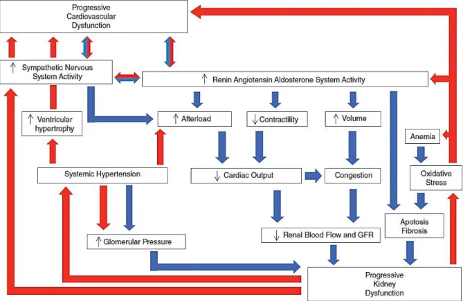
The pathophysiology for CRS is not completely understood (Liang et al., 2006). The pathophysiologic mechanisms responsible for this syndrome are mainly due to hemodynamic alterations, such as, reduced renal pressure, increase to venous pressure, as well as the activation of various neurohormonal systems (Liang et al., 2006). Most of them are caused by compensatory mechanisms to HF, that end up contributing to worsen cardiac and renal function and at the end, lead to CRS (Liu et al., 2012).
A diminished cardiac output, caused by cardiac function alteration in CHF, will result in lower blood flow and renal hypoperfusion. Baroreceptors are triggered, initiating renal vasoconstriction, activating the sympathetic nervous systems (SNS), and releasing catecholaminergic hormones. Fluid retention occurs, since SNS activation will increase renal vasoconstriction (Bock and Gottlieb 2010).
Renal ischemia, oxidative stress, hypoxia and intrinsic renal diseases often activate renal sensory afferent signaling to hypothalamus, which combined with elevated central sympathetic tone are related with the development of CRS (DiBona, 2005; Sobotka et al., 2011).
4 The renin-angiotensin-aldosterone system (RAAS) also plays an important role in CRS development. RAAS activation due to CHF or CKD will cause endothelial dysfunction, inhibition of the fibrinolytic system and also development of atherosclerosis. There is also an increase to blood pressure caused by angiotensin II, which will promote aldosterone secretion, triggering increased sodium and water reabsorption, and cause damage to heart and kidneys as a circulating hormone (Brewster & Perazella, 2004).
Although there aren’t specific studies that correlate CRS pathophysiology to CvRD, empiric evidence and literature suggest the existence of similar patterns, such as neurohormonal activation, ROS and most of all, the intricate sequence of hemodynamic changes (Pouchelon et al., 2015).
Figure 1 - Postulated mechanisms underlying the relationship between heart failure (HF) and renal dysfunction. Blue arrows indicate pathways by which HF may lead to RF. Red arrows indicate pathways by which RF may lead to HF. The relative importance of these mechanisms (and additional mechanisms not discussed) is not known (i.e. boxes are not drawn to scale). From (Pouchelon et al., 2015)
2.4.1 Cardiovascular-renal axis disorders - Heart
The indirect evidence to CvRDH in dogs and cats, resides on the renal function decrease in
5 chronic valvular heart disease and 70% in those with a disease more severe (Nicolle et al., 2007). In cats with hypertrophic cardiomyopathy azotemia was found in 59% of the animals (Gouni et al., 2008).
Potential etiologies to the development of this disorder are: systemic hypertension (SHT), cardiogenic shock, low cardiac output, systemic hypotension, systemic arterial thromboembolism, heartworm infection and passive kidney congestion (Pouchelon et al., 2015).
Mechanisms that are considered to be responsible to cause kidney injure as a result from a cardiovascular disease involve the activation of RAAS and SNS, there’s also a reduction in kidney perfusion resultant from diminished cardiac output, kidneys venous congestion and formation of ROS due to injured endothelial tissue (Haase et al., 2013; McCullough et al., 2013). Treatment for CHF can also cause kidney disease due to exposure to diuretics and angiotensin converting enzyme inhibitors (ACEI), known to be potentially nephrotoxic drugs and to activate the RAAS, leading to volume depletion, vasoconstriction and diminished glomerular filtration rate (GFR), affecting renal function (Francis et al., 1990; Lantis et al., 2011; Pouchelon et al., 2015).
2.4.2 Cardiovascular-renal axis disorders - Kidney
Although being less common, primary disease involving the kidney such as kidney-mediated SHT, volume overload, hypokalemia or hyperkalemia, reduced renal clearance or drugs, uremic hypodipsia, uremic pericarditis, activation of RAAS and anemia caused by CKD, can lead to cardiovascular dysfunction (Pouchelon et al., 2015).
Drugs such as enalapril, atenolol and digoxin, used for cardiac disease treatment, undertake renal excretion, causing primary kidney dysfunction, which leads to lower drug clearance and therefore sings of toxicity, like hypotension and arrhythmias, that will result in deteriorating myocardial function (Pouchelon et al., 2015). Patients with kidney disease present abnormal hemodynamic status and fluid volume which can cause congestion resulting from systemic volume overload, mainly in animals suffering from cardiac disease (Much & Wilcox, 1982; Polzin, 2011).
A common side effect to CKD is SHT that can cause dysfunction and hypertrophy in the myocardium of dogs and cats (Carlos Sampedrano et al., 2006; Chetboul et al., 2003; Henik et al., 2004)
2.4.3 Cardiovascular-renal axis disorders - Others
Despite of shortage of published evidence of CvRDO in dogs and cats, it’s considered that
6 2.5. Clinical assessment
Early diagnosis of CRS, is essential when it comes to preventing disease progression. A close monitoring to renal activity parameters in cardiac patients, as well as heart function monitoring when there is evidence of acute kidney injury (AKI) or chronic kidney injury (CKI) are crucial.
In order to obtain essential information for diagnosis it’s vital to perform a detailed anamnesis and physical examination, being attentive to certain symptoms that can indicate renal impairment, such as polyuria and polydipsia, or cardiac malfunction, as coughing, exercise intolerance, dyspnea and syncope, that can point to what further testing must be done, such as radiographic and ultrasonographic imaging, blood and urine testing and assessment of blood pressure (Pouchelon et al., 2015).
A useful tool in monitoring is biomarkers such as N-terminal pro-B-type natriuretic peptide (NT-proBNP), B-type natriuretic peptide (BNP), N-terminal pro-atrial natriuretic peptide (NT-proANP) and cardiac troponin I (cTnI), the most common biomarkers used to assess cardiac function. Biomarkers are “a characteristic that is objectively measured and evaluated as an indicator of normal biologic processes, pathogenic processes, or pharmacologic responses to a therapeutic intervention” (Biomarkers Definitions Working Group, 2001).
2.5.1. Renal function assessment
When it comes to evaluate renal function there are many parameters that need to be taken account, which can be appraised using traditional urine and blood tests. For instance, GFR can be expressed by serum creatinine levels, glomerular permselectivity by serum or urine albumin and total protein concentration, proximal tube function by urine glucose and amino acid concentrations, ability to maintain electrolyte and acid-base balance by serum electrolytes and bicarbonate concentrations, and renal concentrating ability by urine specific gravity (Pouchelon et al., 2015).
We can also resort to imaging to detect morphological abnormalities (Pouchelon et al., 2015). Information on kidney size, position, shape, unilateral or bilateral abnormalities and presence of radiosense uroliths can be obtained through a ventrodorsal abdominal radiography (Bartges, 2012; Polzin, 2011; Rivers & Johnston, 1996). Using echography we can evaluate integrity of structures as renal parenchyma, renal pelvis and urethra, as well as the presence of mineralization, uroliths, cysts, infarcts and abnormalities or renal blood flow (Debruyn et al., 2012; Lamb, 1998; Rivers & Johnston, 1996).
7 Table 2 - International Renal Interest Society grading criteria for AKI in dogs and cats. From (International Renal Interest Society, 2016).
2.5.2. Cardiac function assessment
To evaluate cardiac function imaging presents a crucial role. Combining cardiothoracic radiography and echocardiography with patients history, physical examination, blood and urine analysis and electrocardiogram, we are able to determine the presence of heart disease, as well as cause and severity (Pouchelon et al., 2015).
Performing a cardiothoracic radiography, in at least two different orthogonal projections (e.g. laterolateral and ventrodorsal), we can evaluate changes to the hearts silhouette, pulmonary parenchymal pattern and vascular structures, assessing indicators for cardiac disease or CHF (Pouchelon et al., 2015).
Cardiac morphology and function can be simply assessed by simple echocardiography, but using Doppler echocardiography (spectral Doppler, tissue Doppler, color flow Doppler), and recent
AKI Grade Blood creatinine Clinical Description
Grade I <1.6 mg/dl (<140 mol/l)
Non Azotemic AKI:
a. Documented AKI: (Historical, clinical, laboratory, or imaging evidence of AKI, clinical oliguria/anuria, volume responsiveness‡) and/or
b. Progressive non azotemic increase in blood creatinine; ≥0.3 mg/dl (≥26.4 μmol/l) within 48 hours c. Measured oliguria (<1ml/kg/hr) or anuria over 6 hours
Grade II 1.7 – 2.5 mg/dl (141 – 220 µmol/l)
Mild AKI:
a. Documented AKI and static or progressive azotemia b. Progressive azotemic increase in blood creatinine; ≥0.3 mg/dl ≥26.4 μmol/l) within 48 hours), or volume responsiveness‡
c. Measured oliguria (<1ml/kg/hr) or anuria over 6 hours
Grade III 2.6 – 5.0 mg/dl (221 – 439µmol/l)
Grade IV 5.1 – 10.0 mg/dl (440 – 880 µmol/l)
Moderate to Severe AKI:
a. Documented AKI and increasing severities of azotemia and functional RF
8 modalities as bi-dimensional speckle tracking, strain and strain rate, we can evaluate myocardial motion and blood flows velocity and direction with more detail (Chetboul & Tissier, 2012; Chetboul, 2010).
Analyzing deviations on information collected through imaging methods, we can assess morbidity and expected mortality for heart disease in dogs and cats (Lord et al., 2011; Reynolds et al., 2012)
Another classic tool when evaluating cardiac function are cardiac biomarkers. NT-proBNP, BNP, NT-proANP and cTnI are the most commonly used biomarkers for detection of primary cardiovascular diseases, proving themselves very accurate (Boswood et al., 2008; Connolly, 2010; DeFrancesco et al., 2007; M. A. Oyama et al., 2013).
Although being one of the most useful tools for cardiologists, their applicability to assess cardiac injury resultant from renal dysfunction hasn’t been proven yet. Despite lack of information, cardiac troponin is the only cardiac biomarker that might be applied in this cases, since its concentration is normally low in healthy dogs, and has shown to be increased in dogs and cats with cardiac disease (primary or secondary), indicating subclinical signs of cardiac injury, and helping to predict clinical outcome (Fonfara et al., 2010; Hezzell et al., 2012; Langhorn et al., 2013; M. a Oyama & Sisson, 2004; Pouchelon et al., 2015). It has, on other hand, shown increased serum or plasma concentration, as well as NT-proBNP and BNP, on dogs with AKI or CKI, but that showed normal cardiac function, because they go through renal excretion (Lalor et al., 2009; Miyagawa et al., 2013; Schmidt et al., 2009; Sharkey et al., 2009).
As for NT-proBNP, BNP, and NT-proANP, although having given proves in the assessment of primary cardiac dysfunction, when evaluating cardiac injury resultant from kidney malfunction its applicability has yet to be fully studied, since they present continuous production by the myocardium, helping plasma volume, sodium excretion and vasomotor tone regulation in healthy and diseased patients (Potter et al., 2009), varying on “moment-to-moment basis” and according to disease stages, and showing, as well, variation in individuals and breeds in either healthy or diseased animals. which can lead to inaccurate conclusions (Kellihan et al. , 2009; Pouchelon et al., 2015; Sjöstrand et al., 2014).
2.5.3. Blood pressure monitoring
Blood pressure measurement is very important when monitoring animals with CRS (or CvRD). When assessing these values is essential to take on account the method throughout they’re obtained, and non-invasive methods are preferable (Pouchelon et al., 2015).
9 increasing renal injury rate, decreased GFR, proteinuria and ultimately a higher mortality rate (Jacob et al., 2003).
Systolic pressure under 90 mmHg is considered hypotension, and can cause cardiovascular and renal injury (Pouchelon et al., 2015).
3. Therapeutics
3.1. Therapeutic Challenge
CRS presents a major therapeutic challenge. Although specific studies in the mater are lacking, it’s fairly well known empirically, that patients with cardiac insufficiency are prone to develop renal impairment with continuous cardiac treatment. Kidney failure develops in cardiac patients, either as a result to compensatory mechanisms to cardiac malfunction, either by chronic aggression to the kidneys caused by many of the drugs commonly used in cardiac patient’s treatments (Pouchelon et al., 2015).
When any animal presents with congestive signs of cardiac insufficiency, such as pulmonary edema, the main objective is to perform aggressive diuresis, as well as providing protein supplementation when showing cardiac caquexia (C. Atkins et al., 2009; Borgarelli & Haggstrom, 2010). But when it’s an animal with fully developed CRS, or CvRD, the same objective sustains, but the scenario has totally changed. This time the main goal resides, but with the additional premise of not compromising renal function furthermore and avoid the development of acute kidney failure (Pouchelon et al., 2015).
The same principle applies conversely. In an animal, suffering from acute kidney failure, the main point is to lower uremic toxins concentration, usually performing aggressive fluid therapy. But in cases of CRS, or CvRD, where the animal also has cardiac insufficiency, aggressive fluid therapy will lead to congestive signs to appear (Pouchelon et al., 2015).
Although it may be difficult, it is also crucial to carefully manage the therapeutics in patients with CRS, either in emergency situations as in chronic treatment, as it will directly reflect on the animal life expectancy (Pouchelon et al., 2015).
3.2. Global vision – therapeutic strategies
10 In patients presenting CRS type 1 and 2, or CvRDH, there are three major points to consider:
avoid causing short-term damage whilst providing CHF treatment, increase monitoring to renal function parameters and, ultimately, develop a therapeutic plan that preserves the kidney as much as possible (Tang & Mullens, 2010).
In CRS types 3 and 4, or with CvRDK, the main therapy goal is to combine different uremic
toxins excretion treatments, in order to minimize IV fluid intake necessary, reducing the patient’s chance to develop congestive signs (Pouchelon et al., 2015).
3.3. Management
3.3.1. Diuretics
Diuretics are the first line option when it comes to control CHF. They are used to control pulmonary congestion in patients with volume overload, and maintaining hydrostatic balance (Krum et al., 2006). The most common diuretics used are furosemide and hydrochlorothiazide (C. E. Atkins & Häggström, 2012; C. Atkins et al., 2009; Goutal et al., 2010). Studies in humans show that loop diuretics, such as furosemide, are preferable to hydrochlorothiazide in patients with growing creatinine serum concentration, since the last one is majorly ineffective in patients with advanced states of RF (Attanasio et al., 2010; Fliser et al., 1994).
Patients suffering from CRS tend to acquire resistance to diuretic effect, resulting in persistent pulmonary congestion, needing higher doses in order to achieve therapeutic concentrations in renal tubules, as kidney function decays (Boerrigter & Burnett, 2004). Diuretic therapy can cause pre-renal azotemia, decline of renal function and electrolyte balance. Therefore, the major goal when applying diuretic therapy, is to achieve optimal results at minimum dosage, reducing the total daily dosage of diuretics (Pouchelon et al., 2015).
In order to minimize adverse effects to the kidney, excessive diuresis, maintain a correct electrolyte balance and renal perfusion, it’s advised to keep patients on supplementary IV fluids low on sodium during aggressive diuretic therapy (Pouchelon et al., 2015).
Table 3 - Diuretics dosage for dogs and cats. Adapted from (“Merck Vet Manual,” 2016)
Drug Treatment Administration Dosage for dogs Dosage for cats
Furosemide
Life-threatening cardiogenic pulmonary
edema
IV, IM or SC 2-4 mg/kg Every 1-6 hour
11 Constant rate
infusion IV 0.25-1 mg/kg/hr 0.25-0.6 mg/kg/hr
Long-term
management PO
Start – 2 mg/kg BID
Range – 1-5 mg/kg BID/TID
Start – 1 mg/kg SID Range – 1-2 mg/kg
SID/BID Up to 4-6 mg/kg daily
Hydrochlorothiazide Monotherapy PO 2-4 mg/kg BID 0.5-2 mg/kg SID/BID
Combined with furosemide
PO
Start: 2 mg/kg BID Furosemide dosage lowers to
25-50%
Torsemide Long-term
management PO
0.25-0.4 mg/kg SID/BID
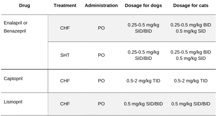
3.3.2. Angiotensin converting enzyme inhibitors
ACEI are one of the most commonly used drugs, when treating patients with heart failure. ACEI, such as benazepril, enalapril, imidapril and ramipril, act as vasodilators, reversing the side effects of chronic activation of the RAAS (excessive fluid retention) that occur in cardiopathic dogs (Guglielmini, 2003). Since they reduce blood pressure, they are considered a “kidney ally” in cardiac patient’s therapy.
12 Table 4 - ACEI dosage for dogs and cats. Adapted from (“Merck Vet Manual,” 2016)
Drug Treatment Administration Dosage for dogs Dosage for cats
Enalapril or
Benazepril CHF PO
0.25-0.5 mg/kg SID/BID
0.25-0.5 mg/kg BID 0.5 mg/kg SID
SHT PO 0.25-0.5 mg/kg
SID/BID
0.25-0.5 mg/kg BID 0.5 mg/kg SID
Captopril CHF PO 0.5-2 mg/kg TID 0.5-2 mg/kg TID
Lisinopril CHF PO 0.5 mg/kg SID/BID 0.5 mg/kg SID/BID
3.3.3. Beta-blockers
Beta-blockers, such as atenolol, are commonly used in animals with left atrial enlargement, and can also be used in order to reduce arrhythmias, infundibular gradient, dynamic left ventricular outflow gradient, syncopal events and to prevent sudden death (Bonagura & Twedt, 2009) . Upon finding alterations of left atrium, either on a primary evaluation or on a control echocardiography, beta-blockers are usually prescribed in order to slow disease progression. Its use it’s not recommended when signs of CHF are present, but it’s many times recommended to add them to the medication regimen, in order to protect myocardium from deterioration (C. Atkins et al., 2009).
Acting by preventing endogenous catecholamines on beta-receptors, its benefit to the patient in humans it’s often compared to ACEI (Liu et al., 2012). Some medical studies associate the use of a beta-blocker, bisoprolol, a highly selective β1-adrenoceptor antagonist, to a good outcome in patients with HF with simultaneous renal dysfunction (Castagno et al., 2010; “The Cardiac Insufficiency Bisoprolol Study II (CIBIS-II): a randomised trial,” 1999).
Table 5 - Beta-blockers dosage for dogs and cats. Adapted from (“Merck Vet Manual,” 2016)
Drug Treatment Administration Dosage for dogs Dosage for cats
Atenolol CHF PO 0.2-1 mg/kg BID
1-2.5 mg/kg BID 6.25-12.5 mg/cat
13
Sotalol CHF PO 1-2.5 mg/kg BID 1-2.5 mg/kg BID
3.3.4. Angiotensin receptor blockers
Presenting similar effects to ACEI, angiotensin receptor blockers are used to prevent the vasoconstriction, SNS stimulation, sodium reabsorption and, as they prevent angiotensin to bind to its receptor, aldosterone secretion and renin synthesis (Siragy, 2000).
Angiotensin receptor blockers, such as temilsartan, in the same way as ACEI, act as “kidney protectors” in cases of heart failure, as they have shown to be able to reduce proteinuria in studies conducted in humans, proving themselves as a valuable alternative to ACEI, but studies in dogs are yet to be considered conclusive (Bugbee et al., 2014)
Table 6 - Angiotensin receptor blockers dosage for dogs and cats. Adapted from (“Merck Vet Manual,” 2016)
Drug Treatment Administration Dosage for dogs Dosage for cats
Temilsartan CKF PO 1 mg/kg BID 1 mg/kg SID
3.3.5. Mineralocorticoid receptor antagonists
Used to prevent hypertension, mineralocorticoid receptor antagonists as spironolactone, play an important in CHF management therapy (Bonagura & Twedt, 2009). They also play an important role in cardiac remodeling, as for studies in mice show that by treating patients with cardiac enlargement in early stages of disease with combined spironolactone with lisinopril, will not only slow disease progression as it will in fact cause improvement to the patients early condition (Rafael-Fortney et al., 2011).
When using in patients that developed CRS (or CvRDH), it’s advised to play a close watch if
14 Table 7 - Mineralocorticoid receptor antagonist dosage for dogs and cats. Adapted from (“Merck Vet Manual,” 2016)
Drug Treatment Administration Dosage for dogs Dosage for cats
Spironolactone CHF PO 1-2 mg/kg BID
2 mg/kg SID 1-2 mg/kg SID/BID
3.3.6. Uremic toxins accumulation treatment
In order to eliminate uremic toxins that accumulate in the organism due to kidney malfunction, as well as to prevent side effects from its accumulation there are two main therapeutic strategies.
The first one is to reduce the concentration of protein bound uremic toxins (Vanholder et al., 2008). We can achieve this by two different means: in humans dialysis treatment with increased frequency, prolonged treatment time or using higher permeability membranes can be useful in uremic toxins removal (Eloot et al., 2008; Fagugli et al., 2002; Mucsi et al., 1998; Vanholder et al., 2008); in veterinary is by using activated charcoal absorbent, as AST-120. In human studies it has been proven to successfully absorb uremic toxins, linked to accelerated renal dysfunction, and also to reduce circulating levels of uremic toxins, serum creatinine and proteinuria in animals presenting CKD (Lekawanvijit et al., 2012; Schulman et al., 2006).
The second option to control uremic toxins accumulation is to control its toxicity, by means of therapeutic strategies, such as ACE-inhibitors, beta-blockers, angiotensin receptor blockers and others (Vanholder et al., 2008).
3.4. Prognosis
15 4. Future perspectives
With CRS being a relatively new area of interest in veterinary medicine, there isn’t much knowledge on the subject so far that can be successfully applied in small animals’ therapy.
Future perspectives for CRS, or CvRD, should go through further investigation, in order to overcome this lack of current knowledge. Besides improving our knowledge on the pathology itself, it is also very important to elaborate new and/or better therapeutic plans, that overcome certain flaws in current ones. On that ground, it would be of great interest to study the applicability in dogs and cats of certain new therapies that are already being used in humans, such as erythropoietin in anemic in CKD and CHF and also the use of inotropes to improve renal function.
Studies in humans, have shown that patients with HF that also have anemia (common in HF and CKD), would have higher mortality rates than those with normal levels of red blood cells and that patients with CKD would present lower concentrations of erythropoietin (Bock & Gottlieb, 2010a). Although anemia should cause an erythropoietin rise, in patients with CRS, erythropoietin levels were also found to be low, which results in further damage to the kidneys, resulting from oxidative stress. This will further affect the kidneys’ ability to produce erythropoietin, aggravating the anemia, leading to a higher mortality rate (Bock & Gottlieb, 2010a).
Despite the fact that the studies already conducted in humans haven’t shown direct clinical benefit of using such drugs as dopamine (Kellum & M Decker, 2001), positive inotropes continue to be used in patients with worsening renal function secondary to decreased blood flow, because they are believed to improve renal blood flow, although their use is not recommended in cases of acute decompensated heart failure (Bock & Gottlieb, 2010a). Much like in human medicine, new drugs are developed every year on the veterinary field, and it would be interesting to study their applicability in dogs and cats.
16 5. Final intakes
Cardiorenal syndrome, or cardiovascular-renal axis disorder, is becoming an increasingly more common and recognized pathology in veterinary practice. With the advances occurred in internal medicine in the field of cardiology, we are able to provide proper treatment to animals suffering from cardiac disease, resulting in an increased life expectancy for such patients. Having cardiac disease controlled with therapy, the problem that presents itself is how will the therapeutic plan affect other organs. CRS, or CvRD, is the most common complication in these cases, affecting a larger percentage of both dogs and cats.
Despite having a great importance and effect on the animal life expectancy, it wasn’t only until recently that this problem gained attention, not only in veterinary but also in human medicine. Because of this, there is serious lack of information regarding this subject. Not only we do not know the exact pathophysiological mechanism behind it, but we also don’t have specific epidemiologic data, nor specific studies that back up the information collected by veterinarians (both cardiologists and nephrologists), making it mostly empirical. Adding to this, most of the information that already exists on the subject has only been studied in humans.
Therefore, by modeling after already existent medical studies, it would be interesting to develop further knowledge on certain important topics in this subject, such as the role of erythropoietin in anemia associated with CKD and CHF.
Regarding diagnosis, there is also interest on further developing our knowledge, by investigating new biomarkers specific to renal and cardiac injury, as they can be helpful on disease staging and also to evaluate responses to certain therapies, which could prove to be useful in therapeutics management.
17 BIBLIOGRAPHIC REFERENCES
Aronson, D. (2012). Cardiorenal syndrome in acute decompensated heart failure.
Atkins, C., Bonagura, J., Ettinger, S., Fox, P., Gordon, S., Haggstrom, J., … Stepien, R. (2009). Guidelines for the Diagnosis and Treatment of Canine Chronic Valvular Heart Disease. Journal of Veterinary Internal Medicine, 23(6), 1142–1150. http://doi.org/10.1111/j.1939-1676.2009.0392.x
Atkins, C. E., & Häggström, J. (2012). Pharmacologic management of myxomatous mitral valve disease in dogs. Journal of Veterinary Cardiology, 14(1), 165–184. http://doi.org/10.1016/j.jvc.2012.02.002
Attanasio, P., Ronco, C., Anker, M. S., Ponikowski, P., & Anker, S. D. (2010). Management of chronic cardiorenal syndrome. Contributions to Nephrology, 165, 129–39. http://doi.org/10.1159/000313751
Bartges, J. W. (2012). Chronic kidney disease in dogs and cats. The Veterinary Clinics of North America. Small Animal Practice, 42(4), 669–92, vi. http://doi.org/10.1016/j.cvsm.2012.04.008
Biomarkers Definitions Working Group. (2001). Biomarkers and surrogate endpoints: Preferred definitions and conceptual framework. Clinical Pharmacology & Therapeutics, 69(3), 89–95. http://doi.org/10.1067/mcp.2001.113989
Bock, J. S., & Gottlieb, S. S. (2010a). Cardiorenal syndrome: New perspectives. Circulation, 121(23), 2592–2600. http://doi.org/10.1161/CIRCULATIONAHA.109.886473
Bock, J. S., & Gottlieb, S. S. (2010b). Contemporary Reviews in Cardiovascular Medicine. http://doi.org/10.1161/CIRCULATIONAHA.109.886473
Boerrigter, G., & Burnett, J. C. (2004). Cardiorenal syndrome in decompensated heart failure: prognostic and therapeutic implications. Current Heart Failure Reports, 1(3), 113–20. Retrieved from http://www.ncbi.nlm.nih.gov/pubmed/16036034
Bonagura, J. D., & Twedt, D. C. (2009). Kirk’s Current Veterinary Therapy XIV. Kirk’s Current Veterinary Therapy XIV (14th Editi). Elsevier/Saunders.
Borgarelli, M., & Haggstrom, J. (2010). Canine Degenerative Myxomatous Mitral Valve Disease: Natural History, Clinical Presentation and Therapy. Veterinary Clinics of North America: Small Animal Practice, 40(4), 651–663. http://doi.org/10.1016/j.cvsm.2010.03.008
Boswood, A., Dukes-McEwan, J., Loureiro, J., James, R. A., Martin, M., Stafford-Johnson, M., … Attree, S. (2008). The diagnostic accuracy of different natriuretic peptides in the investigation of canine cardiac disease. Journal of Small Animal Practice, 49(1), 26–32. http://doi.org/10.1111/j.1748-5827.2007.00510.x
18 http://doi.org/10.1038/nrneph.2013.250
Brewster, U., & Perazella, M. (2004). Cardiorenal effects of the renin-angiotensin-aldosterone system. Hospital Physician, (June), 11–20. Retrieved from http://w.turner-white.com/pdf/hp_jun04_renin.pdf
Bugbee, A. C., Coleman, A. E., Wang, A., Woolcock, A. D., & Brown, S. A. (2014). Telmisartan Treatment of Refractory Proteinuria in a Dog. Journal of Veterinary Internal Medicine, 28(6), 1871– 1874. http://doi.org/10.1111/jvim.12471
Carlos Sampedrano, C., Chetboul, V., Gouni, V., Nicolle, A. P., Pouchelon, J.-L., & Tissier, R. (2006). Systolic and diastolic myocardial dysfunction in cats with hypertrophic cardiomyopathy or systemic hypertension. Journal of Veterinary Internal Medicine / American College of Veterinary Internal Medicine, 20(5), 1106–1115. http://doi.org/10.1111/j.1939-1676.2006.tb00708.x
Castagno, D., Jhund, P. S., McMurray, J. J. V, Lewsey, J. D., Erdmann, E., Zannad, F., … Dargie, H. J. (2010). Improved survival with bisoprolol in patients with heart failure and renal impairment: an analysis of the cardiac insufficiency bisoprolol study II (CIBIS-II) trial. European Journal of Heart Failure, 12(6), 607–16. http://doi.org/10.1093/eurjhf/hfq038
Chetboul, V. (2010). Advanced techniques in echocardiography in small animals. The Veterinary Clinics
of North America. Small Animal Practice, 40(4), 529–43.
http://doi.org/10.1016/j.cvsm.2010.03.007
Chetboul, V., Lefebvre, H. P., Pinhas, C., Clerc, B., Boussouf, M., & Pouchelon, J.-L. (2003). Spontaneous feline hypertension: clinical and echocardiographic abnormalities, and survival rate. Journal of Veterinary Internal Medicine / American College of Veterinary Internal Medicine, 17(1), 89–95. http://doi.org/10.1111/j.1939-1676.2003.tb01328.x
Chetboul, V., & Tissier, R. (2012). Echocardiographic assessment of canine degenerative mitral valve disease. Journal of Veterinary Cardiology : The Official Journal of the European Society of Veterinary Cardiology, 14(1), 127–48. http://doi.org/10.1016/j.jvc.2011.11.005
Connolly, D. J. (2010). Natriuretic peptides: the feline experience. The Veterinary Clinics of North America. Small Animal Practice, 40(4), 559–70. http://doi.org/10.1016/j.cvsm.2010.03.003
Debruyn, K., Haers, H., Combes, A., Paepe, D., Peremans, K., Vanderperren, K., & Saunders, J. H. (2012). Ultrasonography of the feline kidney: Technique, anatomy and changes associated with disease. Journal of Feline Medicine and Surgery, 14(11), 794–803. http://doi.org/10.1177/1098612X12464461
19 Veterinary Internal Medicine / American College of Veterinary Internal Medicine, 21(February 2003), 243–250. http://doi.org/10.1892/0891-6640(2007)21[243:PCEOAE]2.0.CO;2
DiBona, G. F. (2005). Physiology in perspective: The Wisdom of the Body. Neural control of the kidney. American Journal of Physiology. Regulatory, Integrative and Comparative Physiology, 289(3), R633–R641. http://doi.org/10.1152/ajpregu.00258.2005
Eloot, S., Van Biesen, W., Dhondt, A., Van de Wynkele, H., Glorieux, G., Verdonck, P., & Vanholder, R. (2008). Impact of hemodialysis duration on the removal of uremic retention solutes. Kidney International, 73(6), 765–70. http://doi.org/10.1038/sj.ki.5002750
Fagugli, R. M., De Smet, R., Buoncristiani, U., Lameire, N., & Vanholder, R. (2002). Behavior of non-protein-bound and non-protein-bound uremic solutes during daily hemodialysis. American Journal of Kidney Diseases : The Official Journal of the National Kidney Foundation, 40(2), 339–47. http://doi.org/10.1053/ajkd.2002.34518
Fleming, I. (2006). Signaling by the angiotensin-converting enzyme. Circulation Research, 98(7), 887– 96. http://doi.org/10.1161/01.RES.0000217340.40936.53
Fliser, D., Schröter, M., Neubeck, M., & Ritz, E. (1994). Coadministration of thiazides increases the efficacy of loop diuretics even in patients with advanced renal failure. Kidney International, 46(2), 482–488. http://doi.org/10.1038/ki.1994.298
Fonfara, S., Loureiro, J., Swift, S., James, R., Cripps, P., & Dukes-McEwan, J. (2010). Cardiac troponin I as a marker for severity and prognosis of cardiac disease in dogs. Veterinary Journal (London, England : 1997), 184(3), 334–9. http://doi.org/10.1016/j.tvjl.2009.04.004
Francis, G. S., Benedict, C., Johnstone, D. E., Kirlin, P. C., Nicklas, J., Liang, C. S., … Yusuf, S. (1990). Comparison of neuroendocrine activation in patients with left ventricular dysfunction with and without congestive heart failure. A substudy of the Studies of Left Ventricular Dysfunction
(SOLVD). Circulation, 82(5), 1724–9. Retrieved from
http://www.ncbi.nlm.nih.gov/pubmed/2146040
Galle, J. (2001). Oxidative stress in chronic renal failure. Nephrology, Dialysis, Transplantation : Official Publication of the European Dialysis and Transplant Association - European Renal Association, 16(11), 2135–7. http://doi.org/10.1093/ndt/16.11.2135
Gouni, V., Chetboul, V., Pouchelon, J.-L., Carlos Sampedrano, C., Maurey, C., & Lefebvre, H. P. (2008). Azotemia in cats with feline hypertrophic cardiomyopathy: prevalence and relationships with echocardiographic variables. Journal of Veterinary Cardiology : The Official Journal of the European Society of Veterinary Cardiology, 10(2), 117–23. http://doi.org/10.1016/j.jvc.2008.09.002
20 heart failure in dogs and cats: 145 cases (2007-2008). Journal of Veterinary Emergency and Critical Care, 20(3), 330–337. http://doi.org/10.1111/j.1476-4431.2010.00524.x
Guglielmini, C. (2003). Cardiovascular diseases in the ageing dog: Diagnostic and therapeutic problems. Veterinary Research Communications, 27(SUPPL. 1), 555–560. http://doi.org/10.1023/A:1026008010899
Haase, M., M??ller, C., Damman, K., Murray, P. T., Kellum, J. A., Ronco, C., & McCullough, P. A. (2013). Pathogenesis of cardiorenal syndrome type 1 in acute decompensated heart failure: Workgroup statements from the eleventh consensus conference of the acute dialysis quality initiative (ADQI). Contributions to Nephrology, 182(i), 99–116. http://doi.org/10.1159/000349969
Henik, R. A., Stepien, R. L., & Bortnowski, H. B. (2004). Spectrum of M-Mode Echocardiographic Abnormalities in 75 Cats With Systemic Hypertension. Journal of the American Animal Hospital Association, 40(5), 359–363. http://doi.org/10.5326/0400359
Heywood, J. T. (2005). The cardiorenal syndrome: Lessons from the ADHERE database and treatment options. Heart Failure Reviews, 9(3), 195–201. http://doi.org/10.1007/s10741-005-6129-4
Hezzell, M. J., Boswood, A., Chang, Y. M., Moonarmart, W., Souttar, K., & Elliott, J. (2012). The Combined Prognostic Potential of Serum High-Sensitivity Cardiac Troponin I and N-Terminal pro-B-Type Natriuretic Peptide Concentrations in Dogs with Degenerative Mitral Valve Disease. Journal of Veterinary Internal Medicine, 26(2), 302–311. http://doi.org/10.1111/j.1939-1676.2012.00894.x
International Renal Interest Society. (2016). IRIS guidelines.
Jacob, F., Polzin, D. J., Osborne, C. A., Neaton, J. D., Lekcharoensuk, C., Allen, T. A., … Swanson, L. L. (2003). Association between initial systolic blood pressure and risk of developing a uremic crisis or of dying in dogs with chronic renal failure. Journal of the American Veterinary Medical Association, 222(3), 322–9. Retrieved from http://www.ncbi.nlm.nih.gov/pubmed/12564594
Kellihan, H. B., Oyama, M. A., Reynolds, C. A., & Stepien, R. L. (2009). Weekly variability of plasma and serum NT-proBNP measurements in normal dogs. Journal of Veterinary Cardiology : The Official Journal of the European Society of Veterinary Cardiology, 11 Suppl 1, S93–7. http://doi.org/10.1016/j.jvc.2009.03.003
Kellum, J. A., & M Decker, J. (2001). Use of dopamine in acute renal failure: a meta-analysis. Critical Care Medicine, 29(8), 1526–31. Retrieved from http://www.ncbi.nlm.nih.gov/pubmed/11505120
Krum, H., Cameron, P., Slater, J. D. H., Nabarro, J. D. N., Silke, B., Haerer, W., … Bersten, A. D. (2006). Diuretics in the Treatment of Heart Failure: Mainstay of Therapy or Potential Hazard? Journal of Cardiac Failure, 12(5), 333–335. http://doi.org/10.1016/j.cardfail.2006.05.001
21 peptides in normal cats and normotensive and hypertensive cats with chronic kidney disease. Journal of Veterinary Cardiology : The Official Journal of the European Society of Veterinary Cardiology, 11 Suppl 1, S71–9. http://doi.org/10.1016/j.jvc.2009.01.004
Lamb, C. R. (1998). Ultrasonography of the Ureters. Veterinary Clinics of North America: Small Animal Practice, 28(4), 823–848. http://doi.org/10.1016/S0195-5616(98)50080-0
Langhorn, R., Willesen, J. L., Tarnow, I., & Kjelgaard-Hansen, M. (2013). Evaluation of a high-sensitivity assay for measurement of canine and feline serum cardiac troponin I. Veterinary Clinical Pathology, 42(4), 490–498. http://doi.org/10.1111/vcp.12085
Lantis, A. C., Atkins, C. E., DeFrancesco, T. C., Keene, B. W., & Werre, S. R. (2011). Effects of furosemide and the combination of furosemide and the labeled dosage of pimobendan on the circulating renin-angiotensin-aldosterone system in clinically normal dogs. American Journal of Veterinary Research, 72(12), 1646–51. http://doi.org/10.2460/ajvr.72.12.1646
Lekawanvijit, S., Kompa, A. R., Manabe, M., Wang, B. H., Langham, R. G., Nishijima, F., … Krum, H. (2012). Chronic kidney disease-induced cardiac fibrosis is ameliorated by reducing circulating levels of a non-dialysable uremic toxin, indoxyl sulfate. PloS One, 7(7), e41281. http://doi.org/10.1371/journal.pone.0041281
Liang, K. V, Williams, A. W., Greene, E. L., & Redfield, M. M. (2006). Cardiorenal syndrome in acute decompensated heart failure. Clinical Journal of the American Society of Nephrology CJASN, 73 Suppl 2(1 Suppl), 2013–2026. http://doi.org/10.3949/ccjm.73.Suppl_2.S8
Liu, S., Lekawanvijit, S., Kompa, A. R., Wang, B. H., Kelly, D. J., & Krum, H. (2012). Cardiorenal syndrome: pathophysiology, preclinical models, management and potential role of uraemic toxins. Clinical and Experimental Pharmacology & Physiology, 39(8), 692–700. http://doi.org/10.1111/j.1440-1681.2011.05632.x
Lord, P. F., Hansson, K., Carnabuci, C., Kvart, C., & Häggström, J. (2011). Radiographic Heart Size and Its Rate of Increase as Tests for Onset of Congestive Heart Failure in Cavalier King Charles Spaniels with Mitral Valve Regurgitation. Journal of Veterinary Internal Medicine, 25(6), 1312–
1319. http://doi.org/10.1111/j.1939-1676.2011.00792.x
McCullough, P. A., Kellum, J. A., Haase, M., Müller, C., Damman, K., Murray, P. T., … Ronco, C. (2013). Pathophysiology of the cardiorenal syndromes: executive summary from the eleventh consensus conference of the Acute Dialysis Quality Initiative (ADQI).
Merck Vet Manual. (2016). Retrieved May 28, 2016, from http://www.merckvetmanual.com/
22 http://doi.org/10.1016/j.tvjl.2013.02.016
Much, W. E., & Wilcox, C. S. (1982). Disorders of body fluids, sodium and potassium in chronic renal failure. The American Journal of Medicine, 72(3), 536–550. http://doi.org/10.1016/0002-9343(82)90523-X
Mucsi, I., Hercz, G., Uldall, R., Ouwendyk, M., Francoeur, R., & Pierratos, A. (1998). Control of serum phosphate without any phosphate binders in patients treated with nocturnal hemodialysis. Kidney International, 53(5), 1399–404. http://doi.org/10.1046/j.1523-1755.1998.00875.x
Nicolle, A. P., Chetboul, V., Allerheiligen, T., Pouchelon, J.-L., Gouni, V., Tessier-Vetzel, D., … Lefebvre, H. P. (2007). Azotemia and glomerular filtration rate in dogs with chronic valvular disease. Journal of Veterinary Internal Medicine / American College of Veterinary Internal Medicine, 21(1), 943– 949. http://doi.org/10.1892/0891-6640(2007)21[943:AAGFRI]2.0.CO;2
Nodari, S., & Palazzuoli, A. (2011). Current treatment in acute and chronic cardio-renal syndrome. Heart Failure Reviews, 16(6), 583–594. http://doi.org/10.1007/s10741-010-9202-6
Oyama, M. a, & Sisson, D. D. (2004). Cardiac troponin-I concentration in dogs with cardiac disease. Journal of Veterinary Internal Medicine / American College of Veterinary Internal Medicine, 18(6), 831–839. http://doi.org/10.1111/j.1939-1676.2004.tb02629.x
Oyama, M. A., Boswood, A., Connolly, D. J., Ettinger, S. J., Fox, P. R., Gordon, S. G., … Zannad, F. (2013). Clinical usefulness of an assay for measurement of circulating N-terminal pro-B-type natriuretic peptide concentration in dogs and cats with heart disease. Journal of the American Veterinary Medical Association, 243(1), 71–82. http://doi.org/10.2460/javma.243.1.71
Palazzuoli, A., & Ronco, C. (2011). Cardio-renal syndrome: An entity cardiologists and nephrologists should be dealing with collegially. Heart Failure Reviews, 16(6), 503–508. http://doi.org/10.1007/s10741-011-9267-x
Polzin, D. J. (2011). Chronic kidney disease in small animals. The Veterinary Clinics of North America. Small Animal Practice, 41(1), 15–30. http://doi.org/10.1016/j.cvsm.2010.09.004
Potter, L. R., Yoder, A. R., Flora, D. R., Antos, L. K., & Dickey, D. M. (2009). Natriuretic peptides: their structures, receptors, physiologic functions and therapeutic applications. Handbook of Experimental Pharmacology, (191), 341–66. http://doi.org/10.1007/978-3-540-68964-5_15
Pouchelon, J. L., Atkins, C. E., Bussadori, C., Oyama, M. A., Vaden, S. L., Bonagura, J. D., … Van Israel, N. (2015). Cardiovascular-renal axis disorders in the domestic dog and cat: A veterinary consensus statement. Journal of Small Animal Practice, 56(9), 537–552. http://doi.org/10.1111/jsap.12387
23 muscle in duchenne muscular dystrophy mice. Circulation, 124(5), 582–588. http://doi.org/10.1161/CIRCULATIONAHA.111.031716
Reynolds, C. A., Brown, D. C., Rush, J. E., Fox, P. R., Nguyenba, T. P., Lehmkuhl, L. B., … Oyama, M. A. (2012). Prediction of first onset of congestive heart failure in dogs with degenerative mitral valve disease: the PREDICT cohort study. Journal of Veterinary Cardiology : The Official Journal of the European Society of Veterinary Cardiology, 14(1), 193–202. http://doi.org/10.1016/j.jvc.2012.01.008
Rivers, B. J., & Johnston, G. R. (1996). Diagnostic Imaging Strategies in Small Animal Nephrology. Veterinary Clinics of North America: Small Animal Practice, 26(6), 1505–1517. http://doi.org/10.1016/S0195-5616(96)50138-5
Ronco, C., Haapio, M., House, A. A., Anavekar, N., & Bellomo, R. (2008). Cardiorenal Syndrome. Journal of the American College of Cardiology, 52(19), 1527–1539. http://doi.org/10.1016/j.jacc.2008.07.051
Schmidt, M. K., Reynolds, C. A., Estrada, A. H., Prosek, R., Maisenbacher, H. W., Sleeper, M. M., & Oyama, M. A. (2009). Effect of azotemia on serum N-terminal proBNP concentration in dogs with normal cardiac function: a pilot study. Journal of Veterinary Cardiology : The Official Journal of the European Society of Veterinary Cardiology, 11 Suppl 1, S81–6. http://doi.org/10.1016/j.jvc.2009.02.001
Schulman, G., Agarwal, R., Acharya, M., Berl, T., Blumenthal, S., & Kopyt, N. (2006). A multicenter, randomized, double-blind, placebo-controlled, dose-ranging study of AST-120 (Kremezin) in patients with moderate to severe CKD. American Journal of Kidney Diseases : The Official Journal of the National Kidney Foundation, 47(4), 565–77. http://doi.org/10.1053/j.ajkd.2005.12.036
Sharkey, L. C., Berzina, I., Ferasin, L., Tobias, A. H., Lulich, J. P., & Hegstad-Davies, R. L. (2009). Evaluation of serum cardiac troponin I concentration in dogs with renal failure. Journal of the American Veterinary Medical Association, 234(6), 767–770.
http://doi.org/10.2460/javma.234.6.767
Shlipak, M. G., & Massie, B. M. (2004). The clinical challenge of cardiorenal syndrome. Circulation, 110(12), 1514–1517. http://doi.org/10.1161/01.CIR.0000143547.55093.17
Siragy, H. M. (2000). AT(1) and AT(2) receptors in the kidney: role in disease and treatment. American Journal of Kidney Diseases : The Official Journal of the National Kidney Foundation, 36(3 Suppl 1), S4–9. Retrieved from http://www.ncbi.nlm.nih.gov/pubmed/10986153
24 Sobotka, P. A., Mahfoud, F., Schlaich, M. P., Hoppe, U. C., Böhm, M., & Krum, H. (2011).
Sympatho-renal axis in chronic disease. Clinical Research in Cardiology, 100(12), 1049–1057. http://doi.org/10.1007/s00392-011-0335-y
Tang, W. H. W., & Mullens, W. (2010). Cardiorenal syndrome in decompensated heart failure. Heart (British Cardiac Society), 96(4), 255–260. http://doi.org/10.1136/hrt.2009.166256
The Cardiac Insufficiency Bisoprolol Study II (CIBIS-II): a randomised trial. (1999). The Lancet, 353(9146), 9–13. http://doi.org/10.1016/S0140-6736(98)11181-9