Retroperitoneal Laparoscopic Nephroureterectomy for
Tuberculous Nonfunctioning Kidneys: a single-center
experience
_______________________________________________
Xiquan Tian
1, Mingshuai Wang
1, Yinong Niu
1, Junhui Zhang
1, Liming Song
1, Nianzeng Xing
11 Department Of Urology, Affiliated Beijing Chao-Yang Hospital Of Capital Medical University, Beijing, China
ABSTRACT
ARTICLE
INFO
______________________________________________________________ ______________________
Purpose: To present our surgical techniques and experiences of retroperitoneal lapa-roscopic nephroureterectomy for the treatment of tuberculous nonfunctioning kidneys. Materials and Methods: From March 2005 to March 2013, a total of 51 patients with tuberculous nonfunctioning kidney underwent retroperitoneal laparoscopic nephrou-reterectomy at our medical center. The techniques included early control of renal vessels and dissection of the diseased kidney along the underlying layer outside the Gerato’s fascia. The distal ureter was dissected through a Gibson incision and the entire specimen was removed en bloc from the incision. Patient demographics, perioperative characteristics and laboratory parameters as well as postoperative outcome were re-trospectively reviewed.
Results: Retroperitoneal laparoscopic nephroureterectomy was successfully performed in 50 patients, whereas one case required conversion to open surgery due to non-pro-gression of dissection. The mean operating time was 123.0 minutes (107-160 minutes) and the mean estimated blood loss was 134 mL (80-650 mL).The mean postoperative hospital stay was 3.6 days (3-5days) and the mean return to normal activity was 11.6 days (10-14days). Most intra-operative and post-operative complications were minor complications and can be managed conservatively. After 68 months (12-96 months) follow-up, the outcome was satisfactory, and ureteral stump syndrome did not occur. Conclusions: Retroperitoneal laparoscopic nephroureterectomy as a minimally invasive treatment option is feasible for treatment of tuberculous nonfunctioning kidneys.
Key words:
Tuberculosis; Laparoscopy; Nephroureterectomy; Kidney
Int Braz J Urol. 2015; 41: 296-303
_____________________
Submitted for publication: March 09, 2014
_____________________
Accepted after revision: June 23, 2014
INTRODUCTION
Since the initial introduction of laparosco-pic nephrectomy in 1991 (1), laparoscolaparosco-pic surgery has made tremendous progress in the urological field. Currently, many complex kidney surgeries such as radical nephrectomy, partial nephrectomy and living donor nephrectomy can be success-fully completed by laparoscopic approach, and the safety and effectiveness have been confirmed
interpret the overall risk of laparoscopic nephrou-reterectomy for tuberculous nonfunctioning kid-ney due to the lack of cases in these reports, and large trials are required to draw definitive conclu-sions. Moreover, few studies dived into whether it is necessary to remove the distal ureter in the la-paroscopic nephroureterectomy procedure for the treatment of nonfunctioning tuberculous kidneys.
In the present study, we aim to report our eight years’ experience with retroperitoneal lapa-roscopic nephroureterectomy for the management of tuberculous nonfunctioning kidneys.
MATERIALS AND METHODS
Patients who underwent retroperitoneal laparoscopic nephroureterectomy with unilateral nonfunctioning kidney secondary to tuberculosis were included in the study. Patients with a dise-ased kidney less than 10 cm in diameter and wi-thout surgical history at the same anatomical site were selected for this surgery. Patients were ex-cluded from the study if the final histopathologic examination demonstrated that the diseased kid-neys were not associated with tuberculosis. From March 2005 to March 2013, a total of 51 patients were selected for this study. This study was appro-ved by the Ethics Committee of Beijing Chao-Yang Hospital, affiliated with Capital Medical Universi-ty. The advantages and risks of the laparoscopic surgery and the possibility of conversion to open surgery were fully explained to patients; all pa-tients signed a written consent form before sur-gery.
The most common clinical presentation was irritative voiding symptoms (23 cases), follo-wed by recurrent urinary tract infection (18 ca-ses), gross hematuria (6 caca-ses), ipsilateral flank pain (3 cases) and scrotal mass (1 case). Renal tuberculosis was suspected based on symptoms and was confirmed by a positive urine smear, uri-ne polymerase chain reaction for acid-fast bacilli and histopathologic evaluation of bladder biopsy. A computerized tomography was used to
demons-mal or mild impaired function in the contralateral kidney, and the glomerular filtration rate of the diseased side was less than 10 mL/min/1.73 m2.
All patients initially received an anti-tuberculous remedy for 4 weeks to 3 months before opera-tion. The standard regimen was isoniazid 5 mg/kg orally once daily, rifampicin 10 mg/kg orally once daily and ethambutol 15 mg/kg orally once daily. Symptoms of active tuberculosis, such as fever, night sweats, anemia, anepithymia and a rapid erythrocyte sedimentation rate, were controlled before operations. To avoid data deviations due to surgical learning curve, all surgeries were per-formed by the same surgeon (Dr. Nianzeng Xing). The intra-operative complications were analyzed using Satava classification (9), and the postopera-tive complications were graded by modified Cla-vien classifications (10).
All patients were regularly followed up at the second week, third month and sixth month af-ter discharge and every 6 months thereafaf-ter till five years. A physical examination, hematologi-cal examination and ultrasonography were done during regular follow-up. An intravenous uro-graphy, renal scan and cystography were perfor-med according to above examinations. To ensure maximal chance of bacteriologic cure, all patients received at least 6 months of antituberculous tre-atment after surgery.
OPERATION TECHNIQUE
Figure 1 - Positions of the four working trocars.
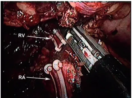
Figure 2 - The renal artery (RA) and renal vein (RV) were clipped with Hem-o-lok clips and divided by LigaSure.
iliac spine, and 2-cm above the iliac crest for the 30º Olympus laparoscope. Finally, a 12 mm trocar was placed in the initial hole. The Gerota’s fascia was cut horizontally from the front edge of the psoas muscle up to the diaphragm and down to the lower pole of the kidney, and the renal hilum
care was taken to avoid puncturing the kidney sac (Figure-3). The ureter was then identified and dis-sected as distally as possible at which point it is clipped with a Hem-O-Lock clip and left in situ.
The retroperitoneal space was inspected with
in-sufflation pressure at 5 mmHg to ensure adequate homeostasis. The trocar incisions were closed and the patient was changed to supine position. An approximately 7-8 cm long Gibson incision was made in the lower abdominal region. The distal ureter was gently dissected until some of the in-tramural ureter was mobilized, and then the bla-dder cuff was cut to free the specimen. After the bladder incision was totally sutured, the entire kidney within Gerota’s fascia and ureter were re-moved en bloc from the Gibson incision.
RESULTS
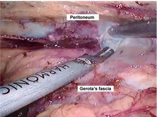
Figure 3 - Dissection of the diseased kidney outside the Gerato’s fascia.
age from 23 to 78 years old. Totally, 30 disea-sed kidneys were at the left side and 21 cases at the right side. The mean disease course was 7.3 months (range: 3-21 mon). Of 51 patients, 6 cases with contracted bladder had mild to moderate
re-nal function impairment (range of serum creatini-ne level: 189-450 mmol/L) and required sigmoid augmentation cystoplasty.
The most common complication was peritoneal tear (7.84%), which can be managed laparosco-pically with no need to conversion. Renal venous injury (1.96%) occurred in one patient due to se-vere adhesion. However, the bleeding was cessfully controlled and did not hamper the suc-cessful completion of the laparoscopic procedure. One diseased kidney was inadvertently punctured (1.96%) during dissection. The working space was thoroughly washed with a copious amount of nor-mal saline plus streptomycin at the end of the
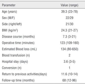
pro-Table 1 - Preoperative and operative characteristics of patients.
Parameter Value (range)
Age (years) 39.3 (23-78)
Sex (M/F) 22/29
Side (right/left) 21/30
BMI (kg/m2) 24.3 (21-27)
Disease course (months) 7.3 (3-21)
Operative time (minutes) 123 (109-160)
Estimated Blood loss (mL) 134 (80-650)
Blood transfusion (n) 2
Hospital stay (days) 3.6 (3-5)
Conversion (n) 1
Return to previous activities(days) 11.6 (10-14)
Follow-up time (months) 68 (12-96)
Table 2 - Peri-operative complication according to Satava classification system.
No. of cases
Satava grade I (incidents without consequence)
Sinus arrhythmia 1 (1.96%)
Satava grade II (incidents repaired intraoperatively)
Renal vein injury 1 (1.96%)
Lumbar vein injury 1 (1.96%)
Peritoneal injury 4 (7.84%)
Spillage of pus 1 (1.96%)
Satava grade III (incidents requiring open conversion)
Inability to dissect pedicle 1 (1.96%)
cedure, and no disseminated or systemic disease was identified during long-term follow-up. The postoperative complications graded by modified Clavien classifications are shown in Table-3. The grade I and II complications were 19.6% in all. One patient was reoperated for a hematoma in retroperitoneum, which was a grade III compli-cation (1.96%).
The histopathologic examination con-firmed the diagnosis of renal tuberculosis in all patients. The common microscopic findings were
Table 3 - Postoperative complications stratified according to the modified Clavien classification.
Clavien Grade n Management
Grade I (11.76%) 6
Fever 1 Antipyretics
Transient elevation in creatinine 2 Observation
Subcutaneous emphysema 3 Observation
Grade II (7.84%) 4
Blood loss anemia 2 Blood transfusion
Pneumonia 1 antibiotics
Wound infection 1 Wound opening and antibiotics
Grade III (1.96%) 1
tuberculous pyelonephritis with caseation, peri-nephritis and tuberculous pyonephrosis. Similar changes were found in the walls of ureters. The normal tissue was completely or partly replaced by caseous substance or fibrous tissue, and the ureter became thicken and stenotic.
After 68 months (range: 12-96 months) regular follow-up, the outcomes were satisfac-tory. All patients achieved improvement in symp-toms and renal function. No local or dissemina-ted recurrence was identified (evaluadissemina-ted by urine polymerase chain reaction for acid-fast bacilli, hematologic examination and imaging examina-tion). The ureteral stump syndrome did not occur in our patients.
DISCUSSION
Benign kidney diseases are the most com-mon indications for laparoscopic tomy (13). However, laparoscopic nephroureterec-tomy for inflammatory renal conditions such as tuberculous nonfunctioning kidney, long-standing renal stone disease and xanthogranulomatous pyelonephritis has always been considered as a relative contraindication because of higher rate of complications and conversion to open surgery. In this study, we retrospectively reviewed 51 patients with tuberculous nonfunctioning kidneys mana-ged by retroperitoneal laparoscopic nephrourete-rectomy in respect to surgical techniques, perio-perative outcome and follow-up, and the results are encouraging.
In our cases, we performed all surgeries by retroperitoneal laparoscopic approach for we have high volume of experience with this approach (12). Current researches have demonstrated that the laparoscopic operation for inflammatory non-functioning kidney can be accomplished through either the transperitoneal or the retroperitoneal approaches (5, 7), and the choice depends mainly on the surgeon’s educational background and la-paroscopic experiences. Although the retroperito-neal approach has some disadvantages such as the
contamination with the pus and potential injury to intraperitoneal organs (14).
Only a few small series have been reported addressing laparoscopic nephroureterectomy for tuberculous nonfunctioning kidneys. The reported rates of successive laparoscopic surgery were be-tween 77.8% and 100% (5-8). The largest cohort included only 31 patients (7). In the present study, a total of 51 patients were enrolled and laparosco-pic nephroureterectomy was successfully perfor-med in 50 patients (98.0%) with only one conver-sion due to dense adheconver-sions. The peri-operative data, including mean operative time, estimated blood loss, hospital stay and complications, were consistent with previous reports (4, 15, 16). These data support that retroperitoneal laparoscopic ne-phroureterectomy is feasible to treat patients with nonfunctioning tuberculous kidneys.
encoun-underlying layer outside the Gerota’s fascia is less affected by inflammation, and a dissection along this layer offers better progression and far less bleeding (19-21). Subcapsular nephrectomy has also been described for the retroperitoneoscopic approach of LN in inflammatory conditions when dense adhesions were encountered (22, 23). For nonfunctioning tuberculous kidneys, we found that the adhesive bundles and fibrosis were main-ly localized inside the Gerota’s fasica after a mi-nimum of 4 weeks of anti-tuberculous chemothe-rapy. Dissection can be performed with minimal difficulty by keeping the plane outside Gerota’s fascia. If difficulty with laparoscopic dissection is encountered in one area, the procedure can be carried out in other areas. The surgeon should try to achieve as much dissection as possible. The safe layer is usually visualized in the dense adhesive areas once the diseased kidney has been mostly dissected. Moreover, the introduction of advance equipments, such as the harmonic scalpel, Liga-sure and bipolar electric coagulation is an excel-lent alternative to avoid bothersome bleeding and makes surgeries easier.
Severe ureteral stump syndrome was re-ported after simple nephrectomy for treatment of nonfunctioning tuberculous kidneys, and power-ful anti-tuberculous regimen failed to completely cure the severe irritative symptoms in these cases (24, 25). To ensure optimal effectiveness, it beco-mes apparent that total ureterectomy should be applied for treatment of nonfunctioning tubercu-lous kidneys. The final histopathological outco-me of all patients in our study demonstrated that ureters were all diffusely invaded by tuberculosis, which supports the essentiality of total ureterec-tomy for nonfunctioning tuberculous kidneys. Multiple techniques have been used to manage the distal ureter during laparoscopic nephroure-tectomy, including open incision and resection via an intravesical or extravesical approach, endos-copic, laparoscopic stapling of the bladder cuff, or combinations of several techniques. Conside-ring the clinical-pathological characteristics of the tuberculosis and our experience, we recommend conventional open surgery to deal with the dis-tal ureter. After completion of the laparoscopic procedure in our series, an approximately 7-8 cm
long Gibson incision was made and the distal ure-ter was dissected till to the bladder wall, and the intact specimen was removed en bloc from the
in-cision. This approach facilitates to remove more tuberculous tissue to eliminate the potentiality of ureteral stump syndrome.
There are several limitations in our study. First, our case number is relatively small. Second, this is a retrospective study of a single center. Randomized controlled trials with large sample size are needed to prove the feasibility and supe-riority of retroperitoneal laparoscopic approach to manage tuberculous nonfunctioning kidneys.
CONCLUSIONS
Our results indicated that retroperitone-al laparoscopic nephroureterectomy is a safe and effective method for the treatment of tuberculous nonfunctioning kidneys with satisfactory long--term follow-up outcomes. However, randomized controlled studies with large sample size and high--quality design are needed to confirm our results.
CONFLICT OF INTEREST
None declared.
REFERENCES
1. Clayman RV, Kavoussi LR, Soper NJ, Dierks SM, Meretyk S, Darcy MD, et al. Laparoscopic nephrectomy: initial case report. J Urol. 1991;146:278-82.
2. Fahlenkamp D, Rassweiler J, Fornara P, Frede T, Loening SA. Complications of laparoscopic procedures in urology: experience with 2,407 procedures at 4 German centers. J Urol. 1999;162:765-70; discussion 770-1.
3. Portis AJ, Yan Y, Landman J, Chen C, Barrett PH, Fentie DD, et al. Long-term followup after laparoscopic radical nephrectomy. J Urol. 2002;167:1257-62.
4. Hemal AK, Gupta NP, Wadhwa SN, Goel A, Kumar R. Retroperitoneoscopic nephrectomy and nephroureterectomy for benign nonfunctioning kidneys: a single-center experience. Urology. 2001;57:644-9.
6. Hemal AK, Gupta NP, Kumar R. Comparison of retroperitoneoscopic nephrectomy with open surgery for tuberculous nonfunctioning kidneys. J Urol. 2000;164:32-5. 7. Lee KS, Kim HH, Byun SS, Kwak C, Park K, Ahn H.
Laparoscopic nephrectomy for tuberculous nonfunctioning kidney: comparison with laparoscopic simple nephrectomy for other diseases. Urology. 2002;60:411-4.
8. Zhang X, Zheng T, Ma X, Li HZ, Li LC, Wang SG, et al. Comparison of retroperitoneoscopic nephrectomy versus open approaches to nonfunctioning tuberculous kidneys: a report of 44 cases. J Urol. 2005;173:1586-9.
9. Satava RM. Identification and reduction of surgical error using simulation. Minim Invasive Ther Allied Technol. 2005;14:257-61.
10. Dindo D, Demartines N, Clavien PA. Classification of surgical complications: a new proposal with evaluation in a cohort of 6336 patients and results of a survey. Ann Surg. 2004;240:205-13.
11. Hemal AK, Mishra S. Retroperitoneoscopic nephrectomy for pyonephrotic nonfunctioning kidney. Urology. 2010;75:585-8.
12. Ping H, Xing NZ, Zhang JH, Yan Y, Kang N, Niu YN. Application of the Hem-o-lok ligation system in laparoscopic nephrectomy. Surg Endosc. 2010;24:1494-7.
13. Raghuram S, Godbole HC, Dasgupta P. Laparoscopic nephrectomy: the new gold standard? Int J Clin Pract. 2005;59:128-9.
14. Gupta NP, Hemal AK, Mishra S, Dogra PN, Kumar R. Outcome of retroperitoneoscopic nephrectomy for benign nonfunctioning kidney: a single-center experience. J Endourol. 2008;22:693-8.
15. Arvind NK, Singh O, Ali Q, Gupta SS, Sahay S. Laparoscopic nephrectomy in xanthogranulomatous pyelonephritis: 7-year single-surgeon outcome. Urology. 2011;78:797-801. 16. Ivey BS, Lucas SM, Meyer CA, Emley TE, Bey A, Gardner TA,
et al. Conversions in laparoscopic renal surgery: causes and outcomes. J Endourol. 2011;25:1167-73.
17. Kouba E, Smith AM, Derksen JE, Gunn K, Wallen E, Pruthi RS. Efficacy and safety of en bloc ligation of renal hilum during laparoscopic nephrectomy. Urology. 2007;69:226-9.
18. Lacombe M. Renal arteriovenous fistula following nephrectomy. Urology. 1985;25:13-6.
19. Duarte RJ, Mitre AI, Chambô JL, Arap MA, Srougi M. Laparoscopic nephrectomy outside gerota fascia for management of inflammatory kidney. J Endourol. 2008;22:681-6.
20. Shekarriz B, Meng MV, Lu HF, Yamada H, Duh QY, Stoller ML. Laparoscopic nephrectomy for inflammatory renal conditions. J Urol. 2001;166:2091-4.
21. Tepeler A, Akman T, Tok A, Kaba M, Binbay M, Müslümanoğlu AY, et al. Retroperitoneoscopic nephrectomy for non-functioning kidneys related to renal stone disease.Urol Res. 2012;40:559-65.Erratum in: Urol Res. 2012;40:567.
22. Xu Z, Xin M, Hong-Zhao L, Zhong C, Li LC, Ye ZQ. Retroperitoneoscopic subcapsular nephrectomy for infective nonfunctioning kidney with dense perinephric adhesions. BJU Int. 2004;94:1329-31.
23. Kapoor R, Vijjan V, Singh K, Goyal R, Mandhani A, Dubey D, et al.Is laparoscopic nephrectomy the preferred approach in xanthogranulomatous pyelonephritis? Urology. 2006;68:952-5.
24. Benchekroun TS, Kriouil A, Belkacem A, Jorio-Benkhraba M, el Fakir Y, Benhammou M, et al. Urogenital tuberculosis in children]. Arch Pediatra. 1997;4:857-61.
25. Burghele T. The place of total nephro-ureterectomy in the treatment of urinary tuberculosis. Rev Chir Oncol Radiol O R L Oftalmol Stomatol Chir. 1976;25:81-8.
_______________________ Correspondence address:
Nianzeng Xing, MD, PhD Professor of the Department of Urology, and the Vice President of Beijing Chao-Yang Hospital,