Evaluation of the risk of a stripping
perforation with gates-glidden drills:
serial
versus
crown-down sequences
Abstract: The aim of this study was to evaluate the remaining dentine/ cementum thickness using Gates-Glidden burs in serial and crown-down sequences and to observe which of the two sequences is the safest for preparing mesial roots of molars. Thirty-six left and right human man-dibular irst molars were selected. Standard access cavities were made and initially explored with Flexoiles sizes 10 and 15 until the tip was visible at the apex. The teeth were embedded in a mufle specially de-veloped for this study using a PVC tube with two parallel metal rods in its lid. Each tooth-block was sectioned 3 mm apically to the furca-tion using a low-speed saw with a diamond disc. The tooth-block was examined under a microscope and an initial image was captured by a digital video system with 8 X and 12 X magniications. Finally, the tooth-blocks were reassembled in the mufle so that the canals could be instrumented. After instrumentation the area of each mesial canal as well as the smallest distance to the root furcation were measured again. The mesio-buccal canals (crown-down order) and the mesio-lingual ca-nals (serial sequence) presented an average area of 0.46r0.16 mm2 and 0.88r0.27 mm2 (P0.01), respectively. The mean values of the smallest distance to the furcation for the mesio-buccal and mesio-lingual canals were 0.66r0.19 mm and 0.39r0.13 mm (P0.01), respectively. The remaining dentine/cementum thickness using Gates-Glidden burs was greater in the crown-down sequence than in the serial sequence.
Descriptors: Dental instruments; Endodontics; Molar.
Tauby Coutinho-Filho(a)
Gustavo De-Deus(a)
Eduardo Diogo Gurgel-Filho(b)
Ana Carolina Rocha-Lima(c)
Katia Regina Cervantes Dias(d)
Carlos Augusto Barbosa(a)
(a)PhDs, Department of Endodontics; (d)PhD,
Department of Restorative Dentistry – State University of Rio de Janeiro (UERJ), RJ, Brazil.
(b)PhD, Department of Endodontics, University
of Fortaleza (UNIFOR), CE, Brazil.
(c)Master’s Candidate, Department of
Endodontics, Piracicaba Dental School, State University of Campinas (UNICAMP), SP, Brazil.
Corresponding author
Eduardo Diogo Gurgel-Filho Av. Dom Luis, 1233/311 - Aldeota Fortaleza - CE - Brazil
CEP: 60160-230
E-mail: [email protected]
Introduction
The Gates-Glidden bur plays an important role among the instruments used during root canal prep-aration. Grossman1 (1982) dated its irst utilization in 1885. However, it was only used after studies per-formed during the 70’s when the concepts of Clean-ing & ShapClean-ing were reintroduced in Endodontics.2-4 Its low cost and high cutting power associated with its simplicity of use made it a widely used in-strument for the preparation of the straight portion of root canals. Abou-Rass et al.5 (1980) demonstrat-ed that the mesial canals of inferior molars do not assume a central position in relation to the root. The area between the canal and the root furcation pos-sesses thinner walls and is therefore named the ‘Risk Zone’. Another aspect is related to the transporta-tion of mesial canals in inferior molars towards the furcation. The great laring capacity of these instru-ments may lead to mechanical laws in the root ca-nal, especially at the “Risk Zone”.6
Excessive laring of the cervical and middle thirds in lat roots may lead to a pronounced decrease of the dentinal wall thickness or even result in a strip per-foration towards the furcation.7,8 Thin dentine walls increase root permeability and the possibility of frac-ture, not only during illing, but also during tooth functioning.9 Especially due to their cutting power, Gates-Glidden (GG) burs have suffered criticism re-garding their employment.10 Although the use of GG burs may be regarded as the traditional preparation technique, there are few studies analyzing their ef-fect in the “Risk Zone” of mandibular molars.3
Even after several years in evidence, their opera-tive sequence has still not been well established in literature, remaining, in most cases, up to the opera-tor’s common sense. The serial sequence is normally the most common technique applied.4,11
This study aimed to evaluate the remaining den-tine/cementum thickness using Gates-Glidden burs in serial and crown-down sequences and to observe which of the two sequences is the safest for prepar-ing the mesio-cervical area of molar roots.
Material and Methods
This study was revised and approved by the Eth-ics Committee, Nucleus of Collective Health Studies,
State University of Rio de Janeiro, Rio de Janeiro, RJ, Brazil. A sample of 36 human irst mandibular left and right molars, free from cracks, with similar anatomical characteristics were selected from the tooth bank of the State University of Rio de Janei-ro. Mean root canal curvature of the teeth used in this study was between 17º and 25º. The teeth were autoclaved and kept in 0.5% NaOCl for no longer than 24 h. Standard access cavities were made and all the canal oriices were located and initially ex-plored with ISO size 10 and 15 stainless steel Flexo-iles (Dentsply-Maillefer, Ballaigues, Vallorbe, Switzerland) until the tip was visible at the apex. 5.25% sodium hypochlorite (B Herzog – 8366, Rio de Janeiro, RJ, Brazil) was used to irrigate the canal during all steps.
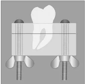
A modiied version of the mufle system12 was used, in which each molar was embedded in epoxy clear resin (Arazyn 1.0 – Araquímica, Araçarigua-ma, SP, Brazil) up to its furcation. The teeth were embedded in a mufle specially developed for this study using a PVC tube (Tigre Ltda., Rio Claro, SP, Brazil) with two parallel metal rods in its lid (Figure 1). Two equidistant nails and screws were
used as metallic guides to permit a safe remounting of the teeth.
Each tooth-block was sectioned 3 mm apically to the furcation using a low-speed saw (Isomet, Bue-hler, Ltd. Lake Bluff, NY, USA) with a diamond disc ( 125 mm x 0.35 mm x 12.7 mm – 330C) while constantly irrigated. Each tooth-block was ex-amined under a microscope (Axiscoppe – Carl Zeiss Vision Gmbh, Hallbergmoos, München, Germany) and an initial image was captured by a digital video system (Carl Zeiss Vision Gmbh, Hallbergmoos, München, Germany) with 8 X and 12 X magniica-tions. Finally, the tooth-blocks were reassembled in the mufle so that the canals could be prepared for instrumentation.
These initial images were submitted to com-puter-assisted evaluation. The KS 400 image sys-tem 3.0 (Carl Zeiss Vision Gmbh, Hallbergmoos, München, Germany) was used for image analysis and processing. The software allowed us to mea-sure the root canal area and the smallest distance to the furcation (SDF) using a semi-automatic process of segmented pixel shades. In order to achieve this, the images were converted to 256 gray tones, as these achieved the best results in the pilot study. Each image captured by the com-puter was gauged, eliminating any possible distor-tion. A protocol (macro) was developed to analyze all images, making the process more eficient and easier to reproduce. The measurements obtained by image analysis were repeated twice to ensure reproducibility.
After capture and initial analysis of the mesial root canals, the GG drills (Dentsply-Maillefer, Bal-laigues, Vallorbe, Switzerland) were used in crown-down order (GG4, GG3 and GG2) in the mesio-buc-cal canal (G1). To operate the drills, a conventional low-speed hand piece operating at 2,500 rpm was used. The movement performed with the rotary in-struments was slight apical pressure, ups and downs with only one penetration with each drill. The depth of the drill was determined by its clinging inside the radicular canal.7 Between the use of each drill, a re-capitulation with the apical ile was used with addi-tional irrigation with 1 ml of 5.25% NaOCl. In the mesio-lingual canal (G2), the GG drills were used
in the same way, although in serial sequence: GG2, GG3 and GG4.
After instrumentation, each tooth-block was re-examined under a microscope and new images of the mesial root canal were captured. At this moment the area of each mesial canal as well as the smallest distance to the root furcation was remeasured.
The initial and inal cross-section areas, as well as the smallest distance to the root furca-tion, were treated and statistically analyzed. Af-ter assessment of the non-normality of the data distribution, the ANOVA analysis was employed. When differences were found, Dunn’s multiple-comparison test was used to isolate the statisti-cally signiicant group. The level of signiicance was set at p<0.05. Origin 6.0 (Microcal Soft-ware, Inc., Northampton, MA, USA) and SPSS (Statistics 4.0 software, SPDD International BV, Gorinchem, The Netherlands) were also used as analytical tools.
Results
Cross-section area
The initial cross-section area values for the me-sio-buccal canals (G1) varied between 0.12 and 0.77 mm2, presenting an average of 0.24r0.10 mm2, while the initial cross-section area values for the mesio-lingual canals (G2) varied from 0.11 to 0.81 mm2, revealing an average of 0.23r0.11 mm2. Because of the high values presented by the standard deviation, it was possible to conirm the existence of great variability of the anatomy of the mesial root canals. However, after statistical treatment, it was possible to determine that no statistical difference (P!0.05) could be observed between the initial cross-section area of the mesio-buccal and the me-sio-lingual canals since their averages were extreme-ly similar.
com-Figure 2 - (A) Initial cross-section area of the mesio-lingual canal of sample 6. (B): After Gates-Glidden preparation in serial sequence, a greater increase of the cross-section area (249.5%) and an excessive drop in the smallest distance to the furcation (70.3%) can be observed.
Figure 3 - (A) Initial cross-section area of the mesio-buccal canal of sample 7. (B): After Gates-Glidden preparation in crown-down sequence, a limited increase of the cross-section area (79.2%) and a moderate reduction in the smallest distance to the furcation (17.3%) can be observed.
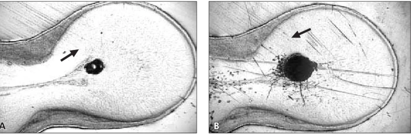
Figure 4 - (A) Cross-section area of a mesio-buccal canal after Gates-Glidden preparation in crown-down sequence. (B): Cross-section area of a mesio-lingual canal after Gates-Glidden preparation in serial sequence. A total rupture of the dentine/ cementum wall, leading to the furcation area, can be observed.
A
A
B
B
A
Cementum
B
parison between groups can be visualized in Fig-ures 2, 3 and 4 and Graph 1.
The SDF values for the mesio-lingual canals (G2) varied between 0.55 and 1.11 mm, present-ing an average of 0.80r0.15 mm. The SDF values for the mesio-buccal canals (G1) varied between 0.53 and 1.29 mm, and demonstrated an average of 0.83r0.17 mm. It was possible to encounter a modest variation when comparing the SDF values found in the mesio-buccal and mesio-lingual canals before instrumentation. No signiicant difference between the mesio-buccal and mesio-lingual canals was found (P!0.05).
After instrumentation, the SDF values for the
mesio-lingual canals (G2) varied from 0 to 0.90 mm, presenting an average of 0.39r0.13 mm. It was possible to determine a signiicant difference be-tween the initial and inal SDF values (P0.01). It is important to point out that two samples suffered stripping perforation, and therefore their SDF values were considered to be zero (Figure 4). After instru-mentation, the SDF values for the mesio-buccal ca-nals (G1) varied from 0.38 to 1.16 mm, presenting an average of 0.66r0.19 mm. It was possible to de-termine statistically signiicant differences between the groups (P0.01). The comparison between groups can be visualized in Graph 2. The relation-ship between the data of the cross-section area and 1.0
0.9
0.8
0.7
0.24
MB bi ML bi MB ai ML ai
0.23 0.46 0.88 0.6 0.5 0.4 0.3 0.2 0.1 0 C ro ss -s ectio n area (mm 2)
P < 0.01
Graph 1 - Cross-section area means before (bi) and after instrumentation (ai), in the MB (mesio-buccal) and ML (me-sio-lingual) canals.
0.80 0.83
0.66
0.39
ML bi MB bi ML ai MB ai
0.9 0.8 0.7 0.6 0.5 0.4 0.3 0.2 0.1 0 SD F (mm)
P > 0.05
P < 0.01
60 50 40 30 20 SD F (%) 10 0
0 100 200
Cross-section area (%)
300 400 500
Graph 2 - Smallest distance to furcation (SDF) means be-fore (bi) and after instrumentation (ai) in the MB (mesio-buc-cal) and ML (mesio-lingual) canals.
Graph 3 - Relationships between the smallest distance to furcation (SDF) and the cross-section area in the mesio-buccal (MB) canal after GG preparation in crown-down sequence.
Graph 4 - Relationships between the smallest distance to furcation (SDF) and the cross-section area in the mesio-lin-gual (ML) canal after GG preparation in serial sequence.
SD F (%) 120 100 80 60 40 20 0
0 200 400 600 800 1,000 1,200
the smallest distance to the furcation can be visual-ized in Graphs 3 and 4.
Discussion
The present work attempted to simplify the muf-le system method. A PVC tube, easily found in the marketplace, was used to create resin-blocks, with-out the necessity of industrial sophistication, there-fore simplifying the process. Two equidistant nails and screws were used as metallic guides to permit a safe remounting of the teeth.
The furcation area of the irst inferior molars, sectioned at a point located between 2 and 3 mm bellow the furcation of the roots, presented a con-cave aspect in 100% of the mesial roots and 99% of the distal roots.13 Several authors have described an area 3 to 4 mm below the entrance of the canals to be the most sensitive location for the perforation of mesial molar roots after the use of rotary instru-ments.5,14,15 Based on these results, the present study opted to section the root samples 3 mm bellow the furcation of the roots.
The cross-section areas of the mesio-buccal and mesio-lingual canals as well as the SDF were pre-cisely measured through a computer-assisted evalu-ation. Calibration of each image captured by the computer decreased the possibility of distortion.
After statistical treatment of the results, statisti-cally signiicant differences were observed between the cross-section area after instrumentation and the GG sequences, as well as in relation to the SDF.
The greatest cross-section area increase was ob-served in sample 27 from G2 (mesio-lingual canal – serial sequence), in which the canal area was 7.4 times bigger, and in sample 7 from G1 (mesio-buc-cal canal – crown-down sequence), in which the ca-nal area was 2.1 times bigger.
Although it was not possible to observe anatom-ic relationship between samples, it was clear that the use of GG drills in the serial sequence tended to re-sult in a stronger increase of the cross-section area.
The increase of the cross-section area tended to be directly associated to the decrease of the distance between the canal and the furcation. The tendency
of greater wear after the use of a serial sequence could be seen in two samples, in which a total rup-ture of the dentine/cementum wall leading to the furcation area could be observed.
Remaining dentine thickness following various in-tra-radicular procedures may be the most important iatrogenic factor correlating to future root resistance against fracture16. Lim, Stock8 (1987) established in their study a limit of 0.2 and 0.3 mm of dentine thick-ness as a measurement in which the resultant forces of condensation during obturation would not result in tooth fracture. Having this in mind, samples in G1 (mesio-buccal canal – crown-down sequence) present-ed wall thickness greater then the limits establishpresent-ed. On the other hand, 9 samples (37.5%) in G2 (me-sio-lingual canal – serial sequence) presented inferior wall thickness, leaving them out of the safety limit. Through the results of the present study it was possi-ble to conclude that GG drills, used in a crown-down sequence, offer the operator a greater safety margin.
Other authors7,17,18 corroborate this statement, believing that the crown-down sequence is safer, since the larger burs tend to penetrate less deeply into the canal, avoiding a great and unnecessary wear of the dentine walls.
Throughout this experiment it was observed that samples always suffer greater wear nearest to the “Risk Zone” rather than the “Safety Zone”, which was also seen in the studies of Abou-Rass et al.5 (1980) and Cunningham, Senia6 (1992). According to Abou-Rass, Jastrab19 (1982), the GG drills are safe to use and the occurrence of instrument separa-tion (of which the removal is usually simple) is often a result of inadequate manipulation. Although this was not the objective of this work, no instrument separation was observed during the instrumentation of the 36 samples.
Conclusion
References
1. Grossman L. A brief history of endodontics. J Endod. 1982;8(1):538.
2. Mullaney TP. Instrumentation of finely curved canals. Dent Clin North Am. 1979;23(4):575-92.
3. Taintor JF. Use of the gates glidden bur in endodontics. J Nebr Dent Assoc. 1978;54(3):10-2.
4. Weine FS, Kelly RF, Lio PJ. The effect of preparation proce-dures on original canal shape and on apical foramen shape. J Endod. 1975;1(8):255-62.
5. Abou-Rass M, Frank AL, Glick DH. The anticurvature filing method to prepare the curved root canal. J Am Dent Assoc. 1980;101(5):792-4.
6. Cunningham CJ, Senia SE. A three-dimensional study of canal curvatures in the mesial roots of mandibular molars. J Endod. 1992;18(6):294-300.
7. Coutinho-Filho T, De-Deus G, Guimarães T, Gurgel-Filho ED, Maniglia-Ferreira C. A computer evaluation of the dentin remaining after cervical preparation in curved canals: Gates-glidden drills vs. orifice shaper. Braz J Oral Sci. 2002;1(3):116-20.
8. Lim SS, Stock CJ. The risk of perforation in the curved canal: anticurvature filing compared with the step-back technique. Int Endod J. 1987;20(1):33-9.
9. Isom TL, Marshall JG, Baumgartner JC. Evaluation of root thickness in curved canals after flaring. J Endod. 1995;21(7):368-71.
10. Gluskin AH, Brown DC, Buchanan LS. A reconstructed com-puterized tomographic comparison of Ni-Ti rotary GT files
versus traditional instruments in canals shaped by novice operators. Int Endod J. 2001;34(6):476-84.
11. Schilder H. Cleaning and shaping the root canal. Dent Clin North Am. 1974;18(2):269-96.
12. Bramante CM, Berbet A, Borges RP. A methodology for evalu-ation of root canal instrumentevalu-ation. J Endod. 1987;13(5):243-5.
13. Bower RC. Furcation morphology relative to periodontal treatment. Furcation entrance architecture. J Periodontol. 1979;50(1):23-7.
14. Kessler JR, Peters DD, Lorton L. Comparison of the relative risk of molar root perforations using various endodontic in-strumentation techniques. J Endod. 1983;9(10):439-47. 15. McCann JT, Keller DL, LaBounty GL. A modification of the
muffle model system to study root canal morphology. J Endod. 1990;16(3):114-5.
16. Pilo R, Corcino G, Tamse A. Residual dentin thickness in mandibular premolars prepared with hand and rotatory in-struments. J Endod. 1998;24(6):401-4.
17. Torabinejad M, Rotstein L, Stabholz A. Effect of preflar-ing on tactile detection of the apical constriction. J Endod. 1995;21(2):92-4.
18. West JD, Roane JB. Limpeza e modelagem do sistema de canais radiculares. In: Cohen S, Burns RC. Caminhos da polpa. 7th ed.
Rio de Janeiro: Guanabara Koogan; 2000. p. 206-36. 19. Abou-Rass M, Jastrab RJ. The use of rotary instruments as