Endoscopic endodontic microsurgery:
2-year evaluation of healing and
functionality
Abstract: This prospective clinical study aimed to evaluate the benei ts of the endoscope as an aid to root-end management, and to assess the treat-ment outcome during 2 years following surgery. Forty-three endodontic surgical procedures in 30 patients were performed with the aid of an endoscope and followed for a period of 2 years. Radiographic criteria and clinical evaluation were used to assess the outcome. All cases were evaluated in terms of healing and functionality. 91.1% and 90.7% of the teeth evaluated after 1 and 2 years, respectively, were classii ed as suc-cessful. We found no statistically signii cant differences for both healing and functionality between the 1- and 2-year evaluations. No difference related to tooth type or tooth location was found at the 2-year follow-up. Fisher’s exact test was used to statistically assess the difference between successful and unsuccessful cases for each of the variables considered. The endoscope can be an aid for endodontic surgical procedures in terms of both periapical healing and functionality up to 2 years follow-up.
Descriptors: Apicoectomy; Endoscopy; Oral surgical procedures. Silvio Taschieri(a)
Massimo Del Fabbro(b)
(a) MD, DDS, Head of the Endodontics
Section; (b)BSc, PhD, Head of the Oral
Physiology Section – IRCCS Istituto Ortopedico Galeazzi, Department of Health Technologies, University of Milan, Milan, Italy.
Corresponding author:
Massimo Del Fabbro
IRCCS Istituto Ortopedico Galeazzi Via R. Galeazzi 4
20161 - Milano - Italy
E-mail: [email protected]
Introduction
Microsurgical instruments and ultrasonic retro-tips signii cantly improved the outcome of perira-dicular surgery when compared with traditional techniques.1 These devices brought advantages for
root-end management.1,2 At the same time, the use
of well-focused illumination and magnii cation de-vices has been recommended as a standard of care in periradicular surgery.3 Rubinstein, Kim4,5 (1999,
2002) have reported very high success rates after periradicular surgery, advocating that the use of a surgical microscope might have been a decisive fac-tor contributing to such an excellent outcome. Re-cently, there has been a growing interest in endos-copy for optimizing visualization.6-11
To date, its use in endodontic surgery has been shown mostly in a few technical reports.6,7,9,10,12
These articles reported advantages using the endo-scope such as: it is a versatile and expandable system, easily transportable, and has excellent illumination and good magnii cation; no mirrors are required; the learning curve is short. Due to its non i xed i eld of vision, the endoscope allows viewing of a treat-ment i eld at various angles and distances without losing depth of i eld and focus. In using the micro-scope, when increasing the magnitude, any move-ment of either the microscope or the patient will cause the surgical i eld to become out of focus. This is one drawback of the microscope when used as an aid to enhance visualization of the surgical i eld. In the authors’ experience using both magnii cation devices, though recognizing the microscope’s use-fulness, they have observed that the depth-of-i eld perception that an operator has using an endoscope is quite similar to the perception that a surgeon has with the naked eye.12
Another advantage in the use of an endoscope is that its control is more tactile than that of a micro-scope. Regulating the endoscope for focusing and zooming using just one i nger is faster and more com-fortable, and the surgeon can hold the endoscope and the camera in one hand with the pen grasp, whereas, with the microscope, it is sometimes necessary to stop and manually adjust the focus and zoom using one or both hands. The operator can examine the morphological aspects of the roots from almost any
angulation in a very short time. This takes longer using a microscope and sometimes it is impossible, especially in the posterior jaws. It is also possible to see behind the roots and ascertain the presence of periradicular lesions and, if necessary, remove them. This is much more complicated when the operator has to use a microscope and the retro-mirror.
The operator can also ascertain the presence of a through-and-through lesion and visualize the size of the bone lesion in the palatal or lingual plate. Using a microscope it is not always possible to achieve the same accurate results.
The rod-lens system of the endoscope allows a good visualization even in the presence of irrigation l uids, so the operator can use retro-tips with ster-ile water irrigation to avoid overheating and at the same time have lens cleaning. In contrast, using the microscope and the retro-mirror the operator has often to stop in order to clean the mirror. During adjustment of the viewing angle, the endoscope lens position can be misplaced. Once the correct position is lost, however, it is easier to i nd it again as com-pared to the microscope.
With regard to the clinical application of endos-copy in periradicular surgery, a few short-term pro-spective studies have been published recently.8,9 In
both studies, the authors reported a better outcome in the group using the endoscope as compared with the control group.
The main objectives of this prospective clinical study were to evaluate the clinical advantages of using an endoscope during root-end management and to compare the outcome of periradicular sur-gery in terms of healing and functionality at 1- and 2-years follow-up.
Material and Methods
All patients requiring surgical treatment were re-cruited during a period of 34 months (from Decem-ber 2001 to OctoDecem-ber 2004) in a university clinic and in a private practice setting. Most of the patients en-rolled in the present study were also included in one arm of a previously reported comparative prospec-tive study that assessed 1-year outcomes only.8 The
in 1983.
Patient selection and inclusion criteria
The following criteria were adopted for case se-lection:
The tooth treated surgically showed a periradic-ular lesion of strictly endodontic origin and the non-surgical retreatment was considered unfea-sible or had previously failed.
The tooth treated surgically exhibited an ad-equate i nal restoration with no clinical evidence of coronal leakage.
The apical root canal had 6 mm or more without the presence of a post.
Periradicular surgery was carried out only in the absence of acute symptoms.
The patient did not have general medical contra-indications for oral surgical procedures (ASA-1 or ASA-2).
The following exclusion criteria were applied: Teeth with pathoses associated with vertical root fracture.
Teeth with perforation of the furcation area or lateral canal walls.
Teeth with traumatic injuries.
Severe periodontal bone loss detected with a periodontal probe (5 mm probing depth).
All teeth with periapical lesions diagnosed radio-graphically were treated with periradicular surgery. Each patient was given written information about the surgical procedure and the necessary follow-up care; they were also given the opportunity to with-draw from the study. A consent form was signed if they agreed.
Surgical procedure and management of the resected root end
Full mucoperiosteal tissue l ap designs were used. The type of l ap design (rectangular, marginal or submarginal) varied according to the periodontal status. The endodontic surgical l ap was rel ected and carefully retracted. The undermining eleva-tion l ap rel eceleva-tion technique was used. Surgical ac-cess to the root was then made through the cortical bone using a round bur. Shaving of the bone was performed with a brush-stroke approach, using 1.
2.
3.
4.
5.
1.
2.
3. 4.
light pressure, low rotation speed, and avoiding pro-longed contact of the bur with the osseous surface. This was done under constant copious sterile water irrigation. The periradicular lesion was removed with sharp bone curettes and angled periodontal curettes. The curetted tissue was placed in 10% formalin solution for pathologic diagnosis. After exposure of the root end, a straight i ssure bur in a hand-piece was positioned perpendicular to the long axis of the root and then, beginning from the apex, cutting coronally, 2.5 to 3 mm of the root end was shaved away. The bur was moved from mesi-al to distmesi-al at the desired angle (little or no bevel), shaving the root-end surface smooth and l at. All of these procedures were performed using magnii -cation loupes (4.3 X) with a headlight. After root-end resection, surgical procedures were performed with an endoscope. In this study, the root ends were examined using a 6-cm-long and 3-mm-wide endoscope with a viewing angle of 70° (Hopkins Tele-Otoscope 70°, Karl Storz GmbH, Tuttlingen, Germany) (Figure 1). Prior to root-end preparation, local haemostasis was achieved using bone wax. Root-end cavities were prepared using zirconium nitrate retro-tips (Figures 2-3) (Dentsply; Maillefer Instruments, Ballaigues, Switzerland) driven by an ultrasonic device unit (Piezon master 400; EMS, Nyon, Switzerland). All root-end cavities were done setting the ultrasonic device unit at no more than
half power, under constant copious sterile water ir-rigation to avoid overheating. The retro-tips had a 3-mm-long tip that allowed a well-dei ned parallel preparation of 2.5- to 3-mm in depth. The root-end cavities were then dried using a paper cone and ex-amined in order to detect root-face alterations in the resected root end, such as marginal chipping. Final-ly, a zinc oxide EBA-reinforced cement (Super Seal; Ogna Pharmaceuticals, Milan, Italy) was used as root-end i lling material (Figure 4). A i ne diamond bur was used to remove the excess of i lling mate-rial. The rel ected tissues were reapproximated to
their original position, compressed and stabilized, and sutured with non absorbable silk 5-0 (Ethicon Inc., Piscataway, NJ, USA).
Endoscope in the surgical room
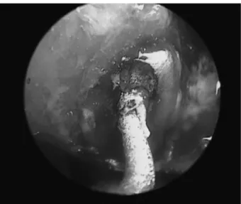
Four main components, all placed on a mobile rack, make up the endoscope: (a) rod-lens system, (b) camera head, (c) camera control unit with a monitor and light source (Figure 5). The rack is fully maneu-verable with its correct position being at the level of the patient’s lower leg region on the opposite side of the surgeon. This allows the surgeon and the whole Figure 3 - Endoscopic view of the retro-tip during root-end cavity preparation.
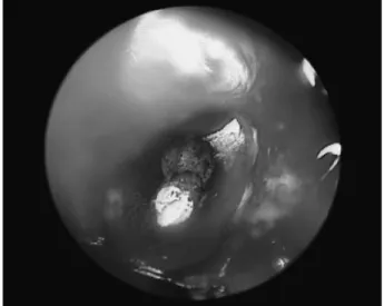
Figure 2 - Endoscopic view of the retro-tip positioned in the orifice of the root canal system.
Figure 4 - Surface of the resected root end after root-end filling.
surgical team to view the surgery on the monitor. The surgeon holds the endoscope and the camera in one hand with a pen grasp. This way, the viewing angle and the position of the endoscope lens can be easily adjusted. For aseptic reasons, the video cam-era and the camcam-era cable are packed in sterile covers and connected to the ocular of the endoscope.
Criteria for healing assessment
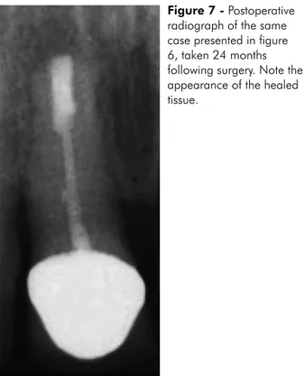
A radiograph of each tooth was taken during the preliminary visit (Figure 6). Further radiographs were taken at each clinical appointment (3, 6, 12 and 24 months). The paralleling technique was ad-opted to ensure reproducibility. The radiographs taken at 12 and 24 months were used to assess heal-ing (Figure 7). The radiographic criteria used to as-sess the outcome were those established by Molven
et al.13,14 (1987, 1996).
At each clinical appointment, a clinical evalua-tion was recorded, following the guidelines of Gut-mann, Harrison15 (1991).
According to modern concepts16 the success of
endodontic treatment was evaluated not only in terms of radiographic and clinical healing but also in terms of functional retention of the tooth. A case
was considered as functionally successful when clin-ical presentation was normal (asymptomatic tooth) regardless of the presence of radiolucency. All cases presenting with clinical symptoms were classii ed as not functional.
Fisher’s exact test was used to statistically as-sess the difference between successful and unsuc-cessful cases for each of the variables considered. The tooth was considered as the unit of analysis. A probability value of p = 0.05 was considered as the level of signii cance.
Results
According to the selection criteria, 50 teeth in 34 patients (20 women, mean age 43 years and 14 men, mean age 37 years) were included in the study and treated with periradicular surgery.
One tooth was extracted during the surgical pro-cedure before root-end resection because of vertical fracture, so the tooth (and the patient as well) was excluded from the study. Two patients accounting for 4 teeth did not come to the 1-year follow-up visit and were therefore excluded from the study. Another patient accounting for two teeth did not come to the 2-year follow-up visit. A i nal total of 43 teeth in 30 Figure 7 - Postoperative radiograph of the same case presented in figure 6, taken 24 months following surgery. Note the appearance of the healed tissue.
patients were evaluated up to 2 years. Among them, 25 were in the maxilla (17 anterior, 6 premolar and 2 molar teeth) and 18 in the mandible (10 anterior, 4 premolar and 4 molar teeth). The maximum di-ameter of the periapical lesion ranged between 2.5 and 15 mm.
After 1 year, 41 teeth out of the 45 evaluated were classii ed as successful (91.1%), 2 as uncertain healing and 2 as failures. Forty-three cases were con-sidered as functional (95.5%). Of the 43 teeth evalu-ated at the 2-year follow-up, 39 (90.7 %) healed suc-cessfully, 1 teeth had uncertain healing, and 3 were classii ed as failures. Forty cases were classii ed as functional (93%) 2 years after surgery. It is notewor-thy that of the two teeth classii ed as uncertain heal-ing (and as functional) at the 1-year follow-up, one developed clinical symptoms during the second year and had to be extracted. The other one was still clas-sii ed as uncertain healing (due to the persistence of radiolucency) at the 2-year follow-up. In agreement with other authors,14,17 the latter case was scheduled
for a further follow-up 2 years later.
No statistically signii cant difference was found in treatment results between the 1- and 2-year eval-uations neither in terms of healing (p = 0.12) nor in terms of functionality (p = 0.31). No difference related to tooth type (p = 0.30) and tooth location (maxilla versus mandible, p = 0.24) was found at the 2-year follow-up.
Discussion
The present study included some patients whose outcomes have been previously reported8 but this
one consists of an expanded cohort followed up for an extended time period.
In addition to evaluating the endoscope benei ts in the surgical endodontic procedure, one of the aims of this study was to investigate a novel aspect of outcome assessment in endodontics, namely tooth functional retention, as proposed earlier by Fried-man, Mor16 (2004).
Many clinical studies have been published on periradicular surgery using microsurgical retro-tips.4,5,18-20 The introduction of ultrasonic retro-tips
in periradicular surgery has brought many advan-tages in the procedure of root-end preparation. They
enable the long axis of the tooth to be followed, while preserving the morphology of the canal. Api-cal cavities can be shaped easily, safely, and with greater precision as compared to using conventional handpieces.3,21 A well shaped root-end cavity, which
is more centrally placed and smaller than that pro-duced by microhandpieces and burs, can also reduce the risk of root perforation in deeply l uted roots and the inability to prepare to a sufi cient depth. In addi-tion, the utilization of ultrasonic retro-tips requires small bony crypts, and the cutting bevel obtained on the resected root end can be quite perpendicular to the canal’s long axis. This fact might be benei cial because it decreases the number of exposed dentinal tubules at the resected root surface, minimizing api-cal leakage.3
Bader, Lejeune22 published in 1998 the i rst
clini-cal study comparing ultrasonic and conventional root-end preparations. The success rates for ultra-sonically treated teeth were signii cantly better than those for teeth treated with microburs. Other stud-ies19,23,24 showed similar differences between the 2
root-end preparations. A number of clinical studies have been published on periradicular surgery us-ing microsurgical retro-tips, reportus-ing success rates ranging between 82% and 96.8%.3,16 In general, the
highest success rates have been reported when mag-nii cation devices were used.4
The use of high quality magnii cation devices in dentistry is becoming more and more common, with the aim of improving treatment quality.15 Many
au-thors showed how these devices provide the visual access necessary to perform microsurgery with a greater degree of coni dence and accuracy.4,5,25-27 A
recent addition to the i eld of visualization devices is the i beroptic endoscope.
Only two short-term clinical prospective studies concerning endoscopy in endodontic surgery were published.8,9 In the i rst published article the authors
compared endodontic surgery with and without en-doscopy. The success rates were not signii cantly different, 88.9% and 75.4%, respectively.9 In the
other study, the endoscope group presented a 94.9% success rate at the one-year follow-up versus 90.6% for the group in which surgical loupes were used.8
the one-year follow-up, and remained substantially unchanged one year later.
The main benei t considered in evaluating the outcome of surgical treatment is the probability of healing. However, the concept of tooth functional retention has been recently proposed as a further in-dicator of treatment success. In a literature review, Friedman, Mor16 (2004) showed that the chance of
teeth with apical periodontitis to completely heal after apical surgery is highly variable among differ-ent studies if the success is evaluated according to conventional clinical and radiographic criteria (from 37% to 85%), mainly due to great heterogeneity. In fact, many variables can affect the prognosis of the surgical treatment, such as: surgical procedure, materials, radiographic and clinical outcome assess-ment, the patient’s systemic condition, type of tooth, quality of previous root canal treatment or re-treat-ment and of coronal restoration. Furthermore, one of the main reasons for the variability of reported outcomes is the different criteria for the evaluation of success and failure of the treatment that have been adopted. If functionality criteria are adopted, a much lower variability occurs (86 to 92%), which
seems to rel ect more closely the generally observed tooth prognosis.16 In the present study, the
differ-ence in treatment outcome as evaluated according to the different criteria was found to be minimal. It can be hypothesized that adherence to a strict end-odontic microsurgical procedure and an accurate case selection might be key factors in maximizing treatment success.
Conclusion
In the present study, the endoscope provided ex-cellent visualization of the surgical i eld during root-end management and can be an aid for root-endodontic surgical procedure in terms of both healing and functionality.
Further clinical trials with standardized proto-cols and a high level of evidence are needed to con-i rm the results of the present study.
Acknowledgements
The Authors wish to thank Mr. Tom Lacey for his support in checking the English language of the manuscript.
References
1. von Arx T, Walker WA. Microsurgical instruments for root-end cavity preparation following apicoectomy: A literature review. Endod Dent Traumatol. 2000 Apr;16(2):47-62. 2. Torabinejad M, Pitt Ford TR. Root-end filling materials: A
review. Endod Dent Traumatol. 1996 Aug;12(4):161-78. 3. Kim S, Kratchman S. Modern endodontic surgery concepts
and practice: A review. J Endod. 2006 Jul;32(7):601-23. 4. Rubinstein RA, Kim S. Short-term observation of the results
of endodontic surgery with the use of a surgical operation mi-croscope and Super-EBA as root-end filling material. J Endod. 1999 Jan;25(1):43-8.
5. Rubinstein RA, Kim S. Long-term follow-up of cases consid-ered healed one year after apical microsurgery. J Endod. 2002 May;28(5):378-83.
6. Bahcall JK, Barss J. Orascopic visualization technique for conventional and surgical endodontics. Int Endod J. 2003 Jun;36(6):441-7.
7. Bahcall JK, Di Fiore PM, Poulakidas K. An endoscopic tech-nique for endodontic surgery. J Endod. 1999 Feb;25(2):132-5.
8. Taschieri S, Del Fabbro M, Testori T, Francetti L, Weinstein RL. Endodontic surgery using two different magnification
devices: Preliminary results of a randomized controlled study. J Oral Maxillofac Surg. 2006 Feb;64(2):235-42.
9. von Arx T, Frei C, Bornstein M. Periradicular surgery with and without endoscopy: A prospective clinical comparative study. Schweiz Monatsschr Zahnmed. 2003;113(8):860-5. 10. von Arx T, Hunenbart S, Buser D. Endoscope- and
vid-eo-assisted endodontic surgery. Quintessence Int. 2002 Apr;33(4):255-9.
11. von Arx T, Montagne D, Zwinggi C, Lussi A. Diagnostic accuracy of endoscopy in periradicular surgery – A compari-son with scanning electron microscopy. Int Endod J. 2003 Oct;36(10):691-9.
12. Taschieri S, Del Fabbro M, Testori T, Francetti L, Wein-stein RL. Combining surgical microscope and endoscope for achieving excellent aesthetic outcome in soft tissue healing and maximizing the success of periradicular surgery. Pract Periodont Esthet Dent. 2006;18:193-8.
14. Molven O, Halse A, Grung B. Incomplete healing (scar tissue) after periapical surgery. Radiographic findings 8-12 years after treatment. J Endod. 1996 May;22(5):264-8.
15. Gutmann JL, Harrison JW. Surgical Endodontics. Boston: Blackwell Scientific Publications; 1991.
16. Friedman S, Mor C. The success of endodontic therapy - heal-ing and functionality. J Calif Dent Assoc. 2004 Jun;32(6):493-503.
17. Rud J, Andreasen JO, Möller Jensen JE. A follow-up study of 1000 cases treated by endodontic surgery. Int J Oral Surg. 1972;1(4):215-28.
18. Chong BS, Pitt Ford TR, Hudson MB. A prospective clinical study of mineral trioxide aggregate and IRM when used as root-end filling materials in endodontic surgery. Int Endod J. 2003 Aug;36(8):520-6.
19. Testori T, Capelli M, Milani S, Weinstein RL. Success and failure in periradicular surgery. A longitudinal retrospective analysis. Oral Surg Oral Med Oral Pathol Oral Radiol Endod. 1999 Apr;87(4):493-8.
20. Zuolo ML, Ferreira MOF, Gutmann JL. Prognosis in peri-radicular surgery: A clinical prospective study. Int Endod J. 2000 Mar;33(2):91-8.
21. Plotino G, Pameijer CH, Grande NM, Somma F. Ultrason-ics in endodontUltrason-ics: A review of the literature. J Endod. 2007 Feb;33(2):81-95.
22. Bader G, Lejeune S. Prospective study of 2 retrograde en-dodontic apical preparations with and without the use of CO2 laser. Endod Dent Traumatol. 1998 Apr;14(2):75-8.
23. Engel TK, Steiman HR. Preliminary investigation of ultrasonic root-end preparation. J Endod. 1995 Sep;21(9):443-5. 24. Mehlhaff DS, Marshall JG, Baumgartner JC. Comparison of
ultrasonic and high-speed-bur root-end preparations using bilaterally matched teeth. J Endod. 1997 Jul;23(7):448-52. 25. Cohen S, Burns RC. Pathways of the Pulp. 8th ed. St. Louis:
Mosby; 2002.
26. Friedman M, Mora AF, Schmidt R. Microscope-assisted precision dentistry. Compend Contin Educ Dent. 1999 Aug;20(8):723-8.