*Correspondence: Layasadat Khorsandi. Cell and Molecular Research Center, Faculty of Medicine, Ahvaz Jundishapur University of Medical Sciences, P. O. Box: 61335, Ahvaz, Iran. E-mail: [email protected]
A
vol. 49, n. 4, oct./dec., 2013
Exendin-4 effects on islet volume and number in mouse pancreas
Layasadat Khorsandi
*, Fereshteh Nejad-Dehbashi
Cell and Molecular Research Center, Ahvaz Jundishapur University of Medical Sciences, Iran
The aim of this study wasto evaluate Exendin-4 (EX-4) effects on islet volume and number in the mouse pancreas. Thirty-two healthy adult male NMRI mice were randomly divided into control and experimental groups. EX-4 was injected intraperitoneally (i. p.) at doses of 0.25 (E1 group), 0.5 (E2 group), and 1 μg/kg (E3 group), twice a day for 7 consecutive days. One day after the inal injection, the mice were sacriiced, and the pancreas from each animal dissected out, weighed, and ixed in 10% formalin for measurement of pancreas and islet volume, and determination of islet number by stereological assessments. There was a signiicant increase in the weight of pancreases in the E3 group. Islet and pancreas volumes in E1 and E2 groups were unchanged compared to the control group. The E3 group showed a signiicant increase in islet and pancreas volume (P < 0.05). There were no signiicant changes in the total number of islets
in all three experimental groups. The results revealed that EX-4 increased pancreas and islet volume in non-diabetic mice. The increased total islet mass is probably caused by islet hypertrophy without the formation of additional islets.
Uniterms: Exendin-4/effects/experimental study. Pancreas/effects fo Exendin-4. Islet/hypertrophy. Stereology.
O objetivo deste estudo foi avaliar os efeitos do Exendin-4 (EX-4) sobre o volume e número de ilhotas no pâncreas. Trinta e dois camundongos NMRI machos saudáveis e adultos foram divididos ao acaso em grupos controle e grupos experimentais. EX-4 foi injetado intraperitonealmente (i. p.) nas doses de 0,25 (grupo E1), 0,5 (grupo E2) e 1 (grupo E3), duas vezes por dia durante 7 dias consecutivos. Um dia após a injeção inal, os camundongos foram sacriicados e o pâncreas de cada animal foi dissecado, pesado e ixado em solução de formaldeído 10% para avaliação do volume do pâncreas e ilhotas e do número de ilhotas por métodos estereológicos. Observou-se aumento signiicativo no peso de pâncreas no grupo E3. O volume do pâncreas assim como das ilhotas não apresentou alterações nos grupos E1 e E2, quando comparados ao grupo controle No grupo E3 houve aumento signiicativo no volume do pâncreas e das ilhotas (P<0,05). Não se observaram alterações signiicativas no número de ilhotas nos três grupos experimentais. Os resultados revelaram que o EX-4 provoca aumento no volume do pâncreas, bem como no volume das ilhotas em camundongos não-diabéticos. O aumento no volume total de ilhotas deve-se, provavelmente, a hipertroia das ilhotas sem a formação de ilhotas adicionais.
Unitermos: Exendina-4/efeitos/estudo experimental. Pâncreas/efeito do Exendina-4. Ilhotas/hipertroia. Estereologia.
INTRODUCTION
Glucagon-like peptide-1 (GLP-1) is a peptide
secreted from the gut in response to food. It acts directly
on β cells, enhancing the effect of glucose in stimulating
insulin secretion from these cells. When administered to diabetic mice, GLP-1 lowers blood glucose levels
and stimulates insulin secretion (Xu et al., 1999). In
addition, GLP-1 increases the β-cell mass by inducing the
differentiation and neogenesis of ductal progenitor cells into islet endocrine cells (Hui et al., 2001; Abraham et al., 2002). In a previous in vitro study, was shown that
GLP-1 is capable of enhancing fetal pig β-cell differentiation
from progenitor epithelial cells as well as initiating their
functional maturation in islet-like cell clusters (Hardikar
et al., 2002).
L. Khorsandi, F. Nejad-Dehbashi 746
potency as GLP-1 (Xu et al., 1999). Exendin-4 (EX-4) is resistant to the enzyme dipeptidyl peptidase 4 (DPT-IV), which is present in serum. GLP-1 is rapidly metabolized by DPP-IV (Kieffer et al., 1995). It has been reported previously that EX-4 is capable of stimulating both the
differentiation of β cells from ductal progenitor cells and proliferation of β cells when given to rats and human
(Zhou et al., 1999; Stoffers et al., 2000; Kastin et al., 2003).
Previous studies on EX-4 action were mostly performed in diabetic rodent models. However, some studies demonstrate that EX-4 has a beneficial effect in non-diabetic animals. It has been reported that EX-4 causes weight loss (De Fronzo et al., 2005; Buse et al., 2004; Kendall et al., 2005). In the placebo-controlled
component of the pivotal trials, which lasted 30 weeks, mean weight reduction ranged from between 1 and 3 kg
compared with placebo. In open-label extensions, weight continued to decline over 2 years of treatment, by up to
5 kg from baseline (Blonde et al., 2006; Buse et al., 2007).
It is known that EX-4 can pass through the blood–
brain barrier (Kastin et al., 2003) and exert central effects, including promotion of neurotrophic or neuroprotective actions (Perry et al., 2000a; Perry et al., 2000b) and enhancement of cognitive functions (During et al., 2003).
These indings suggest that GLP-1 receptor stimulation
in the central nervous system plays a critical role in regulating neuronal plasticity and cell survival. Vella et al. (2003) reported that EX-4 and GLP-1 increase cortisol secretion in human subjects. However, neither of these alter insulin action in non-diabetic human subjects (Vella
et al., 2003). Ranta et al. (2006) demonstrated that EX-4 protects against glucocorticoid-induced mouse beta-cells or INS-1 cell apoptosis. Chen et al. showed that EX-4 can inhibit rat cardiomyocyte apoptosis early after scald injury possibly by suppressing caspase-3 activity in the myocardium.
In spite of numerous experimental studies about EX-4 effects on various tissues in non-diabetic subjects, only one study has investigated its effects on the pancreas (Nachnani et al., 2010). In the present study, the effect of EX-4 on islet volume and number in the mouse pancreas was investigated by using stereological procedures.
MATERIAL AND METHODS
Animals
In this study, 32 healthy adult male NMRI (Naval
Medical Research Institute) mice (6–8 weeks old, 25-30 g)
were used. The animals were obtained from Ahvaz
Jundishapur University of Medical Sciences, Experimental Research Center. This study was approved by the research ethics committee of Jundishapur University and carried out in an ethically proper way by following
the guidelines provided. The animals were kept under standard laboratory conditions (12h-dark and 12 h-light cycle, relative humidity of 50 ± 5% and 22 ± 3 °C) for at
least 1 week before the experiment and these conditions
were maintained until the end of the experiment. Animal
cages were kept clean, and commercial food (pellet) and
water were provided ad libitum.
Experimental design
The mice were randomly divided into control and experimental groups, all of which contained eight animals. EX-4 (Sigma) was dissolved in distilled water and injected intraperitoneally (i.p.) at doses of 0.25 (E1
group), 0.5 (E2 group), and 1 μg/kg (E3 group), twice a
day for 7 consecutive days. The dosage and duration of treatment with EX-4 was selected according to previous
studies that demonstrated the beneicial effect of EX-4 on
focal cerebral ischemia-induced infarction in rats (Briyal
et al., 2012). One day after the inal injection, the mice
were sacriiced by cervical dislocation, and the pancreas from each animal was dissected out, weighed, and ixed in 10% formalin.
Stereological assessments
Histology and sampling of sections
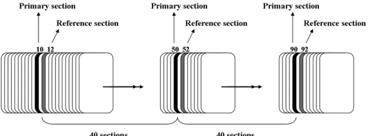
Each pancreas was embedded randomly in parafin and sectioned exhaustively into 5µm-thick sections. Figure
1 illustrates the sampling of sections. Three sections were collected onto each glass slide. With a random start
point among the irst 40 sections, every 40th section was
sampled (the primary sections). In addition, two sections ahead of each primary section were sampled as the
reference section. Because every section was 5 µm thick,
it follows that there was 200 µm between the primary sections and 10 µm between a primary section and the corresponding reference section. All primary and reference sections were stained with hematoxylin and eosin (H & E).
Microscopes and equipment
The sections were analyzed at 400× magniication
onto the monitor so that the microscope projected the image onto the grid. We used two microscopes at the same time for counting the total number of islets in primary and reference sections.
Total volumes of islets and pancreas
Using step-lengths of 950 µm in the x-direction
(Δx) and 750 µm in the y-direction (Δy), all primary sections from each pancreas were systematically
examined. A point-counting grid with 108 points, 1
of them encircled, was applied (Figure 2-A). Moving through all primary sections from the pancreata, the
number of times 1 of the 108 points hit an islet was counted. An islet was deined as a cluster of cells with a
minimum of three visible nuclei displaying the normal characteristics of islet endocrine cells (pale cytoplasm with approximately spherical nuclei). Simultaneously, the number of times the encircled point hit pancreatic tissue (exocrine pancreatic tissue, ducts, vessels, islets, etc.) was counted. The values for the total volume of pancreas and the islets of Langerhans were then calculated based on the Cavalieri principle (Gundersen
et al., 1987; Bock et al., 2003).
1) V (pan) = a/p (pan) × N (p - p) × T × ∑P (pan) = 0.1425 mm3 × ∑P (pan)
where V (pan) is the total volume of pancreas, a/p(pan) is the area per point (in this case Δx×Δ y because only one point in the grid was used to count points that hit pancreas),
N (p – p) is the number of sections between the primary sections (50 sections in this case), T is the section thickness (5 μm), and ∑P(pan) is the total number of points that hit
pancreas.
2) V (isl) = a/p (isl) × N (p - p) × T× ∑P (isl)
= 1.319 × 103 mm3 × ∑P (isl)
where V (isl) is the total volume of islets, a/p (isl) is the area per point (in this case Δx ×Δ y /99 because there were
99 points in the grid used to count points that hit islets),
and ∑P (isl) is the total number of points that hit the islets. Tissue shrinkage influences all stereologic size
estimators whether distance, surface area, or volume. There is no exact unbiased way to obtain information about
tissue deformation during tissue ixation and processing.
The area of a piece of pancreas tissue before and after
fixation/processing may be estimated, and the tissue shrinkage can be calculated as (Nyengaard, 1999):
Total number of islets
In another session, the sampling within the primary sections was performed, but an unbiased counting frame
FIGURE 1- Sampling method of histological sections is shown.
FIGURE 2 - Point counting grade (A) and unbiased counting
L. Khorsandi, F. Nejad-Dehbashi
748
(Figure 2-B) was attached to the monitor. The rules of
the counting frame deine objects completely outside the
frame or objects that touch the exclusion lines (the full lines in Figure 2) as being outside the frame, whereas objects that are completely within the frame or touch only the inclusion lines (the dashed lines in Figure 2)
are deined as being within the frame. We applied the dissector principle (Sterio, 1984) to count the islets. Whenever an islet proile was sampled by the counting
frame, the corresponding position in the reference section was located with the other microscope, and it was then determined whether the islet was also visible in the reference section. An islet was counted if it appeared in the primary section but not in the reference section. Because the sampling of sections, as well as the within
section sampling, were performed with known sampling
fractions, the total number of islets can be calculated according to the fractionator principle (Bock et al., 2003; Gundersen et al., 1987) from:
where N (isl) is the total number of islets in the pancreas,
N (p – p) is the number of sections between the primary sections, N (p – r) is the number of sections between a
primary section and the corresponding reference section
(two in this case), Δ x and Δ y are the step lengths, A(frame) is the area of the counting frame corrected for
magniication (412.674 μm2), and ∑Q- (isl) is the total number of islets counted in one pancreas (Bock et al., 2003).
RNA preparation and Reverse Transcription Polymerase Chain Reaction (RT-PCR)
Isolated pancreases were either used immediately
or snap frozen in liquid nitrogen and stored at −80 °C until use. Using the RNeasy Mini kit (Qiagen), RNA was
isolated from the tissues according to manufacturer’s instructions. RT-PCR was performed using a One Step
RT-PCR kit (Qiagen) which contains reverse transcriptase
to synthesize cDNA from the RNA isolated and DNA polymerase for the PCR. RT-PCR conditions consisted of
a 30 min step at 50 °C to allow the reverse transcriptase activity followed by 15 min at 95 °C to deactivate the
reverse transcriptase and activate the Taq polymerase present in the enzyme mixture. The PCR process consisted
of 6 s at 94 °C (denaturing step), 30 s at the annealing temperature (55 °C), and a 45 s step at 72 °C for extension,
with all steps being repeated for 30 cycles. A final
extension step lasted 10 min at 72 °C.
P r i m e r s e q u e n c e s w e r e a s f o l l o w s w i t h the expected product length: Glut-2, sense 5’ CA G C T G T C T C T G T G C T G C T T G T 3 ’ , a n t i s e n s e 5 ’ GCCGTCATGCTCACATAACTCA3’ (150 bp); Insulin, sense 5’ TCTTCTACACACCCATGTCCC 3’, antisense 5’ GGTGCAGCACTGATCCAC 3’, (149 bp); and GAPDH, sense 5’ CTC TGGTGGACCTCATGGCCTAC 3’, antisense 5’ CAGCAACTGAGGGCCTCTCT 3’ (105
bp) was used as the house keeping gene (Sun et al., 2007).
Statistical analysis
The data were analyzed using one-way ANOVA followed by the Post hoc LSD test and expressed as mean ± SD. P < 0.05 was considered signiicant.
RESULTS
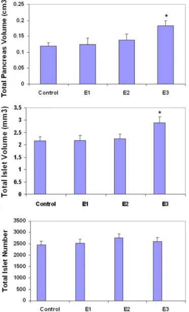
As expected, mean body weight was equal in the four groups. Weight of pancreases in E1 and E2 groups
were similar to the control group. There was a signiicant increase in the relative pancreas weight / body weight in
the E3 group (Figure 3).
The present study conirms a 30% tissue shrinkage in parafin embedding. This shrinkage considered when the inal results were reported.
In the E1 group, total islet volume and total pancreas volume were similar to the control group. Total islet numbers were also similar to the control group. Pancreas tissue showed normal architecture.
In the E2 group, total islet volume and total pancreas volume were slightly increased (P > 0.05). Total islet numbers were similar to the control group. No histopathological changes were observed in this group.
In the E3 group, total pancreas volume was
signiicantly higher than the control group (P < 0.05). Total
FIGURE 3- Relative pancreas weight / body weight in control
and experimental groups. Values expressed as means ± SD for
islet volume was signiicantly increased compared to the
control group (P < 0.05). Total islet numbers were similar to the control group. No histopathological changes were observed in this group. The results for total islet volume, total pancreas volume, and total islet numbers are depicted in Figure 4.
To determine whether EX-4 affects β-cell islet
function, the expression of Glut-2 and insulin genes were assessed using RT-PCR. As illustrated in Figure 5, high expression of Glut-2 and insulin was detected in
EX-4-treated mice. Expression of these genes was markedly
higher in E2 and E3 groups compared to control and E1 groups.
FIGURE 4 - Total pancreas volume, total islet volume and
total islet number of control and experimental groups. Values
expressed as means ± SD for 8 mice. * P < 0.05 compared to control group.
FIGURE 5 - The expression of insulin and Glut-2 genes in
various groups by RT-PCR method is shown.
DISCUSSION
B a s e d o n s t e r e o l o g i c a l m e t h o d s ( s u c h a s fractionator sampling and dissector counting), we found an increase in the total volume of islets in the experimental groups, whereas the total number of islets was equal in the four groups with a reasonably narrow conidence interval for the difference in means.
The importance of GLP-1 for stimulation of islet cell proliferation was originally demonstrated in lean 20-day-old normoglycemic mice (Edvell et al., 1999). Afterwards, several studies using in vivo models showed that GLP-1 can regulate islet growth mainly by controlling β-cell neogenesis (Xu et al., 1999; Stoffers et al., 2000; Perfetti et al., 2000; Tourrel et al., 2001; Tourrel et al.,
2002). Park et al. showed that EX-4 and exercise promotes beta-cell function and mass in islets of diabetic rat. Xu et al. have also reported that EX-4 increases β-cell mass. Fan et al. (1999) reported that EX-4 improves blood glucose control in both young and aging normal non-diabetic mice. The authors showed that EX-4 treatment improved glycemic control in both 3-month and 20 to 22-month-old mice. In both groups of mice, the blood glucose lowering effect was independent of beta cell function as indicated by unchanged beta cell proliferation, insulin secretion or beta cell mass. However, high expression of Insulin 2 and Glut-2 genes in EX-4-treated cells was shown in the
present study. In pancreatic β-cells, the glucose uptake is
controlled by Glut-2, which is essential in the mechanism of glucose-induced insulin secretion (Olson et al., 1996).
Glut-2 is the glucose sensor of β cells that leads to the
L. Khorsandi, F. Nejad-Dehbashi 750
GLP-1 increases insulin secretion and the biosynthesis of
important β-cell products besides insulin: glucokinase and
Glut-2 glucose transporters (Verspohl et al., 2009). The increase in expression of these genes probably induces abnormally elevated secretion of insulin and causes hypoglycemia in non-diabetic animals.
As mentioned above, in this study the volume of islets was higher in EX-4-treated mice. One mechanism responsible for the expansion of islet mass is inhibition of apoptosis (Chen et al., 2011; Farilla et al., 2003; Kwon et al., 2009). It has also been shown that human islets treated with GLP-1 have a down-regulation of caspase-3 at the levels of mRNA of the active protein and up-regulation of the anti-apoptotic protein Bcl-2 (Farilla et al., 2003). A
second mechanism responsible for the expansion of β-cell
mass is enhanced cell proliferation or neogenesis.
Tourrel et al., by using a recognized model of β-cells regeneration (neonatalWistar rats injected with streptozotocin, so-called n0-STZ), showed that GLP-1 and Exendin-4, applied during the neonatal period, strongly stimulated β-cell regeneration mainly by β-cell neogenesis (Tourrel et al., 2001). Furthermore, treatment of diabetic
Goto-Kakizaki (GK) rats with GLP-1 or Exendin-4 from
day 2 to day 6 after birth resulted in stimulation of β-cell neogenesis and proliferation with persistent expansion of β-cell mass detected at adult age (Tourrel et al., 2002). However, the present study revealed that EX-4 caused no change in the number of islets. This indicates that EX-4 has no neogenesis effects on islet cells in non-diabetic adult animals.
It has been stated in a literature review that new islets do develop under certain experimental conditions, such as after partial pancreatectomy, where the formation of new islets has been clearly demonstrated (Nachnani et al., 2010). Other anatomical structures, such as kidney
glomeruli, also lack the ability of hyperplasia and instead
become hypertrophic with an increased demand, probably
because of the highly speciic structure of the neurovascular
and tubular systems necessary for appropriate function. Possibly, the architecture (i.e., the intra-islet vascular structure) of the islets is complex to such a degree that it only allows new islets to be formed during the formation, growth, or regeneration of the pancreas during fetal life or after partial pancreatectomy (Bock et al., 2003).
Nachnani et al. evaluated the histological and biochemical effects of EX-4 on the pancreas in rats. They showed that animals treated with Exendin-4 had pancreatic
acinar inflammation, pyknotic nuclei and weighed signiicantly less than control rats. However, in the present study no evidence of pancreatic acinar inlammation or
histopathological changes were observed.
CONCLUSION
In this study, we demonstrated that EX-4 increased pancreas and islet volume in non-diabetic mice. The increased total islet mass is probably caused by islet hypertrophia without the formation of additional islets. This study also revealed that EX-4 can enhance the expression of insulin and Glut-2 genes, where this may induce hypoglycemia in non-diabetic mice. Further experiments are needed to clarify the exact mechanism of islet hypertrophy induced by EX-4 and other GLP-1 agonists.
ACKNOWLEDGEMENT
This research was supported by a Grant (CM-004) from the research council of the Ahvaz Jundishapur University of Medical Sciences in 2011.
REFERENCES
ABRAHAM, E.J.; LEECH, C.A.; LIN, J.C.; ZULEWSKI,
H.; HABNER, J.F. Insulinotropic hormone glucagon-like
peptide-1 differentiation of human pancreatic islet-derived
progenitor cells into insulin producing cells. Endocrinology,
v.143, p.3152-3161, 2002.
BLONDE, L.; KLEIN, E.J.; HAN, J.; ZHANG, B.; MAC, S.M.; POON, T.H.; TAYLOR, K.L.; TRAUTMANN, M.E.; KIM, D.D.; KENDALL, D.M. Interim analysis of the effects of
exenatide treatment on A1C, weight and cardiovascular risk factors over 82 weeks in 314 overweight patients with type
2 diabetes. Diabetes Obes.Metab., v.8, p.436-447, 2006.
BOCK, T.; PAKKENBERG, B.; BUSCHARD, B.; BUSCHARD, K. Inctrased islet volume but unchanged
islet number in ob/ob mice. Diabetes, v.52, p.1716-1722,
2003.
BRIYAL, S.; GULATI, K.; GULATI, A. Repeated administration of exendin-4 reduces focal cerebral ischemia-induced
infarction in rats. Brain Res., v.1427, p.23-34, 2012.
BUSE, J.B.; HENRY, R.R.; HAN, J.; KIM, D.D.; FINEMAN, M.S.; BARON, A.D. Effects of exenatide (exendin-4)
on glycemic control over 30 weeks in
sulfonylurea-treated patients with type 2 diabetes. Diabetes Care, v.27,
BUSE, J.B.; KLONOFF, D.C.; NIELSEN, L.L.; GUAN, X.; BOWLUS, C.L.; HOLCOMBE, J.H.; MAGGS, D.G.; WINTLE, M.E. Metabolic effects of two years of exenatide
treatment on diabetes, obesity, and hepatic biomarkers in
patients with type 2 diabetes. Clin Ther., v.29, p.139-153,
2007.
CHEN, Y.H.; WANG, J.H.; LI, Z.Q.; YI, Z.H. Effects of
exendin-4 on rat cardiomyocyte apoptosis early after severe
scald injury. Nan. Fang. Yi. Ke. Da Xue.Xue. Bao., v.31,
p.1101-1104, 2011.
DE FRONZO, R.A.; RANTER, R.E.; HAN, J.; KIM, D.D.; FINEMAN, M.S.; BARON, A.D. Effects of exenatide
(exendin-4) on glycemic control and weight over 30 weeks
in metformin-treated patients with type 2 diabetes. Diabetes
Care, v. 28, p.1092-1100, 2005.
DURING, M.J.; CAO, L.; ZUZGA, D.S.; FRANCIS, J.S.; FITZIMONS, H.L.; JIAO, X.; BLAND, R.J.; KLUGMANN, M.; BLANKS, W.A.; DRUCKER, D.J.;
HAILE, C.N. Glucagon-like peptide-1 receptor is involved
in learning and neuroprotection. Nat. Med., v.9,
p.1173-1179, 2003.
EDWEL, A.; LINDSTROM, P.; EDVELL, A.; LINDSTROM, P. Initiation of increased pancreatic islet growth in young
normoglycemic mice (Umea +/?). Endocrinology, v.140,
p.778-783, 1999.
FAN, R.; KANG, Z.; HE, L.; CHAN, J.; XU, G. Exendin-4 improves blood glucose control in both young and aging normal non-diabetic mice, possible contribution of beta cell
independent effects. PloS One,v.6, p.e20443, 2011.
FARILLA, L.; BULOTTA, A.; HIRSHBERG, B.; LI-CALAZI, S.; KHOURY, N.; NOUSHMEHR, H.; BETOLOTTO, C.; DI MARIO, U.; HARLAN, D.M.; PERFETTI, R.
Glucagon-like peptide1 inhibits cell apoptosis and improves
glucose responsiveness of freshly isolated human islets.
Endocrinology, v.144, p.5149-5158, 2003.
GUNDERSEN, H.J. Stereology of arbitrary particles: are view of unbiased number and size estimators and the presentation
of some new ones, in memory of WilliamR. Thompson. J.
Microsc., v.143, n.1, p.3-45, 1986.
GUNDERSEN, H.J.; JENSEN, E.B. The eficiency of
systematic sampling in stereology and its prediction. J.
Microsc., v.14, p.229-263, 1987.
HARDIKAR, A.A.; WANG, X.Y.; WILLIAMS, L.; KWOK, J.; WONG, R.; YAO, M.; TUCH, B.E. Functional maturation
of fetal porcine beta cells by glucagon-like peptide 1 and cholecystokinin. Endocrinology, v.143, p.3505-3514, 2002.
HUI, R.; WRIGHT, C.; PERFETTI, R. Glucagon-like peptide
1 induces differentiation of islet duodenal homeobox-1 positive pancreatic ductal cells into insulin-secretion cells.
Diabetes, v.50, p.785-796, 2001.
KASTIN, A.J.; AKERSTROM, V. Entry of exendin-4 into brain is rapid but may be limited at high doses. Int. J. Obes. Relat. Metab. Disord., v.27, p.313-318, 2003.
KENDALL, D.M.; RIDDLE, M.C.; ROSENSTOCK, J.; ZHUANG, D.; KIM, D.D.; FINEMAN, M.S.; BARON, A.D. Effects of exenatide (exendin-4) on glycemic control
over 30 weeks in patients with type 2 diabetes treated with
metformin and a sulfonylurea. Diabetes Care, v.28,
p.1083-1091, 2005.
KIEFFER, T.J.; MCINTOSH, C.H.; PEDERSON, R.A. Degradation of glucose-dependent insulinotropic
polypeptide and truncated glucagon-like peptide-1 in vitro
and invivo by dipeptidyl peptidase IV. Endocrinology,
v.136, p.3585-3596, 1995.
LOPES DA COSTA, C.; SAMPAIO DE FREITAS, M.; SANCHES MOURA, A. Insulin secretion and
GLUT-2 expression in undernourished neonate rats. J. Nutr.
Biochem., v.15, p.236-241, 2004.
NACHNANI, J.S.; BULCHANDANI, D.G.; NOOKALA, A.; HERNDON, B.; MOLTENI, A.; PANDYA, P.; TAYLOR,
R.; QUINN, T.; WEIDE, L.; ALBA, L.M. Biochemical
and histological effects of exendin-4 (exenatide) on the rat
pancreas. Diabetologia, v.53, p.153-159, 2010.
NYENGAARD, J.R. Stereologic methods and their application
in kidney research. J. Am. Soc. Nephrol., v.10, p.1100-1123, 1999.
OLSON, A.L.; PESSIN, J.E. Structure function, and regulation of the mammalian facilitative glucose transporter gene
family. Annu. Rev. Nutr., v.16, p.235-256, 1996.
PERFETTI, R.; ZHOU, J.; DOYLE, M.E.; EGAN, J. M.
Glucagon-like peptide-1 induces cell proliferation and
pancreatic-duodenum homeobox-1 expression and increases endocrine cell mass in the pancreas of old, glucose-intolerant
L. Khorsandi, F. Nejad-Dehbashi 752
PERRY, T.; HAUGHEY, N.J.; MATTSON, M.P.; EGAN, J.M.; GRIEG, N.H. Protection and reversal of excitotoxic
neuronal damage by glucagon-like peptide-1 and exendin-4.
J. Pharmacol. Exp. Ther., v.302, p.881-888, 2002a.
PERRY, T.; LAHIRI, D.K.; CHEN, D.; ZHOU, J.; SHAW, K.T.; EGAN, J.M.; GRIEG, N.H. A novel neurotrophic property
of glucagon-like peptide 1: a promoter of nerve growth
factor-mediated differentiation in PC12 cells. J. Pharmacol.
Exp. Ther., v.300, p. 958-966, 2002b.
PARK, S.; HONG, S.M.; SUNG, S.R. Exendin-4 and exercise promotes beta-cell function and mass through IRS2 induction in islets of diabetic rats. Life Sci., v.82, p.9-10,
p.503-511, 2008.
RANTA, F.; AYRAM, D.; BERCHTOLD, S.; ADKINS, A.S.; BASU, R.; RIZZA, R.A. Dexamethasone induces cell death in insulin-secreting cells, an effect reversed by exendin-4.
Diabetes, v.55, p.1380-1390, 2006.
STOFFERS, D.A.; KIEFFER, T.J.; HUSSAIN, M.A.; DRUCKER, D.J.; BONNER-WEIR, S.; HABENER, J.F.;
EGAN, J.M. Insulinotropic glucagon-like peptide 1 agonists
stimulate expression of homeodomain protein IDX-1 and
increase islet size in mouse pancreas. Diabetes, v.49,
p.741-748, 2004.
SUN, Y.; ZHANG, L.; GU, H.F.; HAN, W.; REN, M.; WANG, F.; GONG, B.; WANG, L.; GUO, H.; XIN, W.; ZHAO, J.; GAO, L. Peroxisome proliferator-activated
receptor-1 regulates the expression of pancreatic/duodenal
homeobox-1 in rat Insulinoma (INS-1) cells and ameliorates glucose-induced insulin secretion impaired by palmitate.
Endocrinology, v.149, p.662-671, 2007.
TOURREL, C.; BAILBE, D.; LACOME, M.; MEILE, M.J.; KERGOAT, M.; PORTHA, B. Persistent improvement of
type 2 diabetes in the Goto-Kakizaki rat model by expansion
of the beta-cell mass during the prediabetic period with
glucagon-like peptide-1 or exendin-4. Diabetes, v.51, p.1443-1452, 2002.
TOURREL, C.; BAILBE, D.; MEILE, M.J.; KERGOAT, M.;
PORTHA, B. Glucagon-like peptide-1 and exendin-4
stimulate beta cell neogenesis in streptozotocin-treated newborn rats resulting in persistently improved glucose
homeostasis at adult age. Diabetes, v.50, p.1562-1570,
2001.
VELLA, A.; SHAH, P.; REED, A.S.; ADKINS, A.S.; BASU, R.;
RIZZA, R.A. Lack of effect of exendin-4 and glucagon-like
peptide-1-(7,36)-amide on insulin action in non-diabetic
humans. Diabetologia, v.46, p.1589, 2003.
VERSPOHL, E.J. Novel therapeutics for type 2 diabetes:
Incretin hormone mimetics (glucagon-like peptide-1
receptor agonists) and dipeptidyl peptidase-4 inhibitors.
Pharmacol. Ther., v.124, p.113-138, 2009.
XU, G.; STOFFERS, D.A.; HABENER, J.F.; BONNER-WEIR, S. Exendin-4 stimulates both b-Cell replication and neogenesis, resulting in increased b-Cell mass and
improved glucose tolerance in diabetic rats. Diabetologia,
v.48, p.2270-2276, 1999.
ZHOU, J.; WANG, X.; PINEYRO, M.A.; EGAN, J.M.; ZHOU, J.; WANG, Y.; PINEYRO, M.A.; EGAN, J.M.
Glucagon-like peptide 1 and exendin-4 convert pancreatic AR42J cells
into glucagon- and insulin-producing cells. Diabetes, v.48,
p.2358-2366, 1999.
Received for publication on 05th August 2012