cop
yr
ight
© ABE&M todos os dir
eitos r
eser
v
ados
perspectiva
PIERRE PIROT ALESSANDRA K. CARDOZO DÉCIO L. EIZIRIK
Laboratory of Experimental Medicine, Université Libre de Bruxelles (ULB), Brussels, Belgium
Recebido em 19/11/2007 Aceito em 03/12/2007
ABSTRACT
Type 1 diabetes mellitus (T1D) is characterized by severe insulin deficiency resulting from chronic and progressive destruction of pancreatic beta-cells by the immune system. The triggering of autoimmunity against the beta-cells is probably caused by environmental agent(s) acting in the context of a predposing genetic background. Once activated, the immune cells invade the is-lets and mediate their deleterious effects on beta-cells via mechanisms such as Fas/FasL, perforin/granzyme, reactive oxygen and nitrogen species and pro-inflammatory cytokines. Binding of cytokines to their receptors on the beta-cells activates MAP-kinases and the transcription factors STAT-1 and
NFκ-B, provoking functional impairment, endoplasmic reticulum stress and
ultimately apoptosis. This review discusses the potential mediators and
mechanisms leading to beta-cell destruction in T1D. (Arq Bras Endocrinol
Me-tab 2008;52/2:156-165)
Keywords: Pancreatic beta-cell; Diabetes mellitus; Apoptosis; Cytokines; En-doplasmic Reticulum; ER stress
RESUMO
Mecanismos de Destruição e Morte da Célula-beta Pancreática no Diabetes.
O diabetes melito tipo 1 (DM1) tem como característica uma grave deficiência de insulina que resulta da destruição da célula-beta, crônica e progressiva, pelo sistema imune. O desencadeamento da autoimunidade contra a célula-beta é causado, provavelmente, por agentes ambientais que atuam quando existe predisposição genética. Uma vez ativadas, células imunes invadem as ilhotas, e os efeitos deletérios sobre as células-beta são mediados por mecanis-mos relacionados a Fas/FasL, perforina/granzima, espécies reativas de oxigênio e nitrogênio, e a citocinas pró-inflamatórias. A ligação de citocinas a seus receptores na célula-beta ativa MAP-quinase e fatores de transcrição STAT-1 e NFkB, provocando prejuízo funcional, estresse de retículo endoplasmático e, por fim, apoptose. Esta revisão discute os mecanismos e os mediadores
po-tenciais que levam à destruição da célula-beta no DM1. (Arq Bras Endocrinol
Metab 2008;52/2:156-165)
Descritores: Célula-beta pancreática; Diabetes melito; Apoptose; Citocinas; Estresse do retículo endoplasmático
THE GENETIC BACKGROUND OF TYPE 1 DIABETES MELLITUS (T1D)
T
YPE 1 DIABETESMELLITUS (T1D) accounts for about 10-15 % of all cases ofpredisposi-cop
yr
ight
© ABE&M todos os dir
eitos r
eser
v
ados
gens to naïve lymphocytes during their maturation process in the thymus, leading to an inefficient deletion of auto-reactive lymphocytes. In line with this, transge-nic expression of the MHC class II risk allele in mice sensitizes the immune system to beta-cell autoantigens, such as glutamic acid decarboxylase (GAD65) (7). The polymorphisms of the insulin locus that increase diabe-tes risk are characterized by variable numbers of tan-dem repeat (VNTR) regions ~365 bp upstream of the translational start site (3). The number of VNTRs (from 30 to 170 repeats) affects the level of expression of the insulin gene; low amounts of VNTRs (30 to 60 repeats) cause high expression in the pancreas and low expression in the thymus. Low expression of insulin in the thymus may lead to inefficient destruction of self-reactive T-cells during their maturation process (3). The other predisposition loci, PTPN22 and CTLA4, encode for negative regulators of lymphocyte activity. The PTPN22 locus encodes Lyp, a tyrosine phosphata-se that inhibits antigen-specific T-cell activation by de-phosphorylating key components of the T-cell receptor (TCR) signaling cascade (3). CTLA4 is a T-lymphocyte receptor which interacts with a ligand expressed at the surface of B-lymphocytes and dendritic cells, causing inhibition of T-lymphocyte activation (3). The poly-morphisms for those loci associated with T1D are cha-racterized by a loss of function leading to hyperactivity of T-lymphocytes.
ONSET OF T1D: ENVIRONMENTAL TRIGGERS, RECRUITMENT AND ACTIVATION OF IMMUNE CELLS
The earliest sign of autoimmunity against beta-cells, of-ten detectable months or years before the appearance of clinical symptoms, is the presence of circulating anti-bodies against beta-cell antigens (8). These antianti-bodies are used as markers of diabetes risk (2). The most com-mon auto-antibodies in pre-diabetic patients are direct-ed against glutamic acid decarboxylase (GAD65), tyrosine phosphatase-like protein (IA-2) and insulin (IAA). Up to 90 % of newly diagnosed T1D subjects have autoantibodies to one or more of these antigens (8), and autoimmunity detection rates raise to 98% when combining the detection of these antibodies with detection of antibodies against the newly discovered beta-cell autoantigen ZnT8 (9). Epidemiological stud-ies have shown that antibodstud-ies against beta-cell anti-gens can appear after a few months/years of life, tion combines with environmental trigger(s) to induce
the activation of a specific autoimmune destruction of pancreatic beta-cells (1-3).
Several loci, increasing the risk to develop T1D, have been identified (2-5). Among them, the human leukocyte antigen (HLA) locus is by far the most com-mon predisposing polymorphism (2,4). Other well do-cumented predisposing loci include the insulin locus, the cytotoxic T-lymphocyte antigen 4 (CTLA4) locus and the phosphatase non-receptor type 22 (PTPN22) locus (2,3). In addition, a recent genome wide associa-tion study of ~2000 T1D subjects suggested several new candidate loci, encoding for genes involved in im-mune signaling, such as the receptor tyrosine kinase ErbB3 (ERBB3), the SH2B adaptor protein 3 (SH2B3/ LNK), the TRAF-type zinc finger domain containing 1 (TRAFD1) and the protein tyrosine phosphatase non-receptor type 11 (PTPN11) (4,5). Most of these genes are related to the immune system, and may predispose the individual to an exacerbated inflammatory and im-mune response to a given stimulus, potentially increa-sing the risk of autoimmunity. Others, such as the insulin locus and some tyrosine phosphatases, may affect beta-cell function and antigen presentation.
self-anti-cop
yr
ight
© ABE&M todos os dir
eitos r
eser
v
ados
suggesting that the autoimmune process is triggered early in life (2,8). This indicates that the pool of self-reactive naïve T-cells stays under the control of the im-mune system for several years, with many antibody positive individuals never developing insulitis nor dia-betes (10). External events may therefore be required for the activation and expansion of the auto-reactive naïve T-cells. Suspected environmental triggers include viral infection, dietary factors during infancy (e.g. cow milk), vaccination, toxins and the physiological beta-cell turnover occurring in the newborn (11,12). Accu-mulating evidence points to viral infections by enterovirus, rubella, mumps, rotavirus, parvovirus or cytomegalovirus as the most probable candidates for triggering beta-cell autoimmunity (13). Particular at-tention is given to the enteroviruses which have a spe-cific tropism for the pancreas (13). Epidemiological studies investigating the seasonal appearance of beta-cell autoantibodies in a group of risk individuals showed preferential emergence of the self-antibodies during the cold season, correlating with a period of increased enterovirus infections (11). Enterovirus of the coxsakie family, isolated from diabetic patients, were able to in-duce diabetes when injected into susceptible mice (13). More recently, a coxsackie B4 virus was isolated from three out of six samples obtained from T1D patients. This virus was able to infect human islets from non-di-abetic patients in vitro, causing impaired glucose
stim-ulated insulin release (14).
The process by which enteroviruses activate autore-active T-lymphocytes remain elusive, but possible me-chanisms include release of beta-cell antigens via non-T-cell mediated beta-cell cytolysis, expression of vi-ral antigens at the surface of the beta-cells (thus breaking the self tolerance) and activation of self-reactive T-lym-phocytes due to molecular mimicry between viral pro-teins and beta-cell antigens (13). Once activated, the autoreactive T-lymphocytes must be recruited to the pancreatic islets to cause diseases. The initiator events of islet inflammation process, i.e. insulitis, remain to be elu-cidated; this is, at least in part, secondary to the scarcity of human pancreatic samples available for research, and the fact that this material is usually obtained via autopsy or pancreatic biopsy (an ethically debatable practice) of patients with established T1D, when most beta-cells have already been destroyed (15,16). Thus, most of our present knowledge of the early pathogenesis of T1D is based on observations in animal models of T1D, such as the non-obese diabetic (NOD) mice, the BioBreeding
(BB) rat (17) and the more recently established LEW.1AR1/Ztm-iddm rat (18).
The process of T-cell recruitment to the pancreatic islets is not fully understood but it may involve local production of chemokines by specialized APCs, such as the resident dendritic cells (DC) and macrophages, en-dothelial cells and the beta-cells themselves (19) (Figu-re 1A, C, D). Chemokines belong to the cytokine family. They are a group of low molecular weight pep-tides which recruit, activate and co-stimulate cells of the immune system (19). The cytokines TNF-α and IFN-γ induce the release of chemokines by resident macrophages and endothelial cells (19). Transgenic ex-pression of the chemokine macrophage chemoattrac-tant protein-1 (MCP-1; also called CCL2) under the control of the rat insulin promoter induces monocyte infiltration in pancreatic islets (20), while NOD mice deficient for the macrophage inflammatory protein-1α (MIP-1α) has reduced destructive insulitis and are pro-tected against diabetes (21). Transgenic RIP-GP mou-se (a virus-triggered diabetes moumou-se model) deleted for CXC3R, the receptor for the IFN-γ-induced chemoki-nes Mig, IP10 and I-TAC have decreased the incidence of diabetes upon infection with lymphocyte chroriome-ningitidis virus (LCMV) (19). The beta-cells themsel-ves secrete chemokines following viral infection or exposure to cytokines. Exposure of rat beta-cells to IFN-γ in combination with double stranded RNA (a treatment which mimics some of the components of a viral infection) leads to up-regulation of several che-mokines, including IP-10, fractalkine, MCP-1, MIP3α, CINC-1 and RANTES (22). Moreover, the infection of human pancreatic islets with the coxsackie virus B5 induces the expression of the chemokines fractalkine, MIP-2α, MIP-2β, GRO-γ, ENA-78, IP-10 and ITAC (23), suggesting the presence of a virus-triggered “dia-logue” between beta-cells and the immune system.
secre-cop
yr
ight
© ABE&M todos os dir
eitos r
eser
v
ados
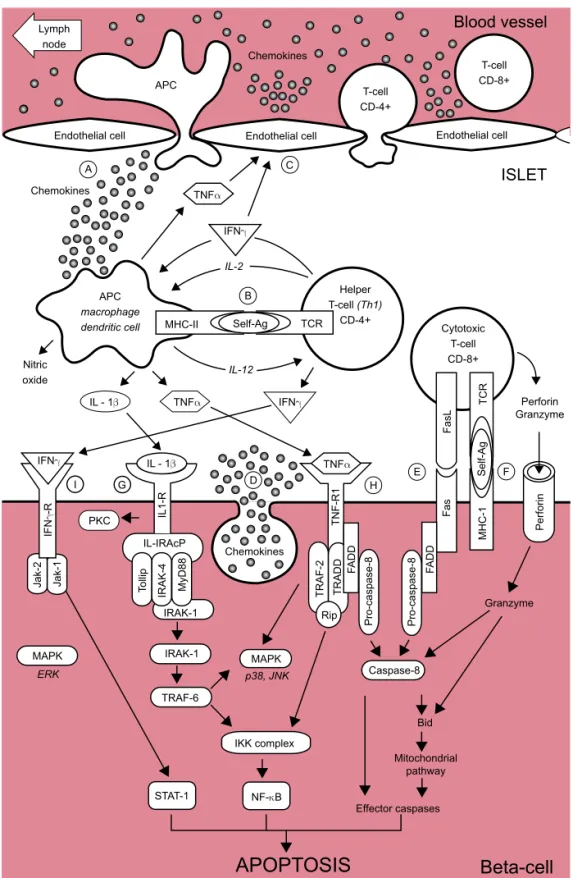
Figure 1. Schematic representa-tion of the auto-immune attack to the beta-cells in T1DM. (A) At the early stages of insulitis, activated local antigen present-ing cells (APC) recruit and acti-vate CD4+ helper T-cells via migration to the pancreatic lymph node, presentation of be-ta-cells antigens and release of chemokines/cytokines.
(B) CD4+ helper T-cells, in turn, stimulate APC secretion of cy-tokines and nitric oxide. (C) Cytokines induce the secre-tion of chemokines by endothe-lial cells which enhance the recruitment of immune cells into the islets and, together with cy-tokines, activate CD8+ cytotoxic T-cells.
(D) The beta-cell themselves also secrete chemokines in response to viral infection or cytokines, fur-ther enhancing the recruitment and activation of immune cells. Activated CD8+ cytotoxic T-cells, in turn, induce beta-cell apopto-sis via
(E) the Fas pathway and (F) the granzyme/perforin sys-tem. Cytokines also bind to re-ceptors at the surface of beta-cells:
(G) Interleukin-1β (IL-1β) acti-vates NF-κB and the kinases PKC, p38 and JNK.
(H) Tumor necrosis factor α (TNFα) activates caspase-8, NF-κB and the MAPK p38 and JNK.
(I) Interferon γ (IFNγ) activates Stat-1 and the kinase ERK. More detailled information is provided in the text.
tion of interleukin-12 (12,24) (Figure 1A). The diffe-rentiated Th1 CD4+ T-cells, in turn, secrete IL-2 and IFN-γ which further stimulate APC to secrete other cytokines, such as IL-1β and TNF-α and free radicals, such as nitric oxide (NO). The secreted cytokines
pro-mote the migration of CD8+ cytotoxic T-cells into the islets and stimulate the beta-cells to release chemokines and IL-15 which further augment attraction and acti-vation of immune cells (25).
APOPTOSIS
Beta-cell
Effector caspases Mitochondrial pathway Bid Granzyme Caspase-8 MAPK p38, JNKNF-κB STAT-1 IKK complex TRAF-6 IRAK-1 IRAK-1 ERK MAPK Ja k-2 Ja k-1 PKC Pro -ca sp a se -8 Pro -ca sp a se -8 F AD D F a s MH C -1 Pe rf o ri n Perforin Granzyme F a sL T C R F E H D T R AD D F AD D T R AF -2 Rip Chemokines T N F -R 1 TNFα TNFα TNFα
IL - 1β IL - 1β
IF N γ -R IFNγ IL-12 TCR Helper T-cell (Th1)
CD-4+ Cytotoxic T-cell CD-8+ IL-2 ISLET C A B I G Nitric oxide IL 1 -R T o lli p IR AK-4 IL-IRAcP MyD 8 8 Chemokines Chemokines IFNγ MHC-II Self-Ag Se lf -Ag APC macrophage dendritic cell APC Blood vessel T-cell CD-4+ T-cell CD-8+
Endothelial cell Endothelial cell Endothelial cell Lymph
node
cop
yr
ight
© ABE&M todos os dir
eitos r
eser
v
ados
MEDIATORS OF BETA-CELL DESTRUCTION IN T1D
As already discussed above, T1D is a slowly-progressing autoimmune disease, and the process causing beta-cell death in humans usually evolves over several years. Clinical symptoms of diabetes are usually present once more than 70% of the beta-cell population is destroyed (26). The main form of beta-cell death observed in T1D rodent models and in human islets from T1D pa-tient is apoptosis (26,27). The mechanisms of beta-cell destruction in T1D have not yet completely clarified, but probably involve the following: 1) Expression of Fas ligand (FasL) and its receptor Fas at the surface of the activated CD8+ T-cells and pancreatic beta-cells re-spectively; 2) Release of perforin and granzyme by acti-vated CD8+ T-lymphocytes; 3) Secretion of cytokines, including interleukin-1β (IL-1β), tumor necrosis factor α (TNF-α) and interferon-γ (IFN-γ), by the diverse im-mune cells infiltrating the islet; 4) Production of reac-tive oxygen species such as nitric oxide (NO) by macrophages, dendritic cells and the beta-cells them-selves (24,26,27).
Fas, also known as APO-1 or CD95, is a member of the TNF receptor surperfamily that is activated via binding of FasL (Figure 1E) (12,27). Fas and FasL were detected respectively at the surface of beta-cells and on a high percentage of T-cells infiltrating the islet (28). In addition, it was shown that Fas expression can be induced in rodent beta-cells upon stimulation with IFN-γ and IL-1β (28). Once activated, Fas trimerizes and recruits the Fas-associated death domain (FADD) protein at its cytoplasmic part (12,27). FADD then re-cruits pro-caspase-8 leading to its activation by auto-cleavage. Activated caspase-8 subsequently cleaves the effector caspase-3 and/or activates the mitochondrial death pathway via cleavage of the BH3 protein Bid. The role of Fas in T-cell mediated beta-cell destruction
in vivo has remained controversial for a long time, but
targeted overexpression of a dominant negative form of Fas (FADD) in beta-cells delays the onset of diabetes in NOD mice, implicating a role for Fas in the early stages of autoimmune beta-cell destruction (24,29,30).
Perforin and granzyme are contained in granules inside the CD8+ T-cells (12). These cytotoxic molecu-les are probably released in the extra-cellular milieu via exocytosis in response to TCR recognition of self-Ag presented at the surface of beta-cells by MHC class-I molecules (Figure 1F). Perforin is involved in pore
for-mation across the membranes via a Ca2+ dependent
me-chanism. This pore enables the entry of the serine protease granzyme inside the cell, causing the cleavage and activation of several targets, such as effector caspa-ses and the BH3 only protein Bid. Several studies point to the perforin/granzym system as an important me-diator of T-cell-induced beta-cell death. Thus, perforin deficient RIP-LCMV mice (a virus-triggered diabetes mouse model) are protected against diabetes upon in-fection with lymphocytic choriomeningitis virus (LCMV), while NOD mice lacking the perforin gene develop severe insulitis but rarely became diabetic (12,24).
In addition to these two CD8+ specific killing me-chanisms, beta-cell death can also be induced by pro-inflammatory cytokines such as IL-1β, TNF-α and IFN-γ (26,27). These pro-inflamatory cytokines are detectable during early insulitis in NOD mice and BB rats (24,27).
cop
yr
ight
© ABE&M todos os dir
eitos r
eser
v
ados
(PKCδ) via an IRAK-independent mechanism which might involve the up-regulation of phospholypase D-1 (27). Evidence for the role of IL-1β in in vivo beta-cell
killing was provided in IL-1R deficient NOD mice, which present a delayed onset of diabetes (34). Moreo-ver, blocking IL-1 signaling with an IL-1 receptor an-tagonist delays the onset of diabetes in NOD mice, prevents the induction of diabetes by multiple low doses of streptozotocin, prolongs mouse islet allograft survival and prevents the recurrence of hyperglycemia in diabetic NOD mice following islet transplantation (27). Besides its pro-apoptotic effects, IL-1β also affects beta-cell function. Thus, IL-1β induces an early release of insulin followed by a progressive inhibition of glucose-stimula-ted insulin secretion in rodent beta-cells (35). This latest effect of IL-1β is due to a decrease in glucose metabo-lism and a reduction in the number of insulin granules docked to the plasma membrane (35,36). In human beta-cells IL-1β, in combination with IFN-γ, impairs conversion of pro-insulin into mature insulin and, after 3-7 days, triggers beta-cell apoptosis (37,38).
The activation of the TNF receptor 1 (TNF-R1) upon TNF-α binding leads to its trimerization and re-cruitment of the adaptator protein TNF receptor-asso-ciated death domain protein (TRADD) (27) (Figure 1H). TRADD, in turn, recruits TRAF-2 and the serine threonine kinase Rip. Similar to TRAF-6, TRAF-2 acti-vates both the NF-κB and MAPK pathways. TRAF-2 acts in cooperation with Rip to induce NF-κB via acti-vation of the IKK complex. TRAF-2 probably activates the MAPK pathway in a similar way as TRAF-6, since it interacts with the MAP3Ks Ask-1 and MEKK-1 (27). TNF-α preferentially phosphorylates and activates p38 and JNK in rat insulinoma cells (33). In addition to TRAF-2-mediated events, the TNF-R1 recruits FADD which in turn leads to pro-caspase-8 recruitment and activation. Caspase-8 then activates effector caspases in the same way as described above for the Fas signaling pathway (27). An important role for TNF-α in beta-cell killing was demonstrated in TNF-R1 null mutant NOD mice, which fail to develop spontaneous diabetes (24). In line with this, the development of diabetes in NOD mice by T-cell adoptive transfer is blocked by anti-TNF-α antibodies (39).
IFN-γ binding to its receptor induces its oligomeri-zation and the cytoplasmic recruitment of two mem-bers of the Janus kinase (Jak) family, Jak1 and Jak2 (27) (Figure 1I). Once activated by transphosphorylation, Jak1 and 2 recruit Stat-1 and trigger its activation by
phosphorylation. Stat-1 then homodimerizes and mi-grates to the nucleus where it regulates the expression of genes containing γ-activated sequence (GAS) ele-ments in their promoter, such as caspases, Fas and iNOS for instance (40). In addition to the Stat-1 pa-thway, the Jaks also activate a member of the MAPK family, namely the extracellular signal-regulated kinase (ERK) (27). Experiments using IFN-R null mutant NOD mice has provided discordant results, with one study showing a marked inhibition of insulitis and dia-betes while the other has not detected changes in the prevalence of insulitis and diabetes (41,42). Recent stu-dies indicate that blocking Stat-1, the main signaling pathway of IFN-γ, prevents multiple-low dose strepto-zotocin-induced diabetes by an action at the beta-cell level (43,44).
cop
yr
ight
© ABE&M todos os dir
eitos r
eser
v
ados
their binding capacity (48). In line with this, nearly 50% of the cytokine-modified genes in insulin-produ-cing cells, as observed by microarray analysis, are se-condary to NO formation (50).
Of note, NO seems to have a less relevant role for cytokine-induced beta cell death in humans and mice as compared to rats. Thus, iNOS blockers do not prevent IL-1β + IFN-γ + TNF-α-induced human beta cell DNA strand breaks and death (38,47), while islets obtained from an iNOS KO mouse are only partially protected against death induced by IL-1β + IFN-γ (51).
DOWNSTREAM MECHANISMS OF CYTOKINE-INDUCED BETA-CELL DEATH: A ROLE FOR ER STRESS?
The downstream mechanisms involved in cytokine-in-duced beta-cell death still remain to be fully clarified. Available evidence suggests a role for mitochondrial mediators of apoptosis, NO, oxygen free radicals and endoplasmic reticulum (ER) stress, among others. These mechanisms have been addressed in previous re-views from our group (26,52), and we will presently and briefly discuss the role for ER stress.
The endoplasmic reticulum (ER) is the organelle re-sponsible for synthesis and folding of secreted and mem-branous protein and lipid biosynthesis. It also functions as one of the main cellular calcium stores. Alterations of ER homeostasis lead to accumulation of misfolded pro-teins and activation of an adaptive response named the unfolded protein response (UPR) (52,53). The UPR is transduced via 3 ER transmembrane proteins, namely PERK, IRE-1 and ATF6. The signaling cascades acti-vated downstream of these proteins: a) induce expres-sion of ER resident chaperones and protein foldases, increasing the protein folding capacity of the ER; b) at-tenuate general protein translation which avoid over-loading the stressed ER with new proteins; c) upregulate ER-associated degradation (ERAD) genes, which de-creases the unfolded protein load of the ER (52,53). In severe cases, failure by the UPR to solve the ER stress leads to apoptosis. The mechanisms linking ER stress to apoptosis are still poorly understood, but potential me-diators include the transcription factors Chop and ATF3, pro-apoptotic members of the Bcl-2 familly, the caspase 12 and the kinase JNK (52-54).
As discussed above, cytokines contribute to beta-cell autoimmune destruction in T1D. Cytokines, via
NO formation, induce a 50% decrease in the expression of the ER calcium pump SERCA2 pump, depleting ER Ca2+ and triggering ER stress (45). In line with this,
SNAP, a chemical NO donor, increases beta-cell cyto-solic Ca2+ concentrations secondary to the release of
calcium from the ER depletion, while Chop knock-out islets are resistant to the toxic effects of SNAP (55). The molecular mechanisms leading to SERCA2 down-regulation in response to NO are still unknown. NO inhibits the binding of zinc finger transcription factors (48), and the SERCA2 promoter contains a conserved binding site for the zinc finger transcription factor SP-1. SP-1 is involved in the basal expression of SER-CA2 in cardiomyocytes and C2C12 muscular cell line; inhibition of SP1 activity by NO may thus contribute for the observed decrease in SERCA2 mRNA expres-sion (56). Besides transcriptional regulation, NO also affects the function of SERCA2 at a post-transcriptio-nal level, and nitration inhibits SERCA2 pump activity (48). IL-1β + IFN-γ-induced ER stress is characterized by eIF2α phosphorylation and subsequent induction of ATF4, activation of Xbp-1 alternative splicing and up-regulation of the pro-apoptotic transcription factor Chop (45,57). Cytokine-induced ER stress in beta-cells seems to be defective for the activation of the ATF-6 branch of the UPR, as suggested by a failure to activate an UPRE luciferase construct which responds to ATF-6 and Xbp-1 and lack of BiP (an ATF6 target) induction. On the other hand, free fatty acids and two chemical ER stress inducers (i.e. thapsigargin and cyclo-piazonic acid) activate all three UPR pathways in beta-cells, including ATF6 (45,58,59,60). This partial induction of ER stress, with defective up-regulation of the chaperone BiP (which may play a protective role against beta-cell death) and the effects of IFN-γ (see below), may explain why beta-cells are particularly sen-sitive to cytokine-induced apoptosis.
cop
yr
ight
© ABE&M todos os dir
eitos r
eser
v
ados
Direct evidence of ER stress markers expression in beta-cells from diabetic patients is still limited. Immu-nostaining of islets from T2D patients (autopsy mate-rial) showed an increased expression of Chop, BiP and p58 in comparison to non-diabetic control subjects (61,58). Another recent study investigated the perinu-clear expression of Chop protein and its nuperinu-clear trans-location in beta-cells obtained from autopsy of T1D and T2D patients (62). The perinuclear expression of Chop was increased in beta-cells from T2D and obese non-diabetic patients when compared to lean non-dia-betic patients. The presence of Chop in the nucleus was observed 6 times more frequently in T2D than in obe-se non-diabetic patients. T1D samples rarely preobe-sented expression of Chop around the nucleus and the fre-quency of Chop positive beta-cell nuclei was compara-ble to the control group. The authors thus concluded that apoptosis is initiated by different mechanisms in T1D and T2D and that ER stress is probably not invol-ved in the initiation of beta-cell apoptosis in T1D. The main limitation of this study is that nuclear Chop is not a specific marker of ER stress, since Chop can be indu-ced independently of ER stress by other stress stimuli (52). Moreover, insulitis is heterogenous in the pancre-as of T1D patients, and another study detected ATF3 expression (similarily to Chop ATF3 is also induced do-wnstream of ATF4) only in islets affected by insulitis (54). Specific determination of Chop expression in is-lets affected by insulitis was not performed in the study by Huang and co-workers (62). Additional studies are thus required to evaluate the involvement of ER stress in human T1D pathogenesis.
The evidence described in the present review, and in the other articles of this issue, indicates that our kno-wledge of the pathogenesis of T1D has greatly increa-sed in recent years. Unfortunately this increaincrea-sed knowledge has not yet been translated into safe and innovative therapies to prevent the disease. Prevention of human T1D will probably require interventions at multiple levels, including “re-education” of the immu-ne system to render it tolerant to the beta-cells, stimu-lation of beta-cell defense/regeneration and prevention of the activation of pro-apoptotic signaling inside them. This is an enormous task, which will require concerted and long-term efforts by basic and clinical researchers. For this purpose, it is imperative that we redouble our efforts to better understand the mechanisms of beta-cell death in diabetes and, based on this knowledge, succeed in designing rational and targeted therapies to prevent beta-cell loss.
ACKNOWLEDGEMENTS
We apologize for not quoting many relevant original papers due to space limitations in the reference list, we quoted preferentially review articles. Work by the au-thors mentioned in the article was supported by grants from the European Union (STREP Savebeta, contract n° 036903; in the Framework Programme 6 of the Eu-ropean Community), the Fonds National de la Recher-che Scientifique (FNRS), Actions de ReRecher-cherRecher-che Concertées de la Communauté Française, Belgium, the Belgium Program on Interuniversity Poles of Attrac-tion initiated by the Belgian State (IUAP P5/17 and P6/40) and the Juvenile Diabetes Foundation Interna-tional (JDRF). Alessandra K. Cardozo is presently a ResearchAssociate of the Fonds National de la Recher-che Scientifique(FNRS; Belgium).
REFERENCES
1. Daneman D. Type 1 diabetes. Lancet. 2006;367:847-58. 2. Jahromi MM, Eisenbarth GS. Cellular and molecular
pathoge-nesis of type 1A diabetes. Cell Mol Life Sci. 2007;64:865-72. 3. Kim MS, Polychronakos C. Immunogenetics of type 1
diabe-tes. Horm Res. 2005;64:180-8.
4. Todd JA, Walker NM, Cooper JD, Smyth DJ, Downes K, Plag-nol V et al. Robust associations of four new chromosome re-gions from genome-wide analyses of type 1 diabetes. Nat Genet. 2007;447:661-78
5. The Wellcome Trust Case Control Consortium. Genome-wide association study of 14,000 cases of seven common diseases and 3,000 shared controls. Nature. 2007;447:661-78.
6. Turner D. The human leucocyte antigen (HLA) system. Vox Sang. 2004;87(Suppl1):87-90.
7. Wong FS,Wen L. What can the HLA transgenic mouse tell us about autoimmune diabetes? Diabetologia. 2004;47:1476-87. 8. Pihoker C, Gilliam LK, Hampe CS, Lernmark A. Autoantibodies
in diabetes. Diabetes. 2005;54:S52-61.
9. Wenzlau JM, Juhl K, Yu L, Moua O, Sarkar SA, Gottlieb P et al. The cation efflux transporter ZnT8 (Slc30A8) is a major au-toantigen in human type 1 diabetes. Proc Natl Acad Sci USA. 2007;104:17040-5.
10. In't Veld P, Lievens D, De Grijse J, Ling Z, Van der Auwera B, Pipeleers-Marichal M et al. Screening for insulitis in adult au-toantibody-positive organ donors. Diabetes. 2007;56:2400-4. 11. Knip M, Veijola R, Virtanen SM, Hyoty H, Vaarala O, Akerblom
HK. Environmental triggers and determinants of type 1 diabe-tes. Diabediabe-tes. 2005;54(Suppl2):S125-36.
12. Pearl-Yafe M, Kaminitz A, Yolcu ES, Yaniv I, Stein J, Askenasy N. Pancreatic islets under attack: cellular and molecular effec-tors. Curr Pharm Des. 2007;13:749-60.
13. van der Werf N, Kroese FG, Rozing J, Hillebrands JL. Viral in-fections as potential triggers of type 1 diabetes. Diabetes Me-tab Res Rev. 2007;23:169-83.
natu-cop
yr
ight
© ABE&M todos os dir
eitos r
eser
v
ados
ral killer cell insulitis in recent-onset type 1 diabetic patients. Proc Natl Acad Sci USA. 2007;104:5115-20.
15. Imagawa A, Hanafusa T, Tamura S, Moriwaki M, Itoh N, Yama-moto K et al. Pancreatic biopsy as a procedure for detecting in situ autoimmune phenomena in type 1 diabetes: close correla-tion between serological markers and histological evidence of cellular autoimmunity. Diabetes. 2001;50:1269-73.
16. Uno S, Imagawa A, Okita K, Sayama K, Moriwaki M, Iwahashi H et al. Macrophages and dendritic cells infiltrating islets with or without beta-cells produce tumour necrosis factor-alpha in patients with recent-onset type 1 diabetes. Diabetologia. 2007;50:596-601.
17. Yang Y, Santamaria P. Lessons on autoimmune diabetes from animal models. Clin Sci (Lond). 2006;110:627-39.
18. Lenzen S, Tiedge M, Elsner M, Lortz S, Weiss H, Jorns A et al. The LEW.1AR1/Ztm-iddm rat: a new model of spontaneous insulin-dependent diabetes mellitus. Diabetologia. 2001;44:1189-96.
19. Rotondi M, Chiovato L, Romagnani S, Serio M, Romagnani P. Role of chemokines in endocrine autoimmune diseases. Endo-cr Rev. 2007;28 :492-520
20. Grewal IS, Rutledge BJ, Fiorillo JA, Gu L, Gladue RP, Flavell RA et al. Transgenic monocyte chemoattractant protein-1 (MCP-1) in pancreatic islets produces monocyte-rich insulitis without diabetes: abrogation by a second transgene expressing sys-temic MCP-1. J Immunol. 1997;159:401-8.
21. Cameron MJ, Arreaza GA, Grattan M, Meagher C, Sharif S, Burdick MD et al. Differential expression of CC chemokines and the CCR5 receptor in the pancreas is associated with pro-gression to type I diabetes. J Immunol. 2000;165:1102-10. 22. Liu D, Cardozo AK, Darville MI, Eizirik DL. Double-stranded
RNA cooperates with interferon-gamma and IL-1 beta to indu-ce both chemokine expression and nuclear factor-kappa B-dependent apoptosis in pancreatic beta-cells: potential mechanisms for viral-induced insulitis and beta-cell death in type 1 diabetes mellitus. Endocrinology. 2002;143:1225-34. 23. Ylipaasto P, Kutlu B, Rasilainen S, Rasschaert J, Salmela K,
Teerijoki H et al. Global profiling of coxsackievirus- and cytoki-ne-induced gene expression in human pancreatic islets. Dia-betologia. 2005;48:1510-22.
24. Yoon JW, Jun HS. Autoimmune destruction of pancreatic be-ta-cells. Am J Ther. 2005;12:580-91.
25. Cardozo AK, Proost P, Gysemans C, Chen MC, Mathieu C, Eizirik DL. IL-1beta and IFN-gamma induce the expression of diverse chemokines and IL-15 in human and rat pancreatic is-let cells, and in isis-lets from pre-diabetic NOD mice. Diabetolo-gia. 2003 ;46:255-66.
26. Cnop M, Welsh N, Jonas JC, Jorns A, Lenzen S, Eizirik DL. Mechanisms of pancreatic beta-cell death in type 1 and type 2 diabetes: many differences, few similarities. Diabetes. 2005;54(Suppl 2):S97-107.
27. Eizirik DL, Mandrup-Poulsen T. A choice of death - the signal-transduction of immune-mediated beta-cell apoptosis. Diabe-tologia. 2001;44:2115-33.
28. Kawasaki E, Abiru N, Eguchi K. Prevention of type 1 diabetes: from the view point of beta-cell damage. Diabetes Res Clin Pract. 2004;66(Suppl 1):S27-32.
29. Allison J, Thomas HE, Catterall T, Kay TW, Strasser A. Trans-genic expression of dominant-negative Fas-associated death domain protein in beta-cells protects against Fas ligand-indu-ced apoptosis and reduces spontaneous diabetes in non-obe-se diabetic mice. J Immunol. 2005;175:293-301.
30. Savinov AY, Tcherepanov A, Green EA, Flavell RA, Chervonsky AV. Contribution of Fas to diabetes development. Proc Natl Acad Sci USA. 2003;100:628-32.
31. Ortis F, Cardozo AK, Crispim D, Storling J, Mandrup-Poulsen T, Eizirik DL. Cytokine-induced proapoptotic gene expression in insulin-producing cells is related to rapid, sustained, and no-noscillatory nuclear factor-kappaB activation. Mol Endocrinol. 2006;20:1867-79.
32. Eldor R, Yeffet A, Baum K, Doviner V, Amar D, Ben-Neriah Y et al. Conditional and specific NF-kappaB blockade protects pan-creatic beta-cells from diabetogenic agents. Proc Natl Acad Sci USA. 2006;103:5072-7.
33. Saldeen J, Lee JC, Welsh N. Role of p38 mitogen-activated protein kinase (p38 MAPK) in cytokine-induced rat islet cell apoptosis. Biochem Pharmacol. 2001;61:1561-9.
34. Thomas HE, Irawaty W, Darwiche R, Brodnicki TC, Santamaria P, Allison J et al. IL-1 receptor deficiency slows progression to diabetes in the NOD mouse. Diabetes. 2004;53:113-21. 35. Sandler S, Eizirik DL, Svensson C, Strandell E, Welsh M, Welsh
N. Biochemical and molecular actions of interleukin-1 on pan-creatic beta-cells. Autoimmunity. 1991;10:241-53.
36. Ohara-Imaizumi M, Cardozo AK, Kikuta T, Eizirik DL, Nagamat-su S. The cytokine interleukin-1beta reduces the docking and fusion of insulin granules in pancreatic beta-cells, preferen-tially decreasing the first phase of exocytosis. J Biol Chem. 2004;279:41271-4.
37. Hostens K, Pavlovic D, Zambre Y, Ling Z, Van Schravendijk C, Eizirik DL et al. Exposure of human islets to cytokines can re-sult in disproportionately elevated proinsulin release. J Clin Invest. 1999;104:67-72.
38. Delaney CA, Pavlovic D, Hoorens A, Pipeleers DG, Eizirik DL. Cytokines induce deoxyribonucleic acid strand breaks and apoptosis in human pancreatic islet cells. Endocrinology. 1997;138:2610-4.
39. Suk K, Kim S, Kim YH, Kim KA, Chang I, Yagita H et al. IFN-gam-ma/TNF-alpha synergism as the final effector in autoimmune diabetes: a key role for STAT1/IFN regulatory factor-1 pathway in pancreatic beta-cell death. J Immunol. 2001;166:4481-9. 40. Battle TE, Frank DA. The role of STATs in apoptosis. Curr Mol
Med. 2002;2:381-92.
41. Wang B, Andre I, Gonzalez A, Katz JD, Aguet M, Benoist C et al. Interferon-gamma impacts at multiple points during the progression of autoimmune diabetes. Proc Natl Acad Sci USA. 1997;94:13844-9.
42. Thomas HE, Parker JL, Schreiber RD, Kay TW. IFN-gamma ac-tion on pancreatic beta-cells causes class I MHC upregulaac-tion but not diabetes. J Clin Invest. 1998;102:1249-57.
43. Gysemans CA, Ladriere L, Callewaert H, Rasschaert J, Flamez D, Levy DE et al. Disruption of the gamma-interferon signaling pathway at the level of signal transducer and activator of transcription-1 prevents immune destruction of beta-cells. Diabetes. 2005;54:2396-403.
44. Callewaert HI, Gysemans CA, Ladriere L, D'Hertog W, Hagen-brock J, Overbergh L et al. Deletion of STAT-1 pancreatic islets protects against streptozotocin-induced diabetes and early graft failure but not against late rejection. Diabetes. 2007;56:2169-73.
45. Cardozo AK, Ortis F, Storling J, Feng YM, Rasschaert J, Tonne-sen M et al. Cytokines downregulate the sarcoendoplasmic reticulum pump Ca2+ ATPase 2b and deplete endoplasmic
reti-culum Ca2+, leading to induction of endoplasmic reticulum
cop
yr
ight
© ABE&M todos os dir
eitos r
eser
v
ados
46. Darville MI, Eizirik DL. Regulation by cytokines of the inducible nitric oxide synthase promoter in insulin-producing cells. Dia-betologia. 1998;41:1101-8.
47. Eizirik DL, Sandler S, Welsh N, Cetkovic-Cvrlje M, Nieman A, Geller DA et al. Cytokines suppress human islet function irres-pective of their effects on nitric oxide generation. J Clin Invest. 1994;93:1968-74.
48. Pacher P, Beckman JS, Liaudet L. Nitric oxide and peroxynitri-te in health and disease. Physiol Rev. 2007;87:315-424. 49. Welsh N, Eizirik DL, Bendtzen K, Sandler S. Interleukin-1
beta-in-duced nitric oxide production in isolated rat pancreatic islets re-quires gene transcription and may lead to inhibition of the Krebs cycle enzyme aconitase. Endocrinology. 1991;129:3167-73. 50. Kutlu B, Cardozo AK, Darville MI, Kruhoffer M, Magnusson N,
Orntoft T et al. Discovery of gene networks regulating cytoki-ne-induced dysfunction and apoptosis in insulin-producing INS-1 cells. Diabetes. 2003;52:2701-19.
51. Liu D, Pavlovic D, Chen MC, Flodstrom M, Sandler S, Eizirik DL. Cytokines induce apoptosis in beta-cells isolated from mice lacking the inducible isoform of nitric oxide synthase (iNOS-/-). Diabetes. 2000;49:1116-22.
52. Eizirik DL, Cardozo AK, Cnop M. The role for endoplasmic reti-culum stress in diabetes mellitus. Endocr Rev. 2008;in press: 53. Ron D, Walter P. Signal integration in the endoplasmic
reticu-lum unfolded protein response. Nat Rev Mol Cell Biol. 2007;8:519-29.
54. Hartman MG, Lu D, Kim ML, Kociba GJ, Shukri T, Buteau J et al. Role for activating transcription factor 3 in stress-induced beta-cell apoptosis. Mol Cell Biol. 2004;24:5721-32.
55. Oyadomari S, Takeda K, Takiguchi M, Gotoh T, Matsumoto M, Wada I et al. Nitric oxide-induced apoptosis in pancreatic beta-cells is mediated by the endoplasmic reticulum stress pa-thway. Proc Natl Acad Sci USA. 2001;98:10845-50.
56. Wankerl M, Boheler KR, Fiszman MY, Schwartz K. Molecular cloning and analysis of the human cardiac sarco(endo)plas-mic reticulum Ca2+-ATPase (SERCA2) gene promoter. J Mol
Cell Cardiol. 1996;28:2139-50.
57. Pirot P, Ortis F, Cnop M, Ma Y, Hendershot LM, Eizirik DL et al. Transcriptional regulation of the endoplasmic reticulum stress gene chop in pancreatic insulin-producing cells. Diabetes. 2007;56:1069-77.
58. Laybutt DR, Preston AM, Akerfeldt MC, Kench JG, Busch AK, Biankin AV et al. Endoplasmic reticulum stress contributes to beta-cell apoptosis in type 2 diabetes. Diabetologia. 2007; 50:752-63.
59. Pirot P, Eizirik DL, Cardozo AK. Interferon-gamma potentiates endoplasmic reticulum stress-induced death by reducing pan-creatic beta-cell defence mechanisms. Diabetologia. 2006;49: 1229-36.
60. Kharroubi I, Ladriere L, Cardozo AK, Dogusan Z, Cnop M, Eizirik DL. Free fatty acids and cytokines induce pancreatic beta-cell apoptosis by different mechanisms: role of nuclear factor-kappaB and endoplasmic reticulum stress. Endocrino-logy. 2004;145:5087-96.
61. Cnop M, Ladriere L, Hekerman P, Ortis F, Cardozo AK, Dogusan Z et al. Selective inhibition of eukaryotic translation initiation factor 2 alpha dephosphorylation potentiates fatty acid-induced endoplasmic reticulum stress and causes pancreatic beta-cell dysfunction and apoptosis. J Biol Chem. 2007;282:3989-97. 62. Huang CJ, Lin CY, Haataja L, Gurlo T, Butler AE, Rizza RA et al.
High Expression Rates of Human Islet Amyloid Polypeptide Induce Endoplasmic Reticulum Stress-Mediated Beta-Cell Apoptosis, a Characteristic of Humans with Type 2 but Not Type 1 Diabetes. Diabetes. 2007;56:2016-27
Address for correspondence:
Décio L. Eizirik
Laboratory of Experimental Medicine, Université Libre de Bruxelles