Fabrício Bitu SOUSA(a) Malena Regina de Freitas e SILVA(b)
Clarissa Pessoa FERNANDES(a) Paulo Goberlânio de Barros SILVA(a)
Ana Paula Negreiros Nunes ALVES(a)
(a) Departamento de Clínica Odontológica, Faculdade de Farmácia, Odontologia e Enfermagem, Universidade Federal do Ceará - UFC, Fortaleza, Ceará, Brasil.
(b) Curso de Odontologia, Universidade Federal do Ceará - UFC, Sobral,Ceará, Brasil.
Corresponding Author: Clarissa Pessoa Fernandes E-mail: [email protected]
Oral cancer from a health promotion
perspective: experience of a diagnosis
network in Ceará*
Abstract: The aim of this study is to share the experience of
implement-ing a network for the diagnosis of oral cancer by integratimplement-ing primary, secondary, and tertiary oral health care centers and identifying the pos-sible weaknesses of the process. The study also investigated the risks of exposure to the main risk factors for oral and lip cancer and their most common potentially malignant lesions (PML). A quantitative cross-sec-tional study was conducted in two different regions, with patients seen at a primary health care facility from August 2010 to July 2011. Patients with oral lesions were referred to dental specialty centers for biopsy. Pa-tients with PML were treated in dental specialty centers, and paPa-tients with squamous cell carcinoma (SCC) were referred to tertiary health care facilities. The dentists’ knowledge of PML and SCC was assessed by an objective questionnaire. A total of 3,965 individuals were examined, 296 lesions were found, and 73 biopsies were performed, of which 13.7%
were diagnosed as PML and 9.6% as SCC. Tobacco use and sunlight
ex-posure were associated with SCC (85.7%) and PML (80%), respectively.
In total, 55 dentists were assessed. The lesions most commonly recog-nized as PML were leukoplakia (74%), erythroplakia (57%), and actinic
cheilosis (56%). Most dentists (74%) felt incapable of performing
biop-sies, most likely because of an anxiety towards oral cancer, and 57% had
never performed one. The integration of primary and secondary health care enables the diagnosis of PML and SCC and establishes a diagnosis network. However, the inability of most primary care dentists to identify PML and perform biopsies is a weakness of the diagnostic process.
Descriptors: Carcinoma, Squamous Cell; Mouth Neoplasms; Health Promotion; Diagnosis; Precancerous Conditions.
Introduction
The term oral cancer refers to tumors of various origins. Approxi-mately 90% are squamous cell carcinoma originating in the oral
epithe-lium.1 Etiologically, oral squamous cell carcinoma arises from potentially
malignant lesions (PML), which are lesions with different malignancy potentials. The lesions that best represent this group are erythroplakias, non-homogeneous leukoplakia, and actinic cheilosis, with malignant transformation rates of 85%, 30%, and 30%, respectively.2
In 2012, the Instituto Nacional de Câncer - INCA (National Can-cer Institute) estimated 14,170 new cases of oral canCan-cer in Brazil,
cor-Declaration of Interests: The authors certify that they have no commercial or associative interest that represents a conflict of interest in connection with the manuscript.
Submitted: Nov 24, 2013
Accepted for publication: Apr 07, 2014 Last revision: Apr 28, 2014
http://dx.doi.org/10.1590/1807-3107BOR-2014.vol28.0018 Epub - XQ, 2014
* Paper presented at the “Equity, Social Inclusion and Oral Health Promotion: Major Challenges” International Symposium, Held at the 18th Congress of the Brazilian
responding to an estimated risk of 10 new cases per 100 thousand men and 4 new cases per 100 thou-sand women. In the Brazilian Northeast, oral can-cers are the fourth and eighth most common cancan-cers in men and women, respectively, when nonmelano-ma skin cancers are excluded.3
The main risk factors for oral squamous cell car-cinoma are tobacco and alcohol consumption,3,4,5,6
which are also risk factors for other head and neck malignancies, such as throat and maxillary sinus cancers.7 However, the risk factors for squamous
cell carcinoma of the lip are similar to those of skin cancer, with chronic sunlight exposure being the main risk factor.8
Of the risk factors associated with oral squa-mous cell carcinoma, oral human papillomavirus (HPV) infection has gained great attention. Oral HPV is mainly associated with oropharynx malig-nancies.9
In Brazil, the prognosis of oral cancer is poor.10
Although the oral cavity can be easily explored vi-sually and tactilely, facilitating the detection of ear-ly lesions,5 the diagnosis of oral cancer, especially
squamous cell carcinoma, during the asymptomatic stage is unusual.1
Two aspects in particular contribute to the ef-fective diagnosis of oral cancer. First, oral cancer is a preventable disease10 that is directly associated
with potentially malignant lesions11 in an anatomic
region that is easily accessed during physical exami-nation.12 Second, the national oral health policy
in-cluded the diagnosis of oral lesions in the scope of practice of primary health care facilities in 2004.13
Therefore, primary health care professionals should perform preventive oral examinations routinely to detect cancers early, giving patients access to treat-ment, regardless of diagnosis.
Torres-Pereira et al.14 believe that knowing the
risk factors and lesion malignancy potential enables primary, secondary, and tertiary health care profes-sionals to make diagnoses and propose preventive interventions.
Primary health care personnel can promote an awareness of risk factors by developing campaigns and actions that discourage smoking and drinking and encourage the use of lip sunscreen while
out-doors. Secondary health care personnel focus on the early diagnosis of the disease, relying directly on the steady effectiveness of the diagnosis network, which consists of primary health care dentists who exam-ine the patients and refer them to secondary health care experts who work in dental specialty centers. Tertiary health care personnel are responsible for containing the disease and improving the patient’s quality of life during their treatment in specialized medical centers.14
To improve outcomes, in 2011, the Ministério da Saúde - MS (Ministry of Health) launched the
Programa Nacional de Melhoria do Acesso e da Qualidade da Atenção Básica - PMAQ-AB (Na-tional Program for Improving Access to and Quality of Primary Health Care) to guarantee, among other things, a nationally, regionally and locally compa-rable standard of quality. The PMAQ-AB also in-cluded oral mucosal changes in the list of oral health markers, encouraging dentists to search for oral le-sions and consequently promoting early cancer di-agnosis.15
Historically, the lack of early diagnoses for oral malignancies has been associated with dificult ac-cess to specialized services, especially for people who live far from state capitals, and with the inabil-ity of primary health care providers to make these diagnoses, resulting in low early diagnosis rates. The establishment of a diagnosis network that al-lows primary health care providers to identify po-tentially malignant lesions and oral squamous cell carcinoma is an important step towards reducing the number of individuals who only seek specialized services when their oral squamous cell carcinoma is already in an advanced stage.
This study aimed to share the experience of im-plementing a network for the diagnosis of oral can-cer by integrating primary, secondary, and tertiary oral health care centers and identifying the possible weaknesses of the process. In addition, the study also investigated the patients’ exposure to the main risk factors for oral and lip cancer and their most common potentially malignant lesions.
Methodology
con-maxillofacial surgery, who decided whether a bi-opsy was necessary. At the dental specialty centers, the patients’ sociodemographic and behavioral data, the presence of risk factors, and clinical information about the oral lesions were collected again for con-irmation. The tissues obtained by incisional biopsy were ixed in 10% formalin and sent to the Oral
Pa-thology Laboratory of the Universidade Federal do Ceará - UFC (Federal University of Ceara’s) School of Dentistry for histopathological analysis.
The patients who were diagnosed with potential-ly malignant lesions were treated and followed up at the dental specialty centers (secondary health care), while the patients who were diagnosed with squa-mous cell carcinoma were referred to tertiary health care centers for treatment.
Before data collection, an objective question-naire was administered to the dentists in the Fam-ily Health Strategy teams and those working in the dental specialty centers to assess their knowledge about oral cancers and potentially malignant lesions and to determine whether the lack of information could impair the diagnosis of potentially malignant lesions and squamous cell carcinoma. The question-naire included questions about the dentists’ feelings towards patients with oral cancer and reasons for their perceived inability to perform biopsies.
The data were organized using Epi Info 3.5.1 software (CDC, Atlanta, USA) and analyzed using GraphPad Prism 5.0 software (GraphPad Software Inc., San Diego, USA) for Windows (Microsoft, New Mexico, USA). The study used the chi-square and Fisher’s exact tests to compare the data, setting the signiicance level at 5% (p < 0.05).
All the patients and dentists signed a free and in-formed consent form. The study was approved by the Research Ethics Committee of the UFC, under protocol number 77/09.
Results
The primary care facilities included in this study examined 3,965 patients from May 2010 to Septem-ber 2012. Most of the patients were female (56.4%),
29.6% of the patients were in Group 1 and 70.4%
were in Group 2. The dentists found 296 lesions in 7.4% of the patients, with 153 (51.6%) of the
le-ducted from August 2010 to July 2011 and com-prised individuals from two regional health districts of the state of Ceará covered by the Estratégia Saúde da Família - ESF (Family Heath Strategy) program. The largest cities in the two regions had mately 65,000 inhabitants at the time, and approxi-mately 45% of their population beneited from this
program. Each regional health district had a dental specialty center. The study population was divided into the following two groups:
• Group 1 consisted of individuals living in the coastal health district, and
• Group 2, of individuals living in the inland health district.
The authors investigated these two populations to compare how sunlight exposure inluences the occurrence of PML and oral and lip cancer.
The study enrolled individuals who spontaneous-ly visited a primary health care facility to schedule an appointment with a dentist, agreed to participate in the study, and met at least one of the inclusion criteria. The inclusion criteria were as follows:
• patients aged 40 years or more who consumed tobacco or alcohol,
• had an oral lesion, were frequently exposed to sunlight, and
• had or had had a sexually transmitted disease (STD).
The following data were collected for all pa-tients:
• gender,
• age,
• address,
• presence of risk factors (consumption of tobacco and/or alcohol, sunlight exposure, or history of STD),
• presence or absence of oral lesion, and
• lesion diagnosis.
sions being in 13% of the patients of Group 1 and
143 (48.4%) of the lesions in 5.1% of the patients
of Group 2.
The prevalence of lesions tended to increase.with age, signiicantly so for males between the irst and second decades of life (p < 0.005).
Of the 296 oral lesions found by primary health care dentists, only 73 (24.6%) were biopsied by
sec-ondary health care dentists. Most lesions were be-nign (76.7%), 13.7% were potentially malignant,
and 9.6% were oral squamous cell carcinoma. The
most common location of all biopsied lesions was the lower lip.
The most common histopathological diagnosis was inlammatory ibrous hyperplasia, accounting for 30 cases (41%). Of these, 8 (28.5%) were
possi-bly associated with HPV and 2 (6.6%) were
dysplas-tic. Actinic cheilosis was the second most common diagnosis (11%), and squamous cell carcinoma
cor-responded to 9.6% of the biopsied lesions.
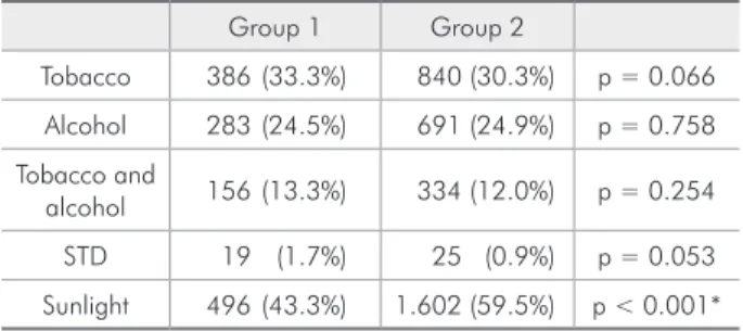
Table 1 shows the participants’ main risk factors for oral cancer, which differed signiicantly between the groups (*p < 0.0001) because more participants
in Group 2 (59.5%) were chronically exposed to
sunlight.
Ten potentially malignant lesions and seven squamous cell carcinomas were found in the study sample. Table 2 shows the distribution of potentially malignant and malignant lesions according to re-gion.
All potentially malignant lesions were found in participants aged 33 to 54 years, with four in men
and 6 in women. Eight potentially malignant lesions were in the lower lip, one was in the buccal mucosa, and one was in the alveolar ridge.
Squamous cell carcinomas were found in individ-uals aged 59 to 85 years, of which 3 were men and 4 were women. Again, the lower lip was the most common site (57.1%). Other sites included the lateral
border of the tongue, hard palate, and buccal mu-cosa, with one lesion each.
Most (85.7%) patients diagnosed with squamous
cell carcinoma were smokers, and 80% of those with
potentially malignant lesions were exposed to sun-light daily. Table 3 shows the participants’ main risk factors.
A total of 55 dentists participated in the study on the knowledge of dentists about potentially malig-nant lesions and squamous cell carcinoma. Of these, 42 were members of the Family Health Strategy teams and 9 worked at the dental specialty centers. Information about working place was absent in four questionnaires. Thirty-two dentists were in Group 1 and 23 in Group 2. The professionals had 4 months to 42 years of practice, with a mean of 6.37 years.
Approximately half (53%) of the dentists had
al-ready seen patients with oral squamous cell carci-noma, and most (83%) had seen patients with
possi-bly potentially malignant lesions. Table 4 shows the dentists’ answers to the questionnaire.
Discussion
The presence of oral lesions in 296 (7.4%)
pa-tients stresses the importance of primary health care personnel performing complete oral examinations, including teeth, soft tissues, bones, and adjacent
Table 1 - Main risk factors for oral cancer of patients exam-ined by primary health care personnel according to group.
Group 1 Group 2
Tobacco 386 (33.3%) 840 (30.3%) p = 0.066
Alcohol 283 (24.5%) 691 (24.9%) p = 0.758
Tobacco and
alcohol 156 (13.3%) 334 (12.0%) p = 0.254
STD 19 (1.7%) 25 (0.9%) p = 0.053
Sunlight 496 (43.3%) 1.602 (59.5%) p < 0.001*
SDT = sexually transmitted diseases; *p < 0.05 according to the chi-square test. Odds ratio = 1.92 (1.67–2.22). The data are expressed as number and (percentage).
Table 2 - Distribution of potentially malignant lesions and squamous cell carcinomas diagnosed by secondary health care personnel according to group.
Group 1 Group 2
Potentially malignant
lesions 6 (0.5%) 4 (0.1%) p = 0.044*
Squamous cell
carcinomas 1 (0.1%) 6 (0.2%) p = 0.337
Risk factor Cases Control
Squamous cell carcinoma
Tobacco 6 (0.48%) 1 (0.03%) p = 0.004*
Alcohol 2 (0.20%) 5 (0.17%) p = 0.686
Tobacco and alcohol 2 (0.40%) 5 (0.14%) p = 0.210
STD 1 (2.32%) 5 (0.13%) p = 0.067
Sunlight 5 (0.24%) 2 (0.11%) p = 0.467
Potentially malignant lesions
Tobacco 1 (0.08%) 9 (0.42%) p = 0.188
Alcohol 2 (0.20%) 7 (0.23%) p = 1.000
Tobacco and alcohol 0 (0.00%) 10 (0.28%) p = 0.623
STD 0 (0.00%) 9 (0.24%) p = 1.000
Sunlight 8 (0.38%) 2 (0.11%) p = 0.124
STD = sexually transmitted diseases; *p < 0.05 according to Fisher’s exact test. Odds ratio = 13.27 (1.60–294.63). The data are expressed as number and (percentage).
Table 3 - Exposure of patients with potentially malignant lesions and squamous cell carcinoma diagnosed by secondary health care personnel to the risk factors for oral cancer.
structures, because this is the only way individuals gain access to higher levels of care. The great dif-ference in size between Groups 1 and 2 relect one of the main dificulties regarding public policies for oral cancer. Speciically, in the absence of political and governmental control, diagnosis, prevention, and treatment depend entirely on the individual actions of the professionals involved.14 In the
pres-ent study, for example, it was observed that the professionals in Group 2 were more involved with the prevention of oral lesions than those in Group 1, which can explain the difference in size between the two groups. Moreover, the number of biopsies performed by secondary health care centers was very low compared with the great number of lesions identiied by primary health care centers, revealing a weakness in the diagnostic process because most of the lesions were in fact developmental defects of the oral and maxillofacial region.
Potentially malignant lesions were more preva-lent in Group 1 (60%), but this group also had more
dentists capable of recognizing leukoplakia, eryth-roplakia, and actinic cheilosis as potentially malig-nant lesions and of identifying the main characteris-tics of an early malignant lesion, namely, a painless, indurated ulcer with rolled edges. Thus, knowledge about potentially malignant lesions was positively associated with their identiication, contributing to the prevention of squamous cell carcinoma, given that the treatment of potentially malignant lesions
may prevent the development of malignant lesions.5
Additionally, the lack of knowledge about potential-ly malignant lesions may be noted as a weakness of the diagnosis network.
Sunlight was the most prevalent risk factor in both study regions, and Group 2 (59.5%) was
sig-niicantly more exposed than Group 1 (p < 0.001).
Sunlight was also the most and second-most com-mon risk factor acom-mong individuals with potentially malignant (71.4%) and malignant lesions (80%),
re-spectively. This reinforces the National Oral Health Policy’s recommendation for knowing the most prevalent diseases in a region, as this allows the de-velopment of region-speciic oral health practices that are effective and curative. Actions to prevent sunlight-related squamous cell carcinoma of the lip are as important as those directed towards tobacco and alcohol consumption, which increase the risk of oral squamous cell carcinoma by 5.49 and 4.62 times, respectively.16
The percentage of malignant lesions (9.4%) found
by the present study was similar to those found by other studies conducted in the Brazilian Northeast.17
An exception was the predominance of lip cancer (57.1%) over intraoral cancer, which was most likely
due to the large number of study participants who were exposed daily to ultraviolet radiation.
tion between sunlight and skin cancer, the associa-tion between sunlight and lip cancer is unknown to most, contributing to people’s negligence towards their lips.8
The dentists surveyed in this study believed that smokers are at the highest risk of oral cancer. Ac-cordingly, smoking was the only factor signiicantly associated with oral cancer (p = 0.004). Squamous cell carcinoma was 13.27 times more common in smokers. The concomitant use of alcohol and tobac-co, observed in 12.3% of the individuals of Groups
1 and 2, increases the risk of oral squamous cell carcinoma. Accordingly, 74.3% of the study
partici-pants diagnosed with squamous cell carcinoma were tobacco and alcohol consumers. Education actions are needed to increase the awareness of these associ-ations and to encourage individuals to give up habits involved in the pathogenesis of oral cancer.18
Although the National Oral Health Policy in-cluded the diagnosis of oral lesions, with special emphasis on oral cancer,13 in the scope of practice
of primary health care facilities in 2004, and the PMAQ-AB launched in 201115 included an indicator
of the incidence of mucosal changes, primary health care dentists still have little knowledge of stomatol-ogy. For example, most dentists (74.1%) considered Table 4 - Dentists’ knowledge of potentially malignant lesions, feelings towards patients with oral cancer, and reasons for their perceived inability to perform biopsies by group.
Knowledge of potentially malignant lesions
Which of these lesions are considered
potentially malignant? n % Group 1 Group 2 p-value* OR
Leukoplakia 40 74.1% 21 (67.7%) 14 (60.9%) 0.6010
Erythroplakia 31 57.4% 16 (51.6%) 12 (52.2%) 0.9674
Actinic cheilosis 30 55.6% 17 (54.8%) 11 (47.8%) 0.6100
Angular cheilitis 11 20.4% 7 (22.6%) 3 (13.0%) 0.4886
Lichen planus 08 14.0% 5 (16.1%) 3 (13.0%) 1.0000
Inflammatory fibrous hyperplasia 07 13.0% 6 (19.4%) 1 (4.3%) 0.2178
Candidiasis 02 3.7% 1 (3.2%) 1 (4.3%) 1.0000
Aphthous ulcers 01 1.9% 1 (3.2%) 0 (0.0%) 1.0000
I do not know 03 5.6% 2 (6.5%) 1 (4.3%) 1.0000
-Feelings towards patients with oral cancer
What do you feel when you see a patient
with oral cancer? n % Group 1 Group 2 p-value* OR
Anxiety 32 59.3% 21 (67.7%) 10 (43.5%) 0.0745
Fear 02 3.7% 0 (0.0%) 2 (8.7%) 0.1767
Pity 16 29.6% 0 (0.0%) 5 (21.7%)* 0.0106 - †
Prejudice 01 1.9% 7 (22.6%) 1 (4.3%) 0.1186
-Oral biopsies
n % Group 1 Group 2 p-value* OR
Does not perform biopsies 40 74.1% 24 (77.4%) 14 (60.9%) 0.1878
-Reasons not to perform a biopsy
Lack of technical knowledge 10 18.5% 9 (29.0%)* 1 (4.3%) 0.0318 9.0 (1.0 - 206.0)
Has never performed one 31 57.4% 20 (64.5%) 11 (47.8%) 0.2200
Fear of erring - - -
Fear of uncontrolled bleeding 4 7.4% 2 (6.5%) 2 (8.7%) 1.0000
themselves incapable of performing biopsies, which are essential for the early diagnosis of oral lesions.
Public health managers have proposed many ac-tions to prevent oral cancer, but few are based on scientiic evidence that can actually decrease its in-cidence or promote its early diagnosis. In 2006, the state of Ceará launched a public policy to reduce the incidence of oral cancer that was approved by the State Board of Health. The policy lists actions to be implemented by the state to ight oral cancer. How-ever, at present, there is no scientiic evidence that any policy, anywhere in the world, has been capable of reducing the incidence of oral cancer.7
The diagnoses of potentially malignant and ma-lignant oral lesions in the sample relect the suc-cessful integration of primary and secondary health care. However, as unprepared primary care profes-sionals were incapable of providing diagnoses and performing biopsies, all the biopsies were performed at secondary health care facilities. The existence of regional dental specialty centers in Ceará reduces the distance barrier between patient and medical care, increasing access to treatment. The lack of experts in other Brazilian states has already been described as a dificulty in the early diagnosis of oral cancer.19
Conclusion
The early diagnosis of potentially malignant le-sions and squamous cell carcinoma is promoted by integrating primary and secondary health care, forming a diagnosis network. However, most pri-mary care dentists were incapable of identifying po-tentially malignant lesions and performing biopsies, most likely because of their anxiety towards oral cancer, which may be a weakness in the diagnostic process.
The present study found a few challenges and possibilities associated with the implementation of an oral cancer diagnosis network. The integration of dentists’ knowledge of oral lesions and risk factors for oral cancer, region-speciic preventive policies, and integration of primary, secondary, and tertiary care may actually reduce the incidence of oral cancer.
Acknowledgments
The authors certify that they have no conlicts of interest. This study was sponsored by the Pro-grama Pesquisa para o Sistema Único de Saúde
(PPSUS/2009) (Brazilian Uniied Health Care Sys-tem Research Program) under protocol number 09100058-0/09.
References
1. Van der Waal I, Bree R, Brakenhoff R, Coebergh J. Early diagnosis in primary oral cancer: is it possible?. Med Oral Patol Oral Cir Bucal. 2011 May;16(3):e300-5.
2. Scully C, Bagan J. Oral squamous cell carcinoma overview. Oral Oncol. 2009 Apr-May;45(4–5):301-8.
3. Ministério da Saúde. Instituto Nacional de Câncer [Inter-net]. Estimativa 2012: incidência do câncer no Brasil. Rio de Janeiro: INCA; 2011 [cited 2013 May 20]. Available from: http://www.inca.gov.br/estimativa/2012/index.asp?ID= 5. 4. Souza RL, Fonseca-Fonseca T, Oliveira-Santos CC, Correa
GT, Santos FB, Cardoso CM, et al. Lip squamous cell car-cinoma in a Brazilian population: epidemiological study and clinicopathological associations. Med Oral Patol Oral Cir Bucal. 2011 Sep;16(6):e757-62.
5. Byakodi R, Shipurkar A, Byakodi S, Marathe K. Prevalence of oral soft tissue lesions in Sangli, India. J Community Health. 2011 Oct;36(5):756-9.
6. Hirota SK, Braga FP, Penha SS, Sugaya NN, Migliari DA. Risk factors for oral squamous cell carcinoma in young and older Brazilian patients: a comparative analysis. Med Oral Patol Oral Cir Bucal. 2008 Apr;13(4):E227-31.
7. Torres-Pereira C. Oral cancer public policies: is there any evidence of impact?. Braz Oral Res. 2010;24 Suppl 1:37-42. 8. Busick TL, Uchida T, Wagner RF Jr. Preventing ultraviolet
light lip injury: beachgoer awareness about lip cancer risk factors and lip protection behavior. Dermatol Surg. 2005 Feb;31(2):173-6.
9. Hennessey PT, Westra WH, Califano JA. Human papillo-mavirus and head and neck squamous cell carcinoma: re-cent evidence and clinical implications. J Dent Res. 2009 Apr;88(4):300-6.
10. Warnakulasuriya S. Global epidemiology of oral and oropha-ryngeal cancer. Oral Oncol. 2009 Apr-May;45(4-5):309-16. 11. Warnakulasuriya S, Johnson NW, van der Waal I. Nomencla-ture and classification of potentially malignant disorders of the oral mucosa. J Oral Pathol Med. 2007 Nov; 36(10):575-80. 12. Lopez Jornet P, Velandrino Nicolas A, Martinez Beneyto Y,
Fernandez Soria M. Attitude towards oral biopsy among gen-eral dentists in Murcia. Med Oral Patol Oral Cir Bucal. 2007 May;12(2):E116-21.
2004. [cited 2013 May 20]. Available from: http://bvsms. saude.gov.br/bvs/publicacoes/politica_nacional_brasil_sor-ridente.pdf.
14. Torres-Pereira CC, Angelim-Dias A, Melo NS, Lemos Jr CA, Oliveira EM. Strategies for management of oral cancer in pri-mary and secondary healthcare services. Cad Saude Publica. 2012;28 Suppl:s30-9.
15. Ministério da Saúde. Portaria no 1.654, de 19 de julho de 2011. Institui, no âmbito do Sistema Único de saúde, o Pro-grama Nacional de Melhoria do Acesso e da Qualidade da Atenção Básica (PMAQ-AB). Saúde Legis; 2011. [cited 2013 May 20]. Available from: http://bvsms.saude.gov.br/bvs/ saudelegis/gm/2011/prt1654_19_07_2011.html.
16. Szymanska K, Hung RJ, Wunsch-Filho V, Eluf-Neto J, Curado MP, Koifman S, et al. Alcohol and tobacco, and the risk of
can-cers of the upper aerodigestive tract in Latin America: a case-control study. Cancer Causes Control. 2011 Jul;22(7):1037-46.
17. Carvalho MV, Iglesias DP, Nascimento GJ, Sobral AP. Epi-demiological study of 534 biopsies of oral mucosal lesions in elderly Brazilian patients. Gerodontology. 2011 Jun;28(2):111-5.
18. Seoane J, Warnakulasuriya S, Varela-Centelles P, Esparza G, Dios PD. Oral cancer: experiences and diagnostic abilities elicited by dentists in North-western Spain. Oral Dis. 2006 Sep;12(5):487-92.