*Correspondence: J.B.Silva. Centro de Desenvolvimento da Tecnologia Nuclear – CDTN/CNEN. Av. Presidente Antônio Carlos, 6.627, Campus da UFMG – Pampulha - 31270-901 – Belo Horizonte, MG – Brasil. E-mail: [email protected]
A
rti
Pharmaceutical Sciences vol. 46, n. 3, jul./set., 2010
Synthesis, quality control and dosimetry of the
radiopharmaceutical
18F-sodium luoride produced at the Center
for Development of Nuclear Technology - CDTN
Marina Bicalho Silveira
1, Marcella Araugio Soares
1, Eduardo Sarmento Valente
1, Samira Soares
Waquil
1, Andréa Vidal Ferreira
2, Raquel Gouvêa dos Santos
1, Juliana Batista da Silva
1,*1Serviço de Aplicação das Radiações na Saúde, 2Serviço de Reator e Irradiações, Centro de Desenvolvimento da Tecnologia
Nuclear/Comissão Nacional Energia Nuclear CDTN/CNEN
18F-Sodium luoride (Na18F) is a radiopharmaceutical used for diagnosis in nuclear medicine by
positron emission tomography (PET) imaging. Bone scintigraphy is normally performed using 99m
Tc-MDP. However, 18F PET scans promise high quality imaging with increased resolution and improved
sensitivity and speciicity. In order to make available a tool for more speciic studies of tumors and non-oncological diseases of bone tissue, the UPPR/CDTN team undertook the production and quality control of Na18F injectable solution with the physical-chemical, microbiological and biological characteristics
recommended in the U.S. Pharmacopeia. Na18F radiochemical purity was 96.7 ± 1.3 %, with Rf= 0.026
± 0.006. The product presented a pH of 5.3 ± 0.6, half life of 109.0 ± 0.8 minutes, endotoxin limit < 5.0 EU.mL-1 and no microbial contaminants. The biodistribution of Na18F was similar to that described in
the literature, with a clearance of 0.19 mL.min-1 and distribution volume of 18.76 mL. The highest bone
concentration (5.0 ± 0.5 %ID.g-1) was observed 20 minutes after injection. Na18F produced at the UPPR
presented all the quality assurance requirements of the U.S. Pharmacopeia and can be safely used for clinical bone imaging.
Uniterms: Radiopharmaceuticals/biodistribution. Sodium luoride 18F/production. Sodium luoride 18F/
quality control. Sodium luoride. 18F/dosimetry. Bone imaging.
O Fluoreto de sódio 18F (Na18F) é um radiofármaco empregado para diagnóstico através da Tomograia
por Emissão de Pósitrons (PET). Cintilograias ósseas são normalmente obtidas utilizando-se 99mTc-MDP.
Entretanto, o interesse pelo Na18F é crescente, principalmente devido à obtanção de imagens de elevada
resolução. Com o objetivo de tornar disponível uma ferramenta mais especíica para estudos de tumores e doenças não-oncológicas do tecido ósseo, o grupo da UPPR/CDTN implementou a produção e o controle de qualidade da solução injetável de Na18F com as características físico-química, microbiológica
e biológica preconizadas pela farmacopéia. Sua pureza radioquímica foi de 96,7 ± 1,3 %, com Rf= 0,026 ± 0,006. O produto apresentou pH igual a 5,3 ± 0,6, tempo de meia-vida de 109,0 ± 0,8 minutos, limite de endotoxinas < 5,0 EU.mL-1 e ausência de microrganismos. O peril de biodistribuição em camundongos
foi semelhante ao disponível na literatura, com depuração igual a 0,19 mL.min-1 e volume de distribuição
igual a 18,76 mL. A concentração máxima (5,0 ± 0,5 % DI.g-1) foi observada no osso 20 minutos após
a injeção. O Na18F produzido na UPPR do CDTN apresentou os parâmetros de qualidade deinidos na
farmacopéia americana e pode ser usado com segurança para uso clínico em cintilograia óssea.
Unitermos: Radiofármacos/biodistribuição. Fluoreto de sódio. 18F/produção. Fluoreto de sódio. 18F/
INTRODUCTION
18F-Sodium luoride injection (Na18F), a
positron-emitting radiopharmaceutical containing no carrier-added (NCA), is used for diagnostic purposes in conjunction with positron emission tomography (PET) imaging. This radiopharmaceutical became widely used for bone scintigraphy after its introduction by Blau et al. (1962) in the early 1960s and was approved for clinical use by the U.S. Food and Drug Administration in 1972. The use of
99mTc-MDP has been brought into question given the
ad-vantages of PET imaging and the limited global supply of
technetium-99m (99mTc). Moreover, bone uptake of Na18F
is 2-fold greater than that of 99mTc-MDP and 18F-luoride
PET is more accurate than planar imaging or SPECT for
localizing and characterizing bone lesions (Blau et al.,
1962).
The radiopharmaceutical Na18F has the desirable
pharmacokinetics properties of high and rapid bone uptake coupled with very rapid blood clearance, which results in a high bone-to-background ratio within a short time frame.
After intravenous administration, 18F-fluoride diffuses
through the bone capillaries into the bone’s extracellular
fluid (ECF). From the bone ECF, 18F-fluoride ions
ex-change with hydroxyl groups in the hydroxyapatite at the surface of bone crystals forming luoroapatite mainly at sites of bone remodelling with high turnover. Therefore,
uptake of 18F-luoride relects blood low and osteoblastic
activity (Grant et al., 2008).
18F-luoride PET is a highly sensitive imaging
modal-ity for detection of benign and malignant osseous abnor-malities and allows the regional characterization of lesions in metabolic bone diseases. Using hybrid PET/CT systems
improves the specificity of 18F-fluoride PET in cancer
patients by accurately differentiating between benign and malignant sites of uptake (Grant et al., 2008). The clinical
usefulness of 18F-luoride PET has been demonstrated for
monitoring the response to therapy against a wide range of clinical indications such as osteoporosis, Paget’s disease, compression fractures, marrow lesion, stress injuries and many other abnormal bone activities (Bridges et al., 2007).
Given the applicability of Na18F for bone imaging
has been conirmed, our objective was to obtain a high quality product. The commercial delivery system used
for 18F-FDG can also be used for the eficient delivery of
other 18F-labeled radiopharmaceuticals, including Na18F.
Microbiological, physical-chemical and biological
qual-ity control tests were performed, and the Na18F injectable
solution met the quality control requirements contained in the United States Pharmacopeia (USP 31). It is now
feasible to perform high-quality Na18F bone scans in most
nuclear medicine departments, contributing to bone dis-order and tumour diagnoses. This radiopharmaceutical is available for clinical use in PET/CT imaging.
All drug manufacture and quality control complied with Current Good Manufacturing Practice regulations described in the regulatory requirements and general guidance published by the Brazilian Regulatory Agency for Medicines (ANVISA).
MATERIALS AND METHODS
Na18F Production
18F-Fluoride was produced on a 16.5 MeV Cyclotron
PETtrace® (GE Healthcare) by the 18O (p,n)18F nuclear
reaction. The niobium target (with yield of 215 mCi/μASat)
was illed with 2.2 mL of enriched O18 water, which was
irradiated with protons for 10 minutes at an intensity of
25 µA. The solution containing 18F- was transferred into
an automatic synthesis module, TracerLab® MX
FDG (GE
Healthcare), modiied and prepared with reagents kit and accessories for Na18F production (Figure1). 18F-luoride ions
were trapped in a SepPak Light Accell, Plus QMA, anion exchange column and were then eluted with NaCl 0.9% so-lution. Finally, the resulting 15 mL of Na18F was dispensed
into sterile, pyrogen-free vials, through a 0.22 μm ilter,
in an automated dispensing unit (Theodorico, Comecer®).
Physical-chemical quality control
The pH value of Na18F was measured using pH paper
(0-14) which displayed a different colour depending on the pH range of the samples. The results were compared with standard pH buffer and the estimated value was registered.
Radiochemical identity and purity of Na18F were
conirmed either by thin layer chromatography (TLC)
ac-cording to Nandy et al. (2007) or using a high-performance
liquid chromatography (HPLC) system.
The TLC stationary phase was silica gel and the mobile phase was acetonitrile : water (95:5% v/v). The ra-dioactivity was determined by scanning the TLC SG plate with a suitable collimated radiation detector (Minigita star beta detector, Raytest®)
. The main peak was analyzed to
deine the retention factor (Rf) value and the radioactivity related to the sample, that must lie between 0.0 - 0.12 and be no less than 95%, respectively.
HPLC analyses were performed on a
chromatogra-phic system (Agilent®), connected in serie with a gamma
detector (Raytest®). The anion-exchange column
(Shi-madzu, IC.A1) (size: 46 mm x 10 cm) temperature was
kept constant at 25 °C. The low rate was 1.0 mL.min-1,
the sample volume was 20 µL and the solvent system was NaOH 0.1M (pH = 4.0).
Radionuclidic purity was evaluated by gamma-ray spectrometry (Camberra Multichannel Analyzer). Half-life of 18Fwas calculated after measuring the radioactivity
de-cay of the sample over a 20-minute period in a radioisotope
dose calibrator (Capintec CRC®-25R). The equation used
is showed below:
(1)
Where: A0= initial activity; A = activity measured
after 20 minutes; t = time interval (in minutes) between the two measures (tA-tAo); t½ = half-life.
Microbiological quality control
Bacterial endotoxins were quantiied by the
chromo-genic method, using an Endosafe® Portable Test System
– PTS. This device includes a pumping system, a portable spectrophotometer and internal software to calculate
sam-ple data. Na18F samples in duplicate (previously diluted)
were applied inside cartridges in parallel with positive control testing. The product was considered apyrogenic
when the endotoxins level was no more than 11.6 EU.mL-1
The sterility of the Na18F solution was assayed
perfor-ming a test by direct inoculation of Na18F solution in
trypti-case soy broth and luid thioglycollate medium. The test was performed in duplicate and a negative control test was also performed. The culture media were incubated at 25 °C and 37 °C, respectively, and veriied daily over a fourteen-day period. The product was considered sterile when there was no evidence of microbiological growth.
Physiological quality control
The Na18F physiological quality control was done by
biodistribution studies in animals. Female Swiss mice with a body weight of around 25 g purchased from the Animal House Center (CEBIO) of the Biological Sciences Insti-tute (ICB/UFMG). Mice were maintained on a light-dark
cycle (12-h light, 12-h dark) at a room temperatureof 25
°C and given food and water ad libitum. Animal experi-Animal experi-ments were performed in compliance with the Institute of Laboratory Animal Resources – Commission on Life Sciences – National Research Council, Washington, D.C. and the Brazilian society in science of Laboratory Animals (SBCAL).
Doses of 70 kBq of Na18F were injected by
in-travenous route into Swiss mice. After different time intervals (2.5 - 60 minutes), the animals were sacriiced. Blood samples were taken, and the thyroid, heart, lungs, liver, spleen, pancreas, kidneys, stomach, intestine, bone, muscle, brain and bladder removed and weighed. Their respective radioactivity was measured in an automatic gamma spectrometer (1480 Wizard 3’’– Wallac – Count- Wizard 3’’– Wallac – Count-ing eficiency for 0.511MeV: 48%). The biodistribution was evaluated after calculating the percentage uptake of
injected dose per gram of organ (% DI.g-1).
Pharmacokinetics parameters were estimated by the Biexp Pharmacokinetics program (Murphy,R., Inaoe tonantzindra, Puebla, México 1991).
Radiation dosimetry
The absorbed radiation dose for each mouse organ
was calculated applying MIRD formalism (Snyderet
al., 1978) to the biodistribution results, according to the
relations:
(2)
(3)
Where: Ãhis the cumulated activity in the target
organ; S(rk←rh) is the mean absorbed dose in the target
organ per unit of cumulated activity in the source organ;
∆is the mean energy emitted; ϕi(rk←rh) is the absorbed
energy fraction; m is the target tissue mass.
A value of ϕ = 1 (non-penetrating radiation) was
adopted for the 18F positron emission radionuclide. The
experimental results of the cumulated activity in the mice organs were extrapolated to humans, assuming a similar concentration ratio among various tissues between mouse
and patient. This extrapolation converted mouse %ID.g-1
and patient (Kirschner et al., 1975 andShen et al., 2005):
(4)
Where: [A0]HO is the activity in the human organ;
[A0]MO is the activity in the mouse organ; MHO is the human
organ mass; MH is the human mass; mM is the mouse mass;
mMO is the mouse organ mass.
This extrapolation was based on the organ masses of mouse used in the experiments and the adult human organ masses of the Cristy-Eckerman phantom (Cristy, Eckerman, 1987).
RESULTS AND DISCUSSION
Na18F production
Enriched water H218O was successfully irradiated on
the Cyclotron. 18F- was produced by the nuclear reaction
180 (p,n) 18F, by irradiation with protons for 10 minutes at
the intensity of 25 µA. Na18F was produced in high yields
of 87.3 ± 6.1%. Samples of the inal product were sent to the quality control laboratories in appropriately shielded containers.
Physical-chemical and microbiological quality control
Na18F physical-chemical and microbiological
characteristics were evaluated. The analyses described earlier determined its radiochemical identity and purity, radionuclidic identity and purity, pH, bacterial endotoxins and sterility.
The quality requirements used for Na18F were in
accordance with those found in the United States
Pharma-copeia (USP 31), supplemented with literature data (Nandy et al.., 2007) (Table I).
Radiochemical purity of the Na18F solution was
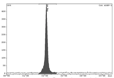
as-sessed by thin-layer chromatography (TLC). The product migration proile was determined by scanning the chroma-togram plate with a suitable collimated radiation detector (the radiochromatogram is represented in Figure 2).
The main peak on the radiochromatogram obtained
using HPLC for the Na18F test solution had approximately
the same retention time (about 2 minutes) as the peak for 18F
described in literature data (Olberg et al., 2008). More than 99% of the radioactivity was related to 18F peak (Figure 3).
The inal product, analyzed by gamma spectrometry, presented high radionuclidic purity. The spectrum obtained (Figure 4) shows one main peak at 0.511 MeV, an usual energy level for a positron annihilation product.
Energy
The results of the tests performed on the PTS were
TABLE I - Results obtained after testing Na18F microbiological, physical-chemical and biological properties (1 USP 31; 2 Nandy et
al., 2007)
Tests Quality Requirements
(USP 31 and Nandy et al., 2007) Results pH1 Between 4.5 and 8.0 5.3 ± 0.6 Radiochemical Identity2 0.00 < Rf ≥ 0.12 0.026 ± 0.006 Radiochemical Purity1 ≥ 95.0 % 96.7 ± 1.3% Radionuclidic Identity1 Between 105 and 115 minutes. 109.0 ± 0.8 minutes Radionuclidic Purity1 Main peak = 0.511 MeV Main peak = 0.511 MeV Bacterial endotoxins1 ≤ 11.6 EU.mL-1 < 5.0 EU.mL-1
Sterility1 Sterile Sterile
FIGURE 2 - TLC scan of Na18F in 95:5 ACN: H2O mobile phase:
FIGURE 3 - HPLC chromatogram of Na18F solution showing its radiochemical purity, using NaOH 0.1M as mobile phase.
FIGURE 4 - Gamma spectrum obtained with a suitable detector
to determine Na18F radionuclidic purity. Main peak can be observed at 0.511 MeV.
in compliance with the requirements established by the USP 31, showing that the samples contained less than 5.0 EU.mL-1.
All the samples assayed for sterility were observed for 14 days and none showed evidence of microbiological
growth. Therefore, the Na18F solutions were considered
sterile.
Biological quality control
The biological quality control of Na18F was carried
out through biodistribution studies in Swiss female mice.
70 kBq of Na18F were injected in mice via the intravenous
route and, after different time intervals (2.5 - 60 minutes), the animals were sacriiced and radiopharmaceutical dis-tribution in the organs was calculated as percentage uptake
of injected dose per gram of organ (%ID.g1).
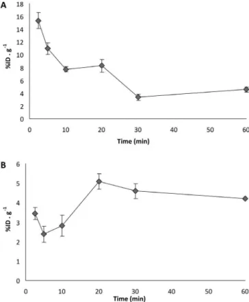
The biodistribution proile is shown in Figure 5 and
FIGURE 5 - Na18F biodistribution proile in Swiss female mice.
70 kBq of Na18F were injected into mice (i.v). After different time intervals, the animals were sacriiced, blood samples were taken, and thyroid, heart, lungs, liver, spleen, pancreas, kidneys, stomach, intestine, bone, muscle, brain and bladder were removed. Organ radioactivity was measured in an automatic gamma spectrometer and data were expressed as the percentage of total injected dose by tissue weight (% ID.g-1).
the results indicated that the blood concentration of the radiopharmaceutical was quickly reduced in the irst hour
post injection (4.50 ± 0.35 % ID.mL-1) (Figure 6a). 18
F-Sodium luoride presented biexponential blood clearance with half-life of 5.1 minutes in the fast distribution phase
and 87 minutes in the slow elimination phase. The Na18F
clearance was 0.19 mL.min-1 and the distribution volume
was 18.76 mL.
The Na18F kinetics in the main vital organs followed
blood kinetics, and the radiopharmaceutical was rapidly cleared out. The only exception was bone kinetics, where
the Na18F concentration increased during the time period,
reaching a peak (5.0 ± 0.5 % ID.g-1) 20 minutes after the
injection (Figure 6b).
Radiation dosimetry
Dosimetric studies aimed to evaluate the safety of
the 18F-Sodium luoride produced in the UPPR/CDTN for
bone scintigraphy. The results (Table II) demonstrated that the highest absorbed doses in the mice were in bladder
(11.5 mGy.70 KBq-1) and bone (7.1 mGy.70 KBq-1).
Ab-sorbed doses for the other organs were signiicantly lower,
ranging from 1.3 - 4.0 mGy.70 KBq-1. The radiation doses
extrapolated to patients indicated that radiation doses in
respectively. These results are quite similar to those
pu-blished by ICPR53 and show that Na18F produced at the
UPPR/CDTN can be safely used for clinical bone imaging.
CONCLUSIONS
The automated synthesis of Na18F and its availabil- and its
availabil-ity was successfully accomplished using the commercial
18FDG synthesizer TracerLab® MX
FDG. The inal solution
presented high quality in accordance with the requirements
of the USP 31. The biodistribution of Na18F was similar
to that described in the literature and absorbed doses in organs were lower than radioprotection limits.
Considering the clinical importance of 18F-Sodium
luoride and the high resolution PET images provided by it,
the UPPR group wish to start Na18F production and make
it widely available. This study showed that the product presents good quality and meet the needs of hospitals, clinics, and research facilities. The radiopharmaceutical
Na18F was produced according to the regulatory
require-ments and general guidance of Good Manufacturing Practices, published by the Brazilian Regulatory Agency for Medicines (ANVISA).
ACKNOWLEDGEMENTS
The authors wish to thank the whole team at the UPPR/ CDTN for technical support on production and quality control of Na18F. In addition, the authors are
grate-ful to PRO da Silva, LM Gabriel and FAS Vilas Boas for technical assistance on biodistribution experiment. This research was supported by Centro de Desenvolvimento da Tecnologia Nuclear (CDTN/CNEN-MG), INCT de
FIGURE 6 - Na18F kinetics in blood (A) and bone (B).
TABLE II - Organ absorbed dose in mice and extrapolated to patients. The results were compared with literature data (ICRP 53).
*Values were converted to clinically used doses (370 MBq)
Organ Absorbed doses in mice
(Gy.70 Kbq-1) Extrapolated dose to patients (Gy.370 MBq-1) Patient dose ICRP data*
Thyroid 2.70x10⁻³ 5.14x10⁻³
-Heart 4.06x10⁻³ 7.73 x10⁻³ 1.44 x10⁻³
Lungs 2.27x10⁻³ 4.33 x10⁻³ 1.51 x10⁻³
Liver 1.32x10⁻³ 2.52 x10⁻³ 1.48 x10⁻³
Spleen 3.49x10⁻³ 6.65 x10⁻³
-Pancreas 3.08x10⁻³ 5.86 x10⁻³ 1.77 x10⁻³
Kidneys 3.04x10⁻³ 5.80 x10⁻³ 7.03 x10⁻³
Stomach 1.63x10⁻³ 3.10 x10⁻³ 1.40 x10⁻³
Intestine 2.54x10⁻³ 4.84 x10⁻³ 2.44 x10⁻³
Bone 7.14x10⁻³ 1.36 x10⁻² 2.22 x10⁻²
Brain 1.56x10⁻³ 2.98 x10⁻³ 2.01 x10⁻³
Medicina Molecular and Conselho Nacional de Desen-volvimento Cientíico e Tecnológico (CNPq).
REFERENCES
BLAU, M.; NAGLER, W.; BENDER, M.A. Fluorine-18: a new isotope for bone scanning. J. Nucl. Med, v. 3, p.332-334, 1962.
BRASIL. Ministério da Saúde. Agência Nacional de Vigilância Sanitária, “Determina a todos os estabelecimentos fabricantes de medicamentos, o cumprimento das diretrizes estabelecidas no Regulamento Técnico das Boas Práticas para a Fabricação de Medicamentos”, Diário Oicial [da] República Federativa do Brasil, Brasília, DF, Resolução – RDC nº 17, de 16 de abril de 2010. Seção 1. p.94-110.
BRIDGES, R.L.; WILEY, C.R.; CHRISTIAN, J.C.; STROHM, A.P. An introduction to Na18F bone scintigraphy: basic principles, advanced imaging concepts, and case examples.
J Nucl. Med. Technol., v.35, p.64-76, 2007.
CRISTY, M.; ECKERMAN, K.Specific absorbed fractions of energy at various ages from internal photons sources. Tennesse: Oak Ridge National Lab., 1987. 100 p.
GRANT F.D.; FAHEY F.H.; PACKARD A.B.; DAVIS R.T; ALAVI A.; TREVES S.T. Skeletal PET with 18F-Fluoride: applying new technology to an old tracer. J. Nucl. Med., v.28, p.68-78, 2008.
INTERNATIONAL COMMISSION ON RADIOLOGICAL PROTECTION. ICRP. Publication 53. Radiation dose to patients from radiopharmaceuticals. Stockholm: International Commission on Radiological Protection, 1987. 388 p.
KIRSCHNER A.S.; ICE R.; BEIERWALTES W. Radiation dosimetry of I-131-iodocholesterol: the pitfalls of using tissue concentration data – author’s reply. J. Nucl. Med.,
v.16, p.248-249, 1975.
NANDY, S.K; RAJAN, M.G.R.; SONI, P.S. Production of sterile [F-18] NaF for skeletal Pet imaging. Indian J. Nucl. Med., v.281, p.16-23, 2007.
OLBERG, D.E; HJELSTUEN, O.K.; SOLBAKKEN, M.; ARUKWE, J.; KARLSEN, H.; CUTHBERTSON, A. A novel prosthetic group for site-selective labeling of peptides for positron emission tomography. Bioconjugate Chem., v.19, p.1301-1308, 2008.
SHEN, S.; KHAZAELI1, M.B., YANCEY GILLESPIE, G.; ALVAREZ V. L. Radiation dosimetry of 131I-chlorotoxin for targeted radiotherapy in glioma-bearing mice. J. Neurooncol., v.71, p.113-119, 2005.
SNYDER, W.; FORD, M.; WARNER, G. Estimates of speciic absorbed fractions for photon sources uniformly distributed in various organs of a heterogeneous phantom. New York: Society of Nuclear Medicine, 1978. 70 p. (MIRD Pamphlet, n.5).
UNITED STATES PHARMACOPEIA. Sodium luorine F 18 injection. 31.ed. The NF. 26.ed. Rockville: The United States Pharmacopeia Convention, 2008. p. 2195.