ABSTRACT: The aim of this study was to evaluate the microleakage of direct and indirect composite inlays by ste-reomicroscopy and scanning electron microscopy (SEM). Thirty bovine incisors were ground to obtain an incisal platform, simulating the occlusal surface of a human molar. Each tooth received two 8° proximal cavities with cervical inishing line prepared in dentine or enamel. One of the cavities was illed with Filtek Z250/Single Bond, using the direct technique, and the other was illed with with Solidex/Rely X ARC/Single Bond, using the indirect technique. The samples were stored in water at 37°C for 24 hours and placed in a 50% silver nitrate solution for 6 hours in a dark container. Next, the samples were washed under running water, immersed in a developing solu-tion and exposed to luorescent light for 12 hours. The teeth were then severed and evaluated for dye penetrasolu-tion by stereomicroscopy and SEM. There were no signiicant differences between the direct and indirect techniques for the cervical inishing line in enamel, but for the inishing line in dentin, the indirect technique allowed less mi -croleakage than the direct technique. SEM analysis showed leakage similar to that observed by stereomicroscopic analysis. The use of stereomicroscopic and SEM evaluations improves microleakage analysis.
DESCRIPTORS: Composite resins; Dental restoration failure; Dental leakage; Marginal adaptation (dentistry). RESUMO: O objetivo deste estudo foi avaliar a microiniltração em restaurações diretas e indiretas em resina com -posta por meio de estereomicroscopia e microscopia eletrônica de varredura (MEV). Trinta incisivos bovinos foram lixados de forma a produzir uma plataforma incisal, simulando a face oclusal de um molar humano. Cada dente recebeu dois preparos proximais com 8º e término cervical em esmalte ou dentina. Uma cavidade foi restaurada pela técnica direta com Filtek Z250/Single Bond, e a outra pela técnica indireta com Solidex/RelyX ARC/Single Bond. As amostras foram armazenadas em água a 37°C por 24 horas e então imersas em solução de 50% de ni-trato de prata por 6 horas em recipiente escuro. As amostras foram lavadas em água corrente, imersas em solução reveladora e expostas à luz luorescente por 12 horas. Os dentes foram seccionados para avaliação da penetração do marcador em lupa estereoscópica e MEV. Não houve diferença entre as técnicas direta e indireta para o término em esmalte, porém, para o término em dentina, a técnica indireta resultou em menor iniltração. MEV mostrou iniltração similar à da lupa estereoscópica. A análise por meio de lupa estereoscópica e MEV melhora a avaliação da microiniltração.
DESCRITORES: Resinas compostas; Falha de restauração dentária; Iniltração dentária; Adaptação marginal (odontologia).
INTRODUCTION
Adequate polymerization of resin composite is considered to be a very important factor for assur-ing appropriate physicaland biological properties. Shrinkage stress, however, is one of the inherent disadvantages that occur when visible
light-ac-tivated resin composites are submitted to light polymerization. Stresses arising from post-gel po-lymerization shrinkage may produce defects in the composite to tooth bond, leading to failure associ-ated with microleakage, postoperative sensitivity,
* PhDs, Professors, Dental School, Federal University of Uberlândia.
Marginal integrity and microleakage of direct and indirect
composite inlays – SEM and stereomicroscopic evaluation
Integridade e microinfiltração marginal de restaurações diretas
e indiretas de compósitos – avaliação por estereomicroscopia
e MEV
Carlos José Soares* Leonardo Celiberto** Paula Dechichi*
Rodrigo Borges Fonseca**
and recurrent caries. These problems are the most frequent consequence of fluid penetrating along cavity walls toward the pulp23. Soft-start
polym-erization with short-pulses of light energy12,15, in
association with glass ionomer as the gingival in-crement17, and incremental techniques have been
used in an endeavor to minimize this effect. Different resin-composite inlay systems were also developed and direct composite resin is one of the most popular3. However, because of the
complexities associated with insertion and finish-ing techniques in large direct composite restora-tions, many clinicians have difficulty in establish-ing proper anatomic form, proximal contour and contact25. Indirect inlay systems became popular
to overcome this limitation of direct restorations. Shrinkage stress should be minimized with indirect restoration, since polymerization occurs before the restoration is cemented. However, indirect resin restorations require internal adjustment, which could result in poor marginal fit25.
Resin composite bonding to the acid-etched enamel surface is a clinically well-established treat-ment procedure in restorative dentistry26. However,
dentin is a complex, heterogenic, hydrated biologi-cal substrate14. From this standpoint, adhesion to
dentin is not as reliable as adhesion to enamel, although the new adhesives have shown high val-ues in laboratorial tests20. When caries occurs in
the proximal surface of a posterior tooth, it is not uncommon to have to place the cavity preparation finishing line in dentin, which is another factor that produces poor adhesion and sealing in Class II composite resin restorations. In addition, some studies have demonstrated poorer marginal fit in proximal surfaces than in buccal or lingual sur-faces25. Studies have shown that indirect
restora-tions exhibited more gap formation and leakage at the gingival margin in dentin than in enamel16.
Achieving a perfect marginal quality with inlay
restorations, when gingival margins are located in dentin, continues to be critical even when the new adhesive systems are used9.
It has been hypothesized that direct or indirect techniques and different cervical margin cavity types would have an effect on the occurrence of microleakage and gap formation in proximal resin composite inlays. The aim of this study was to in-vestigate microleakage in cervical proximal cavities with the finishing line placed in enamel or dentin, restored with direct or indirect composite resins, as well as to observe the micro-morphology of the hybrid layer.
MATERIALS AND METHODS
Thirty freshly extracted bovine incisive teeth were used for microleakage and SEM analysis. The teeth were cleaned, with calculus and soft tissue deposits being removed with a hand scaler, and then stored in a 0.2% thymol solution (Pharmácia Biopharma Ltda., Uberlândia, Brazil). In order to facilitate manipulation, preparation and restora-tion of the proximal cavities, every tooth was indi-vidually fixed in a cylinder so that the roots were resin-embedded up to 5 mm below the apex.
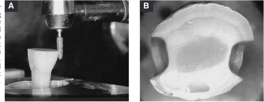
The coronal portion of the tooth was ground with #600 silicon carbide paper (Norton, Recife, Brazil) to create a surface with dimensions similar to those of a human molar, 3.0 mm (margin in dentin) or 6.0 mm (margin in enamel) short of the cementum-enamel junction in the proximal sur -face. The pulp canal was exposed and filled with composite resin. A medium-grained diamond bur with an 8-degree taper was used in a standardized fashion, to make the two proximal inlay prepara-tions with convergent angles. The dimensions of the cavity preparation for the inlays were: 4.0 mm in buccolingual width, 4.0 mm of axial wall height and 2.0 mm of gingival wall width (Figure 1).
FIGURE 1 -A. Diamond bur positioned for preparing proximal cavities in bovine incisor, which had its incisal portion removed and its
pulp canal illed with resin;
B. Proximal cavities with gingival margin in enamel. Each cavity was restored either with direct or indirect technique.
A one-stage impression was taken using a double viscosity polyvinyl-siloxane (Aquasil, Dent-sply DeTrey, Konstanz, Germany) in a stock plastic tray. After 2 hours, the impressions were poured with type IV dental stone (Velmix, Kerr, Romulus, USA). One of these two cavities in the stone dies was randomly selected and used to make the in-direct composite inlay, which was manufactured with laboratory composite resin (Solidex, Shofu, Kyoto, Japan), polymerized in a multi-focal labora-tory source (Solidilate, Shofu, Kyoto, Japan).
Inlay cavities were restored as follows: one of them with the direct composite technique – (Filtek Z250, 3M-ESPE, St. Paul, MN, USA) and Single Bond (3M-ESPE, St. Paul, MN, USA), and the other with the indirect technique – Solidex cemented with Single Bond and dual cure resin cement (Rely X ARC, 3M-ESPE, St. Paul, MN, USA). In the direct procedure, dentin and enamel surfaces were con-ditioned with 36% phosphoric acid gel (3M-ESPE, St. Paul, MN, USA) for 15 seconds, rinsed with wa-ter for 15 seconds and blot-dried, leaving a moist surface. Single Bond was applied according to the manufacturer’s directions and light-polymerized. Z250 composite resin was inserted in three incre-ments and then light-polymerized (XL 3000, 3M Dental Products, St. Paul, USA) for 20 seconds for each restoration. During the first 5 seconds, the light was kept 1 cm away from the restorative ma-terial. The other cavity was restored following the indirect procedure. Both the enamel and dentin surfaces were conditioned with 36% phosphoric acid gel for 15 seconds, rinsed with water for 15 seconds and blot-dried, leaving a moist surface. Single Bond was applied and light-polymerized. The inner surface of the Solidex inlay was treat-ed with aluminum-oxide 50 µm airborne particle abrasion (CVM Ltda., Cachoeirinha, Brazil) for 5 seconds and rinsed with water for 10 seconds. One-bottle pre-hydrolyzed silane (Ceramic Primer, 3M-ESPE, St. Paul, MN, USA) was then applied for 1 minute. Rely X ARC was mixed and applied to the inner surface of the Solidex inlay, which was in-serted in the cavity. Excess material was removed, and then the restoration was light-polymerized for 40 seconds with an XL 3000 light-activation unit. After 15 minutes, the restorations were finished and polished with diamond burs (KG Sorensen, São Paulo, Brazil), and final finishing was done with aluminum oxide disks (Sof-Lex, 3M-ESPE, St. Paul, MN, USA).
The teeth were removed from their cylinders
ed with epoxy resin (Polipox, São Paulo, Brazil). All external surfaces of each tooth were coated with two layers of nail varnish (Risqué, Niasi, Taboão da Serra, Brazil), leaving a 1.0 mm-wide margin around the restoration free of varnish. Specimens were placed in a freshly prepared 50wt% aqueous silver nitrate solution (Quimibrás, Rio de Janeiro, Brazil) for 6 hours and kept in the dark. Next, the teeth were rinsed in tap water for 1 minute and immersed in a photo developing solution (Kodak, Rochester, USA) and exposed to fluorescent light for 12 hours23. Then the specimens were rinsed
in tap water to remove the photo developing so-lution. After that, the teeth were sectioned in a mesiodistal direction through the center of each restoration. The cut surfaces were polished with 6, 3, 1 and 1/4 micrometer diamond pastes (Aro-tec, São Paulo, Brazil). Both halves were evaluated blind and independently by three examiners with a stereomicroscope (Olimpus, Tokyo, Japan) at a 16 X magnification, to determine the microleakage at the gingival margins21. The criteria used are
shown in Table 1.
For SEM analysis, the samples were acid-etched with 10% phosphoric acid (Pharmácia Bio-pharma Ltda., Uberlândia, Brazil) for 5 seconds to remove the smear layer produced by the polishing method. Each specimen was desiccated by im-mersion in a series of different concentrations of alcohol (Super’Sol, Uberlândia, Brazil). Next, the specimen was fixed onto a metal stub, its surface was coated with a thin layer of gold, and then it was observed under a scanning microscope (LEO, Japan), which examined it by means of backscat-tered electron images24. Overall leakage was
calcu-lated based on the method of Sano et al.24 (1995),
who defined that the overall leakage scores could
TABLE 1 - Dye Penetration Scores used as the criterion for microleakage analysis (Saboia et al.22, 2002).
Scores Characterization of dye penetration 0 No dye penetration
1 Dye penetration up to 1/3 of the length of the gingival wall
2 Dye penetration from 1/3 to 2/3 of the gingival wall
3 Dye penetration greater than in score 2, but not including the axial wall
tin surface margins that were penetrated by silver nitrate, using the equation:
Overall Leakage = p/L × 100 Where:
p = length of silver nitrate penetration along the resin dentin interface and
L = total length of the dentinal cavity wall on the cut surface.
The coefficient of agreement among the three examiners was verified by the Kappa estimate21.
Subsequently, data were analyzed by the Krus-kal-Wallis non-parametric test at the 5% prob-ability level. The SEM data were analyzed by one-way ANOVA and Duncan’s multiple range test (p < 0.05).
RESULTS
From the stereomicroscopy microleakage analysis, the Kruskal-Wallis test showed that the gingival margin in enamel obtained significantly lower scores than did the gingival margin in den-tin. No significant difference was shown between direct and indirect techniques when the gingival margin was placed in enamel. However, the direct technique showed greater microleakage than the indirect technique when the gingival margin was placed in dentin.
Table 2 shows the distribution of microleakage scores and Table 3 shows the mean and standard
deviation values of the overall leakage levels and marginal gap openings of each of the restorative techniques and gingival margin type associations. The one-way ANOVA demonstrated that there were differences among these groups. The Duncan test showed SEM results similar to those observed with stereomicroscopic analysis.
DISCUSSION
For many years the dental profession has strived to achieve good adhesion of resin com-posites to tooth substrates, since reliable bond-ing should produce less microleakage and more restoration stability20. In accordance with
Han-nig, Friedrichs10 (2001), a central goal achieved
by adhesive dentistry has been to secure intimate adaptation between the restorative materials and the cavity walls in order to resist microleakage. The occurrence of a gap could promote dentinal fluid percolation, and this phenomenon may cause pulpal sensitivity during functional load on the restoration, in addition to intensifying microleak-age and bacterial invasion whenever the marginal integrity of the restorations fails10.
In this study, as in previous studies2, bovine
teeth were used because it is difficult to obtain large numbers of intact, extracted human teeth for conducting bond and microleakage tests, and bo-vine teeth present a similar number and diameter of coronal dentinal tubules. Unfortunately,
micro-TABLE 3 - Marginal gap openings and overall leakage levels by SEM analysis.
Restorative Method Gingival margin Marginal Gap Leakage score (%) mean ± SD
Ranked using Duncan’s Test (p < 0.05) n total
Direct Enamel 0 15 9.00 ± 3.42 A
Dentin 3 15 31.50 ± 7.31 C
Indirect Enamel 0 15 7.79 ± 2.78 A Dentin 2 15 21.00 ± 4.20 B
TABLE 2 - Distribution of microleakage scores according to the stereomicroscopic analysis.
Restorative Method
Gingival margin
type
Frequency of microleakage scores
0 1 2 3 4
n % N % n % n % n %
Direct Enamel 4 26.67 10 66.66 1 6.670 0 0.00 0 0.00 Dentin 0 0.00 4 26.67 4 26.67 6 40 1 6.67
leakage is still one of the most frequent problems associated with composite restorations, especially in the Class II cervical margin3. Several studies
have reported that indirect inlay composite res-torations result in less microleakage than direct composite resins11,16 depending on the interaction
between the dentin system and the restorative used. Other studies showed similar behavior be-tween direct and indirect composite techniques1.
In this study, the restorative method used pro-duced a significant effect only when the gingi-val margin was placed in dentin. The shrinkage produced by the polymerization process inherent to the composite resin is greater for direct inser-tion in a cavity when the direct technique is used, than the shrinkage of the resinous cement layer used to fix the indirect inlay; this fact resulted in a greater magnitude of stress in the gingival wall, thus facilitating microleakage. Other important aspect is that the volumetric shrinkage of the lut-ing composite resins is compensated by the de-formation of the cavity walls. Alavi, Kianimanesh1
(2002) reported that, when bonding agents are properly applied, there is no advantage to the indi-rect technique in small class V cavities, but when large Class II cavities are restored, the effect of the shrinkage stress at the cervical margin placed in dentin-cementum is most significant. Liberman et al.13 (1997) related that the indirect procedure
resulted in a significantly reduced microleakage when compared to that produced by the semi-di-rect inlay technique. When bonding adhesives are polymerized before inlays are cemented, adhesion between the tooth surface and the composite lut-ing cement can be improved, however this causes an increase in the resin cement thickness between the inlay and the cervical margin8. Several
stud-ies have shown greater marginal gaps after inlay cementation6. However this aspect did not
pro-duce any significant effect when specimens were analyzed by SEM.
Irrespective of the restorative technique used, this study showed a significant difference between dentin and enamel margins, which is in agreement with the findings of Alavi, Kianimanesh1 (2002),
Gerdolle et al.7 (2005). Adhesive bonding of
com-posite to dentinal surfaces is far more complex and less reliable5,19. Dentin is a substrate with a highly
oriented microstructure, dominated by tubules that converge from the dentine-enamel junction in the crown and from the cementum in the root. The orientation of the tubules toward the cavity wall depends on its location4. In the gingival wall
the tubules are perpendicular to the interface, but the influence of their direction on bond strength to dentin is still unclear. The direction of tubules appears to be an important variable in determining bond strength. This may determine the intrinsic wetness of the surface18. Irrespective of the
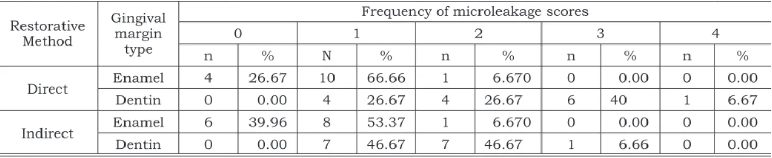
pres-ence of marginal gap (Figures 2 and 3), the pen-etration of silver between the hybrid layer and into the dentin is easily observed, the amount of leak-age being determined by the restorative method used. SEM analysis showed no difference in the formation of gap-free enamel margins with direct or indirect inlays (Table 3), but the microleakage was greater in dentin with the direct technique. In addition, when the overall leakage in the same method was analyzed, microleakage was greater in the dentin (Figure 4) than in the enamel gingival margins (Figure 3).
The professional’s initial intention should always be to analyze the possibility of using the direct technique. However, the indirect composite
FIGURE 2 -A: SEM of the dentin gingival margin cavity restored with direct composite showing the marginal gap and large secondary electron image along the dentin/adhesive interface
(magniication 200 X). B: SEM of the enamel gingival margin cavity restored with indirect composite inlay with intact external margin and absence of the secondary electron image along the dentin/
adhesive interface (magniication
200 X). DC - direct composite restoration; IC - indirect composite inlay; RC - Resinous
DC
D
IC
D
RC
restoration, which has shown a close fit at the margins26, may become an important alternative
for restoring complex and extensive cavities in pos-terior teeth.
CONCLUSION
Within the limits defined in the experimental design, the following conclusions may be drawn:
• Direct and indirect restorations showed simi-lar behavior in relation to leakage when the preparation’s gingival margin was placed in enamel.
• Indirect restorations showed smaller leakage than direct restorations when the prepara-tion’s gingival margin was placed in dentin. • Both evaluation methods showed similar
ca-pacity to analyze marginal leakage, but their combined use increased accuracy.
ACKNOWLEDGMENTS
The authors are indebted to Dr. E. W. Kitajima (NAP-MEPA/ESALQ-USP) for the SEM technical support. This study was supported by grants from FAPESP, SP, and CAPES/Brazil.
DC
D HL
B
RC
HL
D A
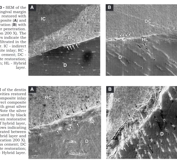
FIGURE 4 - SEM of the dentin gingival margin cavities restored with indirect composite inlay (A) and direct composite restoration (B) with great silver penetration. Note the silver penetration indicated by black arrows between restorative material and top of hybrid layer, and white arrows indicating
the silver iniltrated between
bottom of hybrid layer and
dentin (magniication 200 X).
RC - Resinous cement; DC - direct composite restoration; D - Dentin; HL - Hybrid layer.
IC
RC
D
D
DC
HL
A B
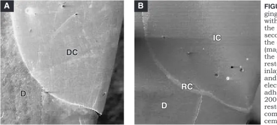
FIGURE 3 - SEM of the enamel gingival margin cavities restored with indirect composite (A) and direct restoration (B) with little silver penetration.
(magniication 200 X). The
white arrows indicate the
silver iniltrated in the
hybrid layer. IC - indirect composite inlay; RC - Resinous cement; DC - direct composite restoration; D - Dentin; HL - Hybrid layer.
REFERENCES
1. Alavi AA, Kianimanesh N. Microleakage of direct and in-direct composite restorations with three dentin bonding agents. Oper Dent 2002;27(1):19-24.
2. Ateyah NZ, Elhejazi AA. Shear bond strengths and mi-croleakage of four types of dentin adhesive materials. J Contemp Dent Pract 2004;5(1):63-73.
3. Beznos C. Microleakage at the cervical margin of composite Class II cavities with different restorative techniques. Oper Dent 2001;26(1):60-9.
for bonding in Class V and Class II preparations. J Dent 1997;25(5):379-89.
5. Eick JD, Gwinnett AJ, Pashley DH, Robinson SJ. Current concepts on adhesion to dentin. Crit Rev Oral Biol Med 1997;8(3):306-35.
6. Gemalmaz D, Ozcan M, Yoruc AB, Alkumru HN. Marginal adaptation of a sintered ceramic inlay system before and after cementation. J Oral Rehabil 1997;24(9):646-51. 7. Gerdolle DA, Mortier E, Loos-Ayav C, Jacquot B, Panighi
MM. In vitro evaluation of microleakage of indirect com-posite inlays cemented with four luting agents. J Prosthet Dent 2005;93(6):563-70.
8. Hahn P, Schaller H, Hafner P, Hellwig E. Effect of different luting procedures on the seating of ceramic inlays. J Oral Rehabil 2000;27(1):1-8.
9. Haller B, Hassner K, Moll K. Marginal adaptation of dentin bonded ceramic inlays: effects of bonding systems and lut-ing resin composites. Oper Dent 2003;28(5):574-84. 10. Hannig M, Friedrichs C. Comparative in vivo and in
vitro investigation of interfacial bond variability. Oper Dent 2001;26(1):3-11.
11. Hasanreisoglu U, Sonmez H, Uctasli S, Wilson HJ. Microleakage of direct and indirect inlay/onlay systems. J Oral Rehabil 1996;23(1):66-71.
12. Kanca J 3rd, Suh BI. Pulse activation: reducing
resin-based composite contraction stresses at the enamel cavo-surface margins. Am J Dent 1999;12(3):107-12.
13. Liberman R, Ben-Amar A, Herteanu L, Judes H. Mar-ginal seal of composite inlays using different polymerization techniques. J Oral Rehabil 1997;24(1):26-9.
14. Linden LA, Kallskog O, Wolgast M. Human dentine as a hydrogel. Arch Oral Biol 1995;40(11):991-1004. 15. Mehl A, Hickel R, Kunzelmann KH. Physical
prop-erties and gap formation of light-cured composites with
and without ‘softstart-polymerization’. J Dent 1997;25(3-4):321-30.
16. Milleding P. Microleakage of indirect composite in-lays. An in vitro comparison with the direct technique. Acta Odontol Scand 1992;50(5):295-301.
17. Miller MB, Castellanos IR, Vargas MA, Denehy GE. Effect of restorative materials on microleakage of Class II composites. J Esthet Dent 1996;8(3):107-13.
18. Ogata M, Okuda M, Nakajima M, Pereira PN, Sano H, Tagami J. Influence of the direction of tubules on bond strength to dentin. Oper Dent 2001;26(1):27-35.
19. Pashley DH, Carvalho RM. Dentine permeability and dentine adhesion. J Dent 1997;25(5):355-72.
20. Perdigao J, Lopes M. Dentin bonding – state of the art 1999. Compend Contin Educ Dent 1999;20(12):1151-8. 21. Rodrigues JA, De Magalhaes CS, Serra MC, Rodrigues
Junior AL. In vitro microleakage of glass-ionomer composite resin hybrid materials. Oper Dent 1999;24(2):89-95. 22. Saboia V de P, Pimenta LA, Ambrosano GM. Effect
of collagen removal on microleakage of resin composite restorations. Oper Dent 2002;27(1):38-43.
23. Sano H, Shono T, Takatsu T, Hosoda H. Microporous dentin zone beneath resin-impregnated layer. Oper Dent 1994;19(2):59-64.
24. Sano H, Takatsu T, Ciucchi B, Horner JA, Matthews WG, Pashley DH. Nanoleakage: leakage within the hybrid layer. Oper Dent 1995;20(1):18-25.
25. Soares CJ, Martins LR, Fernandes Neto AJ, Giannini M. Marginal adaptation of indirect composites and ceramic inlay systems. Oper Dent 2003;28(6):689-94.
26. Swift EJ Jr, Perdigao J, Heymann HO. Bonding to enamel and dentin: a brief history and state of the art, 1995. Quintessence Int 1995;26(2):95-110.