In vitro
evaluation of marginal leakage in
bonded restorations, with mechanical or
chemical-mechanical (Carisolv) removal
of carious tissue
Avaliação
in vitro
da infiltração marginal em
restaurações adesivas, com remoção mecânica
ou químico-mecânica (Carisolv) do tecido
cariado
Abstract: This study carried out an in vitro evaluation and comparison of the occurrence of marginal leakage in bonded restorations using mechanical or chemical-mechanical (Ca-risolv) removal of carious tissue. For that purpose, 40 extracted decayed human molars were divided into 4 groups: GI (burs + Prime & Bond NT + TPH), GII (Carisolv + Prime & Bond NT + TPH), GIII (burs + SBMP + Z100) and GIV (Carisolv + SBMP + Z100). After accomplishment of the restorations and thermal cycling, the teeth were exposed to dye, sectioned and qualitatively evaluated. The results demonstrated that the system of removal of carious tissue did not inluence the results of microleakage at any of the cavity margins. At dentinal margins, use of the Prime & Bond NT + TPH restorative system al-lowed the occurrence of less microleakage than the SBMP + Z100 system.
Descriptors: Dental leakage; Dental caries; Dental restoration, permanent.
Resumo: Este estudo avaliou e comparou in vitro a ocorrência da iniltração marginal em restaurações adesivas, com a utilização de remoção mecânica e químico-mecânica do tecido cariado. Para a proposição, 40 dentes molares humanos extraídos cariados foram divididos em quatro grupos: GI (brocas + Prime & Bond NT + TPH), GII (Carisolv + Pri-me & Bond NT + TPH), GIII (brocas + SBMP + Z100) e GIV (Carisolv + SBMP + Z100). Após a execução das restaurações e termociclagem, os dentes foram expostos a corante, seccionados e avaliados qualitativamente. Os resultados demonstraram que o sistema de remoção de tecido cariado não inluenciou a ocorrência de microiniltração em quaisquer margens cavitárias. Nas margens dentinárias, a utilização do sistema restaurador Prime & Bond NT + TPH demonstrou menor ocorrência de microiniltração do que o sistema SBMP + Z100.
Descritores: Iniltração dentária; Cárie dentária; Restauração dentária permanente.
Ricardo Coelho Okida(a)
Thiago Marchi Martins(b)
André Luiz Fraga Briso(a)
(a) PhDs, Assistant Professors, Discipline
of Restorative Dentistry; (b)Specialist in
Restorative Dentistry – School of Dentistry of Araçatuba, State University of São Paulo.
Corresponding author:
Ricardo Coelho Okida
Faculdade de Odontologia de Araçatuba da Universidade Estadual Paulista (UNESP) Rua José Bonifácio, 1193, Vila Mendonça, Araçatuba - SP - Brazil
CEP: 16015-050
E-mail: [email protected]
Introduction
For years, it has been observed that, despite the efforts in adopting a preventive approach, dental caries is still the main factor leading to tooth loss.6
Thus, after the development of carious lesions, in most cases the dentist must perform surgical restor-ative procedures aiming at reestablishing the shape, function and esthetics of the teeth involved in the carious processes and especially restoring the health status.6
In these situations, when more invasive proce-dures are needed, several types of cavity preparation and removal of carious tissue have been suggested and developed, especially due to fast technologi-cal evolution and to the several materials currently available.6
Most professionals, however, still perform con-ventional removal of carious tissue using excavators and round burs at low speed. Notwithstanding, a chemical-mechanical system has been recently de-veloped as an option for the removal of this tissue, whose main advantage is to provide a less traumatic resource for the patient and allow more selective re-moval of the carious tooth structure.1
The utilization of chemical substances for dis-closure and removal of carious tissue was initiated in 1979 with Fusayama8, who employed a dye
com-posed of 0.5% basic fuchsin in propylene glycol on carious lesions to observe infected dentin. However, due to the carcinogenic potential of this substance, a dye composed of 1% acidic red in propylene gly-col was adopted, which theoretically would disclose only the dentin that should be removed.8
Later, Maltz et al.16 (1998)demonstrated that
dyes such as 1% acidic red in propylene glycol have afinity with areas with lower mineral concentra-tion, such as the dentinoenamel juncconcentra-tion, and deep circumpulpar dentin, and with areas with a high degree of porosity, such as white spot lesions.15 For
that reason, the utilization of these dyes has not been effective for the identiication of only infected dentin; rather, they also stain areas of intact den-tin.
More recently, the current stage of alternatives for the disclosure and removal of carious tissue com-prises a new chemical-mechanical system for caries
removal called Carisolv, which has been presenting good outcomes compared to the conventional sys-tem.9 This product has been the subject of many
his-totomographic studies that revealed that the intact dentinal structure is not signiicantly affected by its utilization, since the penetration of Carisolv into in-tact dentinal tubules is not greater than 50 µm in depth.9 Similarly, while testing microtensile bond
strength, Burrow et al.3 (2003) observed that the
product does not signiicantly affect the bonding of adhesive restorative materials to dentinal tissue, as also observed by Erhardt et al.7 (2004).
However, considering the increased utilization of adhesive restorative techniques, there is a lack of studies in the literature relating the utilization of chemical-mechanical systems with the occurrence of marginal leakage in restorations.
Thus, the present study evaluated the occurrence of microleakage in Class V cavities, with cervical margins in dentin/cementum and occlusal margins in enamel, prepared by the chemical-mechanical method (Carisolv) or by the conventional method for the removal of carious tissue, and restored with different adhesive restorative materials currently employed.
Material and Methods
Before carrying out the experimental part of the study, the project was submitted to and approved by the Institutional Review Board of the Dental School of Araçatuba, UNESP (Process n. 2001-00631). The study was conducted on 40 human teeth present-ing caries at the buccal and lpresent-ingual cervical thirds, extracted for periodontal reasons. The teeth were obtained from emergency dental services, dental of-ices and basic health centers in the city of Araça-tuba, SP, Brazil, and were extracted for reasons not related to the study. After extraction, the surfaces of the teeth were cleaned with scalers and a n. 11 blade for the removal of calculus and remnants of the periodontal ligament. They were then stored in a 2% formalin solution (pH 7.0) for 14 days, and subsequently stored in saline solution.
Group I
In this group, the carious tissue was removed by the conventional method, i.e. beginning with excava-tors and then followed by smooth round burs with sizes adequate to the extension of the carious lesion, at low speed. For observation of the complete re-moval of carious tissue, the clinical-tactile criterion was adopted, and the desirable characteristic was the achievement of a remaining dentin resistant to attri-tion and to the penetraattri-tion of a dental probe. Then, prophylaxis was performed with pumice and water with the aid of a rubber dam at low speed. After washing and drying, acid etching was performed with 37% phosphoric acid gel (Dentsply Caulk, Petrópo-lis, RJ 90915, Brazil). Application of the adhesive sys-tem Prime & Bond NT (Dentsply Caulk, Petrópolis, RJ 90915, Brazil) was then carried out following the manufacturer’s instructions. After obtaining shiny dentin and enamel surfaces, the cavity was consid-ered ready to receive the restorative material.
The cavity was then illed with increments of composite resin TPH (Dentsply Caulk, Petrópolis, RJ 90915, Brazil) which were light-cured for 40 sec-onds each with a light intensity of 450 mW/cm².
Group II
In this group, the infected tissue was removed with the chemical-mechanical system Carisolv (Medi Team, Dentaluveckling I, Goteborg, AB). This sys-tem is available in two plastic syringes, and its acti-vation is initiated by the contact between the gels in each syringe. This step was performed immediately after onset of treatment. The mixture was taken to the carious lesion with the aid of its proper instru-ments and the product was allowed to act for 30 seconds. After staining of the infected tissue was observed, it was removed with a manual cutting in-strument supplied by the manufacturer. The product was applied until complete removal of the infected tissue, as clinically demonstrated by the absence of staining of the dentinal tissue secondary to applica-tion of the dye supplied in the product. In this situ-ation, the Carisolv gel remains clear because it no longer reacts with the infected tissue.
The restorative procedures were then performed as described for Group I.
Group III
For this group, the infected tissue was removed by the conventional method, as described for Group I, yet the adhesive system employed was Scotchbond Multi Purpose (3M Dental Products, Irvine, CA 92614), whose application technique also followed the manufacturer’s instructions. The adhesive system was then light-cured for 20 seconds and composite resin Z100 (3M Dental Products, Irvine, CA 92614) was inserted as described for the other groups.
Group IV
In this group, Carisolv was also employed for the removal of carious tissue, and the product was ap-plied as described for Group II. The restorative pro-cedures were the same as described for Group III.
After completion of the restorations, the speci-mens were kept for 24 hours in a FANEM oven (model 315 SE) at 37°C, and then inished and pol-ished with the Solex Pop-On system (3M Dental Products, Irvine, CA 92614). Then the specimens were submitted to 100 thermal cycles at 5°C and 55°C (± 2) and received two coats of nail enamel; only the interface to be evaluated was kept exposed. The specimens were then immersed in 2% basic fuchsin for 8 hours, washed in tap water for 10 min-utes, individually mounted onto acrylic resin blocks and sectioned in the buccolingual direction with a Buehler metallographic cutter (model Isomet), under water cooling, to produce two sections, which were then evaluated.
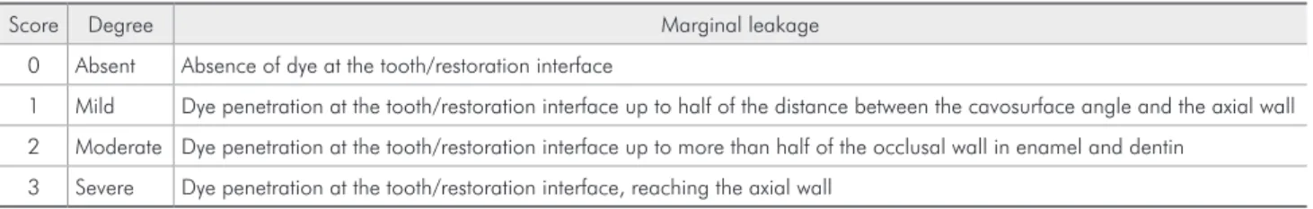
Evaluation of the degree of marginal leakage was performed with the aid of a stereoscopic magnifying glass Stemi SU11 (Zeiss) with 16 X magniication; scores 0 to 3 were assigned to the margins in enamel and dentin/cementum, as described in Chart 1. The data were plotted and submitted to statistical analy-sis by the Mann-Whitney test at 5%.
Results
With regard to the cavity margins in dentin, res-torations performed with TPH and Z100 were not inluenced by the technique for removal of carious tissue. However, the TPH restorative system provid-ed better control of the occurrence of microleakage with both types of preparation, as revealed by com-parison of the medians presented in Table 2.
Discussion
The search for a conservative manner of remov-ing carious tissue has been investigated by several authors since the conventional approach using burs allows a fast procedure, yet may lead to unnecessary wear of tooth structure, due to the effective cutting of these instruments, consequently weakening the remaining tooth structure.2,4,5,19,21,22
According to Inaba et al.11 (1995), Carisolv,
which is made of sodium hypochlorite (NaOCl), is a proteolytic agent that allows effective removal of organic components, leading to rupture of hy-drogen bonds in the dentinal collagen ibers previ-ously degraded by the demineralization secondary to the carious process. These authors further state that this rupture would soften this tissue and con-sequently reduce the pressure required for removal by attrition,19 a characteristic of the ideal action
of instruments especially developed for utilization
with this product. This undoubtedly allows better control of wear when compared to removal with burs following the conventional technique, in which large amounts of dentin are removed in blocks, with higher possibility of pulp exposure.1
It should be highlighted that the interaction of the chloride present in the product with collagen does not occur in mineralized tissues. The chemical-mechanical removal, thus, may not be applied in in-cipient lesions, since the mineral components protect the collagen ibers from the action of sodium hypo-chlorite, assigning selectivity to this technique.1
Fusayama8 (1979) made use of similar solutions
and proposed the removal of only infected dentin, which could not be remineralized, allowing preser-vation of the inner layer, which is not contaminat-ed.8,10 However, more recently, Maltz et al.16 (1998)
conducted histological and bacteriological analyses and demonstrated that chemical-mechanical removal does not assure complete absence of microorganisms in the remaining dentin. Also, according to Kidd et al.14 (1993), morphological and ultrastructural
anal-yses of intact teeth demonstrated that the dyes stain areas with high degree of porosity, such as areas with limited organic content (lesions without cavities) and areas with a large amount of organic matter (denti-noenamel junction and circumpulpar dentin). Thus,
Table 1 - Comparison between the medians obtained for the occurrence of microleakage at the enamel margins.
Conventional Carisolv
Median Min. Max. Median Min. Max.
TPH 0.00 Aa 0.00 1.00 0.00 Aa 0.00 1.00
Z100 0.00 Aa 0.00 1.00 0.00 Aa 0.00 1.00
Medians followed by similar letters (capital letters in lines and lower case letters in rows) are not statistically different according to Mann-Whitney’s test (p > 0.05). Min. = Minimum, Max. = Maximum.
Table 2 - Comparison between the medians obtained for the occurrence of microleakage at the dentin/cementum margins.
Conventional Carisolv
Median Min. Max. Median Min. Max.
TPH 0.50 Ab 0.00 1.00 0.00 Ab 0.00 1.00
Z100 1.00 Aa 0.00 2.00 1.00 Aa 0.00 2.00
Medians followed by similar letters (capital letters in lines and lower case letters in rows) are not statistically different according to Mann-Whitney’s test (p > 0.05). Min. = Minimum. Max. = Maximum.
Chart 1 - Evaluation of the degree of marginal leakage in enamel/dentin.
Score Degree Marginal leakage
0 Absent Absence of dye at the tooth/restoration interface
1 Mild Dye penetration at the tooth/restoration interface up to half of the distance between the cavosurface angle and the axial wall
2 Moderate Dye penetration at the tooth/restoration interface up to more than half of the occlusal wall in enamel and dentin
the chemical-mechanical removal in deep carious le-sions might increase the risk of pulp exposure.
Considering these observations as to the utiliza-tion of Carisolv, the formautiliza-tion of a uniform hybrid layer in all cavity walls becomes fundamental to al-low an hermetic sealing of the tooth/restoration in-terface and thus eliminate the substrate, so that any possible remaining bacteria would reduce their me-tabolism and become inactive.23
It should be highlighted that failure in the inter-action between adhesive system and tooth substrate yields poor marginal sealing with consequent micro-leakage, which may lead to early loss of the restora-tion, postoperative sensitivity, discolorarestora-tion, mar-ginal deterioration and secondary caries, ultimately leading to displacement of the restoration and pulp damage.20
Within this context, after accomplishment of the cavity preparation and removal of the carious tissue, especially using burs, formation of a layer of organic and mineral debris called smear layer occurs. Its re-moval is an important step to allow better interaction with most hydrophilic adhesive systems currently adopted.17 On the other hand, when manual cutting
instruments are used, the evidence demonstrates that this layer is not formed and the surface treatments are applied directly on the dentinal tissue.18
The results of the microleakage test revealed no statistically signiicant difference between the mate-rials evaluated at the enamel margins, regardless of the technique employed for removal of the carious tissue (Table 1), which once again demonstrates the effectiveness of enamel acid etching on the sealing achieved in restorations with enamel margins, as also observed in other studies5,23. As for the decayed
tissue removal, the procedures used did not inlu-ence the occurrinlu-ence of microleakage in any margin, being the restorative system the limiting factor of restoration performance.
However, with regard to the margins in dentinal tissue, the groups restored with the Prime & Bond NT adhesive system and TPH composite resin pre-sented better outcomes when compared to the groups restored with the Z100 composite resin system.
The similar performance of both the convention-al and chemicconvention-al-mechanicconvention-al techniques associated to
the difference in the performance of restorations in dentinal tissue suggest that more importance should be assigned to the restorative system and cavity margins than to the method of removal of carious tissue. Moreover, it can be assumed that the use of phosphoric acid by the evaluated restorative systems produced similar results in terms of hybrid layer formation, with or without the presence of smear layer.
According to Jacobsen, Soderholm13 (1998),
ac-etone-based adhesives present a higher diffusion ability and display more effective penetration of the adhesive into the dentinal structure, either exposed to hypochlorite or not.
On the other hand, Inai et al.12 (1998) assigned
the results observed in dentin with the Prime & Bond 2.1 system to the presence of a phosphoric acid ester in its composition, which would allow a better interaction between the phosphate radicals and the calcium ions in dentin, and this substance is not found in the Scotchbond Multi Purpose adhesive system. The authors further report that, even after removal of collagen by NaOCl, the performance of the Prime & Bond adhesive was better compared to that of Scotchbond Multi Purpose, whose primer contains water and alcohol.
Thus, the present study is expected to contribute to evaluate and compare the conventional and chem-ical-mechanical methods for removal of carious tis-sue, prior to the accomplishment of the restorative procedure. The indication and utilization of Carisolv as a means for the removal of carious tissue has pre-sented encouraging outcomes; however, further stud-ies and a strict analysis of the viability of its routine utilization are recommended. On the other hand, the conventional method for the removal of carious tis-sue, in addition to being an easy one, still provides favorable results from a biological standpoint.
Conclusions
Based on the obtained results and according to the methodology employed, the following conclu-sions could be drawn:
The technique for removal of carious tissue did not inluence the degree of marginal leakage for margins in enamel and dentin/cementum.
The restorative system did not inluence the oc-currence of microleakage in the enamel margins. The restorative system comprising the Prime & Bond NT adhesive and the TPH composite resin 2.
3.
was more effective to control microleakage than the system comprising the SBMP adhesive and the Z100 resin for margins in dentinal tissue.
References
1. Bianchi S, Ciuffreda M, Poggio C, Piacontini C, Paroli R. Sis-tema Caridex per la rimozione della carie: sperimentazione clinica. Dent Cadmos. 1989;57(12):46-52.
2. Black GV. Operative dentistry. Chicago: Medical Dental; 1908.
3. Burrow MF, Bokas J, Tanumiharja M, Tyas MJ. Microtensile bond strengths to caries-affected dentine treated with Cari-solv. Aust Dent J. 2003;48(2):110-4.
4. Çehreli ZC, Yazici AR, Akca T, Özgünaltay G. A morphologi-cal and micro-tensile bond strength evaluation of a single-bottle adhesive to caries-affected human dentine after four different caries removal techniques. J Dent. 2003;31(6):429-35.
5. Conceição EN. Dentística: saúde e estética. Porto Alegre: ART-MED; 2000.
6. Cortes O, Garcia C, Perez L, Perez D. Marginal microleakage around enamel and cementum surfaces of two compomers. J Clin Pediatr Dent. 1998;22(4):307-10.
7. Erhardt MC, Amaral CM, de Castro AK, Ambrosano GM, Pimenta LA. In vitro influence of Carisolv on shear bond strength of dentin bonding agents. Quintessence Int. 2004;35(10):801-7
8. Fusayama T. Two layers of carious dentin: diagnosis and treat-ment. Oper Dent. 1979;4(2):63-70.
9. Gallo J, Xu X, Burgess JO, Re GJ. Dentin bond strength of three composite resins using five adhesives [abstract]. J Dent Res. 1988;77:945.
10. Haak R, Wicht MJ, Noack MJ. Does chemomechanical caries removal affect dentine adhesion? Eur J Oral Sci. 2000;108(5): 449-55.
11. Inaba D, Duschner H, Jongebloed W, Odelius H, Takagi O, Arends J. The effects of a sodium hypochlorite treatment on demineralized root dentin. Eur J Oral Sci. 1995;103(6):368-74.
12. Inai N, Kanemura N, Tagami J, Watanabe LG, Marshal SJ, Marshal GW. Adhesion between collagen depleted dentin and dentin adhesives. Am J Dent. 1998;11(3):123-7.
13. Jacobsen T, Soderholm KJ. Effect of primer solvent, primer agitation, and dentin dryness on shear bond strength to den-tin. Am J Dent. 1998;11(5):225-8.
14. Kidd EA, Ricketts DN, Pitts NB. Occlusal caries diagnosis: a changing challenge for clinicians and epidemiologists. J Dent. 1993;21(6):323-31.
15. Kubo S, Li H, Burrow MF, Tyas MJ. Nanoleakage of dentin adhesive systems bonded to Carisolv-treated dentin. Oper Dent. 2002;27(4):387-95.
16. Maltz M, Henz SL, Volkweis A, Campos CS. Morphologi-cal and ultrastructural evaluation of the specificity of caries detector dye in the identification of carious dentine [abstract]. Caries Res. 1998;32(4):294.
17. Mertz-Fairhurst EJ, Call-Smith KM, Shuster GS, Williams JE, Davis QB, Smith CD et al. Clinical performance of sealed composite restorations placed over caries compared with sealed and unsealed amalgam restorations. J Am Dent As-soc. 1987;115(5):689-94.
18. Naressi SCM. Comparação da infiltração marginal de restau-rações adesivas empregando instrumento rotatório e um siste-ma químico-mecânico na remoção do tecido cariado [Tese de Doutorado]. São José dos Campos: Faculdade de Odontologia da UNESP; 1999.
19. Petruzillo MA, McNierney HD. Chemomechanical caries removal system in pediatric dentistry. NY State Dent J. 1998;54(2):29-32.
20. Pinheiro SL, Aoki CMB, Mendes FM, Bengston AL. Avaliação morfológica da dentina após diferentes métodos de remoção do tecido cariado. Rev Assoc Paul Cir Dent. 2004;58(5):363-8. 21. Robbins A, Ragan MR. Dentist’s influence on patient demand
for local anesthesia with a chemomechanical system. J Pros-thet Dent. 1988;59(2):142-5.
22. Toi CS, Bönecker M, Cleaton-Jones PE. Mutans streptococci strains prevalence before and after cavity preparation during atraumatic restorative treatment. Oral Microbiol Immunol. 2003;18(3):60-4.