Nitric oxide pathway is an important modulator of
heat loss in rats during exercise
Ana Cristina R. Lacerda, Umeko Marubayashi, Cˆandido C. Coimbra
∗Department of Physiology and Biophysics, Institute of Biological Sciences, Federal University of Minas Gerais, 31270-901 Belo Horizonte, Minas Gerais, Brazil
Received 1 June 2005; accepted 3 June 2005 Available online 27 June 2005
Abstract
To assess the role of nitric oxide (NO) in central thermoregulatory mechanisms during exercise, 1.43mol (2L) ofN-nitro-l-arginine
methyl ester (l-NAME,n= 6), a NO synthase inhibitor, or 2L of 0.15 M NaCl (SAL,n= 6) was injected into the lateral cerebral ventricle of
male Wistar rats immediately before the animals started running (18 m min−1, 5% inclination). Core (
Tb) and skin tail (Ttail) temperatures were measured. Body heating rate (BHR), thresholdTbfor tail vasodilation (TTbV), and workload (W) were calculated. During the first 11 min of exercise, there was a greater increase inTbin thel-NAME group than in the SAL group (BRH = 0.17±0.02◦C min−1,
l-NAME, versus
0.09±0.01◦C min−1, SAL,p< 0.05). Following the first 11 min until
∼40 min of exercise,Tblevels remained stable in both groups, but levels remained higher in thel-NAME group than in the SAL group (39.16±0.04◦C,l-NAME, versus 38.33±0.02◦C, SAL,p< 0.01). However,
exercise went on to induce an additional rise inTb in the SAL group prior to fatigue. These results suggest that the reducedWobserved inl-NAME-treated rats (10.8±2.0 kg m,l-NAME, versus 25.0±2.1 kg m, SAL,p< 0.01) was related to the increased BHR inl
-NAME-treated animals observed during the first 11 min of exercise (r= 0.74,p< 0.01) due to the change in TTbV (39.12±0.24◦C,l-NAME, versus
38.27±0.10◦C, SAL,p< 0.05). Finally, our data suggest that the central nitric oxide pathway modulates mechanisms of heat dissipation
during exercise through an inhibitory mechanism. © 2005 Elsevier Inc. All rights reserved.
Keywords: Exercise; Core temperature; Central fatigue; Heat storage; Body heating rate
1. Introduction
Internal body temperature is considered to be a limiting factor during prolonged physical exercise [7,12,16,23,33]. However, the mechanisms responsible for exercise fatigue related to increasing body temperature (Tb) are still not fully
understood, and it is not known whether the increase in tem-perature during exercise is due to a rise in set-point levels or whether it occurs passively through an unwanted accumula-tion of heat[4,12,40,54,55]. Controversy also exists regard-ing whether there is a critical absolute value ofTband/or heat storage (HS) that determines the point of fatigue. Fuller et al.
[12]showed that rats exercising at high room temperatures reached fatigue at the same abdominal and brain
tempera-∗Corresponding author. Tel.: +55 31 34992936; fax: +55 31 34992924.
E-mail address:[email protected] (C.C. Coimbra).
tures (∼40◦C) even after pre-exercise body temperature was
altered. Their results are therefore consistent with the concept that high body core temperature reduces the CNS drive for exercise performance[34,35,54], and with the hypothesis that hyperthermia precipitates feelings of fatigue at a sublethal threshold and establishes a safe guard against heat stroke, pro-tecting the brain from thermal damage[7,23,54]. On the other hand, high body heat storage is also associated with the termi-nation of work in animals[12]and healthy humans[16,27]. Therefore, considering that fatigue is coincident or may be precipitated by high core temperature and/or heat storage, activation of a central mechanism that would increase heat loss and decrease core temperature might improve exercise performance. It has been demonstrated that nitric oxide (NO) acting on the brain exerts thermoregulatory effects produc-ing anapyrexia[18,29]and hypothermia[1,2,3,30,48,52].l
-Arginine (nitric oxide synthase—NOS substrate) analogues
and NOS blockers such asN
-nitro-l-arginine methyl ester
(l-NAME) have been used to investigate the role of NO in
body temperature under resting conditions[1,10,30,51,52]. Several studies have observed that intracerebroventricular injection of l-NAME causes a slight increase in the Tb
of rats, indicating that central NO plays a tonic role in reducingTb[3,10,30,51,52]. Moreover, in the case of fever
induced by interleukins (IL-1), lipopolysaccharide (LPS) or psychological stress, intracerebroventricular l-NAME
enhanced stress fever, supporting the hypothesis that NO in the CNS leads to a reduction inTb[10,30,39,42,50]. Since
hypothermia and increased heat dissipation may be neuropro-tective, activation of central oxide nitrergic transmission may exert important effects on thermoregulation during exercise, influencing running performance. It is important to empha-size that until now, there have been no reports in the literature relating brain nitric oxide and thermoregulation during exer-cise. Therefore, the aim of this study was to assess the effects of the central administration of the NO synthase inhibitorl
-NAME on heat balance and thresholdTbfor tail vasodilation
(TTbV) in untrained rats submitted to exercise until fatigue.
2. Materials and methods
2.1. Animals
Male Wistar rats (250–340 g) were housed individually under 14-h light:10-h dark cycles and had free access to water and rat chow. Following anesthesia achieved using 2,2,2-tribromoethanol (300 mg/kg body weight ip), the rats were fixed to a stereotaxic apparatus (David Kopf Instruments, M-900, Tujunga, CA, USA) and a guide cannula (22 G) was implanted into the right lateral cerebral ventricle using a pre-viously described technique [25,26,40,46,47]. All animals were allowed to recover for at least 1 week before being submitted to the experiments. The rats were familiarized to exercise on the motor-driven treadmill by running at a speed of 18 m min−1at 5% inclination for 5 min per day during 4
consecutive days prior to the experiments. The purpose of this preliminary exercise was to show the animals in which direction to run without becoming entangled in the rectal-probe leads and preventing measurement of the conditioned hyperthermic response. All experiments were approved by the Ethics Committee for the Care and Use of Laboratory Animals of the Federal University of Minas Gerais and were carried out in accordance with the regulations described in the Committee’s Guiding Principles Manual (protocol 012/05).
2.2. Exercise
Exercise was performed on a motor-driven treadmill (Columbus Instruments, OH, USA, Modular Treadmill) between 13:00 and 17:00 h at a room temperature of 21±2◦C. The intensity of exercise (18 m min−1 and 5% inclination) corresponded to an oxygen uptake of∼66% of
VO2max[6,21]. Fatigue was defined as the point at which the
animals were no longer able to keep pace with the tread-mill [40,46,47]. Time to fatigue (minutes) and workload (kilogram meter) were considered indexes of exercise per-formance.
2.3. Experimental protocol
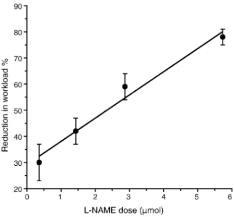
On the day of the experiment, the animals were allowed to rest for 1 h in the rodent treadmill chamber before being sub-mitted to the test. A needle (30 G) protruding 0.3 mm from the tip of the guide cannula was introduced into the right lateral cerebral ventricle by connecting it to a Hamilton syringe. Immediately prior to exercise, 2.0L of 0.15 M NaCl (n= 6) or 2.0L ofl-NAME (1.43mol,n= 6) was injected into
the right lateral ventricle. The dose of brain l-NAME was
based on the results of our previous experiments that showed that the response of reduction in workload percentual, related to SAL group, was clearlyl-NAME dose-dependent
(Fig. 1). Furthermore, according to the literature, the effect ofl-NAME was mediated entirely centrally because of the
inability of low doses ofl-NAME to cross the blood brain
barrier[8,24,31,53]. Rats were randomly assigned to groups receiving either saline or l-NAME solution. Immediately
after the injections, the animals were submitted to running exercise until fatigue was reached. Colonic temperature was taken as core temperature (Tb) index and it was measured
using a thermistor probe (model 401, Yellow Springs Instru-ments, USA). The lubricated thermistor probe was inserted 4 cm past the anal sphincter after fecal pellets had been removed from the colon by gentle, external massage. Skin tail temperature (Ttail) was measured using a probe, series 409-B
(Yellow Springs Instruments), taped to the dorsal surface of
the skin, about 10 mm from the base of the tail.TbandTtail
were used to determine the thresholdTbfor tail vasodilation,
i.e. the core temperature that corresponds to the moment at whichTtailclearly begins to increase (vasodilation).
TbandTtailwere recorded at rest, every minute during the
first 20 min of exercise, every 10 min from then until fatigue, and every 5 min thereafter during the 30 min of recovery.
2.4. Calculations
Body heating rate (BHR;◦C min−1), i.e. rate of increase
in core temperature, was calculated as BHS =Tb/(running
time interval), where Tb is the change in core
tempera-ture (Tf−Ti),Tf: core temperature at fatigue point andTi:
initial core temperature measured prior to exercise. Heat stor-age was calculated [17] as HS = (Tb)·m·c, where “m” is
the body weight in grams and “c” is the specific heat of the body tissues (0.826 cal g−1◦C−1). Workload (W; kg m) was
calculated asW= [body weight (kg)]·[TTF]·[treadmill speed (m min−1)]·[sinθ (treadmill inclination)] [5,6,26], where
TTF is time to fatigue (minutes).
2.5. Statistical analysis
The data are reported as mean±S.E.M. Differences between groups and the effect of time were evaluated using the analysis of variance (ANOVA) test followed by the Newman–Keuls test. The data were also compared using paired or unpaired Student’st-test, as applicable. The asso-ciation between BHR and W or TTbV was assessed using
Pearson’ s correlation coefficient. Significance level was set atp< 0.05.
3. Results
As illustrated inFig. 2, intracerebroventricular injection ofl-NAME in untrained normal rats (l-NAME group,n= 6
Fig. 2. Effect of intracerebroventricular injection of 2L of l-NAME (1.43mol;n= 6) or 0.15 M NaCl (SAL;n= 6) on time to fatigue (TTF) and workload (W) in untrained normal exercising rats. Data are expressed as mean±S.E.M. Significantly different from the control group (*p< 0.05).
Fig. 3. Effect of intracerebroventricular injection of 2L of l-NAME (1.43mol) or 0.15 M NaCl (SAL) on core temperature (Tb) during exercise (A) and during recovery time (B). Time to fatigue is indicated in (A) by the horizontal bar at bottom of the graph: SAL (open bar) andl-NAME (solid bar). Values are expressed as mean±S.E.M.,n= 6 for each group.*p< 0.05 compared with saline-treated group.#p< 0.05 compared with corresponding basal value.+p< 0.05 compared with corresponding fatigue point.
rats) induced a marked decrease in time to fatigue and work-load compared to saline-treated rats (SAL group,n= 6 rats). During the first 11 min of exercise, there was a greater increase inTbin the l-NAME group (Fig. 3A). Following
the first 11 min of exercise until ∼40 min of exercise, Tb
remained stable in both groups, but levels were higher in thel-NAME group than in the SAL group (39.16±0.04◦C, l-NAME, versus 38.33±0.02◦C, SAL,p< 0.01). However,
exercise still induced an additional rise inTb in the SAL
group prior to fatigue. Although rats in thel-NAME group
showed a more accentuated increase inTbduring the dynamic
phase of exercise and voluntarily interrupted the exercise much earlier than the control rats, both groups showed a very similar core temperature at fatigue point (39.32±0.13◦C, l-NAME, versus 39.17±0.11◦C, SAL). On the other hand, during recovery period,l-NAME-treated rats took a longer
Fig. 4. Effect of intracerebroventricular injection of 2L of l-NAME (1.43mol) or 0.15 M NaCl (SAL) on body heating rate (BHR) and heat storage (HS) during the first 11 min of running. Values are expressed as mean±S.E.M.,n= 6 for each group. Significantly different from the con-trol group (*p< 0.05).
Fig. 5. Effect of intracerebroventricular injection of 2L of l-NAME (1.43mol) or 0.15 M NaCl (SAL) on skin tail temperature (Ttail) during exercise (A) and during recovery period (B). Time to fatigue is indicated in (A) by the horizontal bar at bottom of the graph: SAL (open bar) and l-NAME (solid bar). Values are expressed as mean±S.E.M.,n= 6 for each group.*p< 0.05 compared with saline-treated group.#p< 0.05 compared with corresponding basal value.+p< 0.05 compared with corresponding lower value (7 min).++p< 0.05 compared with corresponding fatigue point.
displaying higherTbduring the first 5 min of the recovery
period (Fig. 3B). To compare the total thermal effects of exercise in the two treatment groups, BHR and HS were calculated and are shown in Fig. 4. As may be seen, the BHR and HS of l-NAME-treated animals were,
respec-tively, 53 and 55%, both higher (p< 0.01) than in SAL-treated group during the first 11 min of running (dynamic phase of exercise).
As illustrated inFig. 5A,Ttailincreased within 14–20 min of exercise in both groups, indicating that vasodilation had occurred and heat loss mechanisms had been acti-vated. Thereafter,Ttailremained stable in both groups (SAL:
30.95±0.06◦C; l-NAME: 31.17±0.14◦C, the average
measurement taken between 20 min and fatigue point). Dur-ing the recovery period,Ttailgradually decreased, and
dif-ferences in Ttail between groups were not apparent for up
to 30 min (Fig. 5B). We also observed a close correlation between BHR and TTbV (Fig. 6A,r= 0.905,p< 0.01), and
between BHR andW(Fig. 6B,r= 0.740,p< 0.05).
4. Discussion
In the present experiments,l-NAME-treated rats showed
the same baseline Tb as control animals. However, within
11 min of the onset of exercise, they presented a significantly higher BHR that rapidly produced hyperthermia 0.8◦C
higher than controls. This finding suggests a stronger inhibi-tion of heat dissipainhibi-tion mechanisms inl-NAME-treated rats,
which is reflected both by the marked increase (55%) in heat storage and the 0.85◦C increase in TTbV observed in these
animals. It is important to point out that the reduced exercise performance observed inl-NAME-treated rats was closely
associated with BHR during the first 11 min of exercise. Our results provide evidence that central oxide nitrergic transmis-sion exerts an important effect on thermoregulation during exercise by increasing heat dissipation through peripheral vasodilation and preventing high level of heat storage and excessive hyperthermia. To the best of our knowledge, this is the first study to examine the effect of central NO system on thermoregulation and our results may point towards a possible neuroprotective role in response to prolonged exercise.
The increase in body temperature that occurs in response to continuous exercise results from the temporary imbalance in the rates of heat production and dissipation during the early stage of exercise [14,23,55]. Vasoconstriction mediated by the sympathetic nervous system during this stage of exer-cise [19,28] impairs heat loss. Consequently,Tb increases
rapidly until it reaches the threshold for peripheral thermal vasodilation, thereby improving heat dissipation. Thereafter, the rise inTbplateaus at a high level and remains high until
fatigue.
The dissipation of heat from the body is thought to be more important than the control of heat production in the regulation of body temperature during exercise [15,55]. In exercising rodents, tail skin vasodilation is an important route of heat loss from the body, as rats do not dissipate heat through sweat evaporation[17,45,56]. Therefore, it is reasonable to propose that inhibition of central NO by intracerebroventricularl
-NAME reduces heat dissipation by increasing sympathetic outflow to superficial vascular beds in the tail, promoting vasoconstriction. The increased Tb threshold for tail
ther-mal vasodilation induced byl-NAME treatment, which was
strongly related to the BHR, supports this hypothesis. There-fore, the fact that the tail vasodilation threshold during exer-cise was induced at a higherTbin thel-NAME-treated rats
may have contributed to the increased BHR by delaying heat dissipation through skin tail.
Elevated internal body temperature and increased heat storage have been considered to be limiting factors that
[7,12,16,33]reduce the CNS drive for exercise performance
[34,35]and precipitate feelings of fatigue, thus protecting the brain from thermal damage. The brain nitric oxide pathway improving heat loss mechanisms may act by protecting the brain from excessive hyperthermia and improving physical performance.
Our results agree with the general idea that central NO plays a role in reducing the sympathetic tonus, and the increased tail thermal threshold to vasodilation induced by
l-NAME treatment supports this hypothesis.
The findings of various studies involving intracerebroven-tricular administration of NO blockade [30,32,38,48], or administration within specific sites in the CNS[38,41], are all in general agreement with the view that the central NO sys-tem is inhibitory to overall sympathetic outflow. At the central level, the currently available data suggest the specificity of NO actions on physiological temperature regulation, mainly inducing hypothermia and anapyrexia[1,2,3,13,16,29,50]. In addition, intracerebroventricular (icv) administration of l
-NAME in anesthetized rats produces an increase in heart rate and arterial blood pressure[37]blocked by administration of the adrenergic beta blocker, atenolol. Conversely, administra-tion ofl-arginine icv increases NO synthesis within the CNS
and produces a decrease in abdominal sympathetic nerve dis-charge in rats[36]. Furthermore, administration ofl-NAME
orl-NMMA, another inhibitor of nitric oxide synthase, into
the PVN or medial preoptic area (MPOA) also produced an increase in blood pressure and heart rate [57]that was reversed by central administration ofl-arginine. In summary,
the results of the central administration of modulators on the NO pathways within the cerebral ventricles consistently support the concept of tonic restraint of central sympathetic outflow by NO.
The exact location and precise pathways involved in the nitrergic mediation of normal thermoregulation during exercise still require clarification. However, hypothalamic regions expressing NOS, such as the preoptic area or paraventricular nucleus, are possible sites at which NO may influence thermoregulation during exercise. Therefore, we hypothesized that infusion ofl-NAME into the cerebral
ven-tricle would perfuse the thermoregulatory centers situated in the hypothalamus, inhibiting the heat loss response and accelerating the BHR and HS during prolonged exercise. The POA/AH is thought to be the primary locus for body tem-perature regulation[4,9,22,43,44,56] due to the fact that it contains both warm-sensitive and cold-sensitive neurons that respond to small changes in temperature[22,56]. Moreover, lesions or pharmacologic blockade of the POA/AH have been shown to produce a severe impairment in thermoregulation
[9,11,20,43,44]. It has been established that the POA/AH is an integrative region for the maintenance of metabolic, vasomotor, and thermal homeostasis[9,11,20,22,43,44,56]. It is important to point out that POA/AH cell groups project to the sympathetic outflow of the tail artery involved in heat loss in the rat[49], warming the preoptic area, and producing tail vasodilation in rats[22,56]. In addition, it has recently been shown that inhibition of the POA/AH by local infusion of tetrodotoxin impairs heat loss in running rats[20]. These results indicate that the POA/AH is an important mediator of heat loss as opposed to heat production during exercise and might be one possible site forl-NAME action. However,
of nitrergic mediation involved in normal thermoregulation during exercise.
In summary, icv infusion ofl-NAME induced a significant
increase in BHR that rapidly produced hyperthermia 0.8◦C
higher than in controls with a significant increase in TTbV. In
addition, treatment withl-NAME reduced exercise
perfor-mance that was closely associated with BHR during the first 11 min of exercise. Therefore, our results provide the first evidence that central oxide nitrergic transmission has impor-tant effects on thermoregulation during exercise by increasing heat dissipation through peripheral vasodilation, preventing high levels of heat storage, and protecting the brain against excessive hyperthermia.
Acknowledgements
The authors thank CNPq, CAPES, and FAPEMIG for financial support. The technical assistance of Mr. Andr´e Luis Pimenta de Faria is also acknowledged.
References
[1] M.C. Almeida, L.G.S. Branco, Role of nitric oxide in insulin-induced hypothermia in rats, Brain Res. Bull. 54 (2001) 49–53.
[2] K. Benamar, M.Z. Yondorf, D. Kon, E.B. Geller, M.W. Adler, Role of nitric-oxide synthase isoforms during morphine-induced hyper-thermia in rats, J. Pharmacol. Exp. Ther. 307 (2003) 219–222. [3] L.G.S. Branco, E.C. Carnio, R.C.H. Barros, Role of nitric oxide
pathway in hypoxia-induced hypothermia, Am. J. Physiol. Regul. Integr. Comp. Physiol. 273 (1997) 967–971.
[4] E. Briese, Normal body temperature of rats: the setpoints contro-versy, Neurosci. Biobehav. Rev. 22 (1998) 427–436.
[5] G.A. Brooks, C.M. Donovan, T.P. White, Estimation of anaerobic energy production and efficiency in rats during exercise, J. Appl. Physiol. 56 (1984) 520–525.
[6] G.A. Brooks, T.P. White, Determination of metabolic and heart rate responses of rats to treadmill exercise, J. Appl. Physiol. 45 (1978) 1009–1015.
[7] M. Caputa, G. Feistkorn, C. Jessen, Effect of brain and trunk temper-atures on exercise performance in goats, Pfl¨ugers Arch. 406 (1986) 184–189.
[8] R. Cespuglio, S. Burlet, S. Marinesco, F. Robert, M. Jouvet, Volta-metric detection of cerebral NO in rats. Variations of the signal throughout the sleep–wakefulness cycle, C. R. Acad. Sci. III 319 (1996) 191–200.
[9] C.C. Coimbra, R.H. Migliorin, Cold induced free fatty acid mobi-lization is impaired in rats with lesions in preoptic area, Neurosci. Lett. 88 (1988) 1–5.
[10] D. De Paula, A.A. Steiner, L.G.S. Branco, The nitric oxide pathway is an important modulator of stress-induced fever in rats, Physiol. Behav. 70 (2000) 505–511.
[11] M.L. Ferreira, U. Marubayashi, C.C. Coimbra, The medial preoptic area modulates the increase in plasma glucose and free fatty acid mobilization induced by cold exposure, Brain Res. Bull. 49 (1999) 189–193.
[12] A. Fuller, R.N. Carter, D. Mitchell, Brain and abdominal tempera-tures at fatigue in rats exercising in the heat, J. Appl. Physiol. 84 (1998) 877–883.
[13] R. Gerstlberger, Nitric oxide and body temperature control, News Physiol. Sci. 14 (1999) 30–36.
[14] M. Gleeson, Physiology of body temperature regulation: temperature regulation during exercise, Int. J. Sports Med. 19 (1998) 96–99. [15] C.V. Gisolfi, F. Moura, What’s so important about a body
temper-ature of 37◦C, in: C.V. Gisolfi, F. Mora (Eds.), The Hot Brain:
Survival, Temperature, and Human Body, The MIT Press, Cam-bridge/Massachusetts/London, 2000, pp. 95–119.
[16] J. Gonz´alez-Alonso, C. Teller, S.L. Andersen, F.B. Jensen, T. Hyldig, B. Nielsen, Influence of body temperature on the development of fatigue during prolonged exercise in the heat, J. Appl. Physiol. 86 (1999) 1032–1039.
[17] C.J. Gordon, Temperature Regulation in Laboratory Rodents, Cam-bridge University Press, CamCam-bridge, UK, 1993, pp. 8–11. [18] A.V. Gourine, Pharmacological evidence that nitric oxide can act
as an endogenous antipyretic factor in endotoxin-induced fever in rabbits, Gen. Pharmacol. 26 (1995) 835–841.
[19] L.H. Hartley, J.W. Mason, R.P. Hogan, L.G. Jones, T.A. Kotchen, E.H. Mougey, F.E. Wherry, L.L. Pennington, P.T. Ricketts, Multi-ple hormonal responses to graded exercise in relation to physical training, J. Appl. Physiol. 33 (1972) 602–606.
[20] H. Hasegawa, T. Ishiwata, T. Saito, T. Yazawa, Y. Aiahara, R. Meeusen, Inhibition of the preoptic area and anterior hypothalamus by tetrodotoxin alters thermoregulatory functions in exercising rats, J. Appl. Physiol. 98 (2005) 1458–1462.
[21] S.O. Hussain, J.C. Barbato, L.G. Koch, P.J. Meeting, S.L. Britton, Cardiac function in rats selectively bred for low- and high-capacity running, Am. J. Physiol. 281 (2001) 1787–1791.
[22] T. Ishiwata, H. Hasegawa, T. Yazawa, M. Otokawa, Y. Aihara, Functional role of the preoptic area and anterior hypothalamus in thermoregulation in freely moving rats, Neurosci. Lett. 325 (2002) 167–170.
[23] C. Jessen, Hyperthermia and its effects on exercise performance, in: J.R.S. Hales, D.A.B. Richards (Eds.), Heat Stress: Physical Exertion and Environment, Elsevier, Amsterdam, 1987, pp. 241–249. [24] L. Kap´as, J. Fang, J.M. Krueger, Inhibition of nitric oxide synthesis
inhibits rat sleep, Brain Res. 664 (1994) 189–196.
[25] N.R.V. Lima, C.C. Coimbra, U. Marubayashi, Effect of intracere-broventricular injection of atropine on metabolic responses during exercise in untrained rats, Physiol. Behav. 64 (1998) 69–74. [26] N.R.V. Lima, W. Pereira, A.M. Reis, C.C. Coimbra, U. Marubayashi,
Prolactin release during exercise in normal and adrenodemedul-lated untrained rats submitted to central cholinergic blockade with atropine, Horm. Behav. 40 (2001) 526–532.
[27] J.D. MacDougall, W.G. Reddan, C.R. Layton, J.A. Dempsey, Effects of metabolic hyperthermia on performance during heavy prolonged exercise, J. Appl. Physiol. 36 (1974) 538–544.
[28] R.M. McAllister, T. Hirai, T.I. Musch, Contribution of endothelium-derived nitric oxide (EDNO) to the skeletal muscle blood flow response to exercise, Med. Sci. Sports Exerc. 27 (1995) 1145–1151. [29] S. Moncada, R.M.J. Palmer, E.A. Higgs, Nitric oxide: physiol-ogy, pathophysiology and pharmacolphysiol-ogy, Pharmacol. Rev. 43 (1991) 109–142.
[30] M. Monda, S. Amaro, A. Sullo, B. De Luca, Nitric oxide reduces body temperature and sympathetic input to brown adipose tissue during PGE1-hyperthermia, Brain Res. Bull. 38 (1995) 489–493. [31] R.A. Mueller, R. Hunt, Antagonism of ketamine-anesthesia by an
inhibitor of nitric oxide synthesis, Soc. Neurosci. Abstr. 19 (1993) 923.
[32] T. Nagashima, H. Ohinata, A. Kuroshima, Involvement of nitric oxide in noradrenaline-induced increase in blood flow through brown adipose tissue, Life Sci. 54 (1994) 17–25.
[33] B. Nielsen, Hales JRS, S. Strange, N.J. Christensen, J. Warberg, B. Saltin, Human circulatory and thermoregulatory adaptations with heat acclimation and exercise in a hot, dry environment, J. Physiol. 460 (1993) 467–475.
[35] B. Nielsen, S. Strange, N.J. Christensen, J. Warberg, B. Saltin, Acute and adaptative responses in human to exercise in warm humid envi-ronment, Pfl¨ugers Arch. 434 (1997) 49–56.
[36] M. Nishimura, H. Takahashi, A. Nanbu, M. Sakamoto, M. Yoshimura, Cardiovascular regulation byl-arginine in the brain of rats: role of the brain renin–angiotensin system and nitric oxide, Am. J. Hypertens. 10 (1993) 389–396.
[37] M.L. Nurminen, A. Ylikorkala, H. Vapaatalo, Central inhibition of nitric oxide synthesis increases blood pressure and heart rate in anes-thetized rats, Methods Find. Exp. Clin. Pharmacol. 19 (1997) 35–41. [38] K.P. Patel, Y. Li, Y. Hirooka, Role of nitric oxide in central
sympa-thetic outflow, Exp. Biol. Med. 226 (2001) 814–824.
[39] C.A.A. Perotti, M.S. Nogueira, J. Antunes-Rodrigues, E.C. C´arnio, Effects of a neuronal nitric oxide synthase inhibitor on lipopolysaccharide-induced fever, Braz. J. Med. Biol. Res. 32 (1999) 1381–1387.
[40] A.G. Rodrigues, N.R.V. Lima, C.C. Coimbra, U. Marabayashi, Intracerebroventricular physostigmine facilitates heat loss mecha-nisms in running rats, J. Appl. Physiol. 97 (2004) 333–338. [41] W.A. Saad, I.F.M.S. Guarda, L.A.A. Camargo, G. Garcia, L.I.
Gutier-rez, W.A. Saad, S. Sim˜oes, R. Guarda, Lateral hypothalamus lesions influences water and salt intake, and sodium and urine excretion, and arterial blood pressure induced byl-NAME and FK 409 injections into median preoptic nucleus in conscious rats, Life Sci. 75 (2004) 685–697.
[42] D.B. Sanches, E.C. Carnio, L.G.S. Branco, Central nNOS is involved in restraint stress-induced fever: evidence for a cGMP pathway, Phys-iol. Behav. 80 (2003) 139–145.
[43] G.L. Santos, J.V.P. Leite, C.C. Coimbra, Metabolic adjustments induced by exposure to elevated ambient temperature is impaired in rats bearing lesions in the preoptic area, Braz. J. Med. Biol. Res. 23 (1990) 831–834.
[44] G.L. Santos, J.V.P. Leite, C.C. Coimbra, Metabolic adjustment dur-ing adaptation to high ambient temperature in preoptic-lesioned rats, Braz. J. Med. Biol. Res. 24 (1991) 1169–1172.
[45] F.G. Shellock, S.A. Rubin, Temperature regulation during treadmill exercise in rats, J. Appl. Physiol. 57 (1984) 1872–1877.
[46] D.D. Soares, N.R.V. Lima, C.C. Coimbra, U. Marubayashi, Evidence that tryptophan reduces mechanical efficiency and running perfor-mance in rats, Pharmacol. Biochem. Behav. 74 (2003) 357–362. [47] D.D. Soares, N.R.V. Lima, C.C. Coimbra, U. Marubayashi,
Intrac-erebroventricular increases heating and heat storage rate in exercising rats, Pharmacol. Biochem. Behav. 78 (2004) 255–261.
[48] E. Simon, Nitric oxide as a peripheral and central mediator in tem-perature regulation, Amino Acids 14 (1998) 87–93.
[49] J.E. Smith, A.S.P. Jansen, M.P. Gilbey, A.D. Loewy, CNS cell groups projecting to sympathetic outflow of tail artery: neural circuits involved in heat loss in rat, Brain Res. 786 (1998) 153–164. [50] A.A. Steiner, L.G.S. Branco, Nitric oxide in the regulation of body
temperature and fever, J. Thermal Biol. 26 (2001) 325–330. [51] A.A. Steiner, L.G.S. Branco, Hypoxia-induced anapirexia:
implica-tions and putative mediators, Annu. Rev. Physiol. 64 (2002) 263–288. [52] A.A. Steiner, E.C. Carnio, J. Antunes-Rodrigues, L.G.S. Branco, Role of nitric oxide in systemic vasopressin-induced hypothermia, Am. J. Physiol. Regul. Integr. Comp. Physiol. 275 (1998) 937–941. [53] J. Steward, S.E. Deschamps, S. Amir, Inhibition of nitric oxide synthase does not block the development of sensitization to the behavioral activating effects of amphetamine, Brain Res. 641 (1994) 141–144.
[54] T.J. Walters, K.L. Ryan, L.M. Tate, P.A. Mason, Exercise in the heat is limited by a critical internal temperature, J. Appl. Physiol. 89 (2000) 799–806.
[55] P. Webb, The physiology of heat regulation, Am. J. Physiol. 268 (1995) 838–850.
[56] Y. Zhang, T. Hosono, M. Yanase-Fujiwara, X. Chen, K. Kanosue, Effect of midbrain stimulations on thermoregulatory vasomotor response in rats, J. Physiol. 503 (1997) 177–186.