Hospital Universitário Pedro Ernesto, IBRAG e Geologia - UERJ
Mailing address: Sérgio da Cunha - Rua Aquidaban, 184 - 20720-291 - Rio de Janeiro, RJ, Brazil - E-mail: [email protected]
English version by Stela Maris C. e Gandour
Objective - To analyze the association of thiamin, se-lenium, and copper serum levels with cardiac function in patients with idiopathic dilated cardiomyopathy using diuretics, and also to compare them with levels in control patients with no evidence of disease.
Methods - The study comprised 30 patients with heart disease and 30 healthy control individuals. Thiamin was analyzed by measuring the activity of erythrocytic transke-tolase and the effect of thiamin pyrophosphate. Selenium and copper serum levels were measured by hydride gene-ration and flame atomic absorption spectrophotometry, respectively.
Results - Thiamin deficiency was observed in 10% of the control individuals and in 33% of the patients with heart disease (p=0.02). The mean selenium and copper se-rum levels in control individuals and patients with heart di-sease were, respectively, 73.2±9.9 µg/L (56.5 to 94.5 µg/L) and 72.3±14.3 µg/L (35.5 to 94 µg/L) (p=0.77); 1.1±0.4mg/L (0.6 to 1.8mg/L) and 1.2± 0.4mg/L (0.6 to 2.2mg/L) (p=0.27). No association between the levels of these nutrients and cardiac function was observed.
Conclusion - Thiamin deficiency was significantly more frequent in patients with heart disease. No signifi-cant difference was observed between the mean selenium and copper serum levels in control individuals and in pa-tients with heart disease. The results suggest possible bene-fits with thiamin replacement in patients taking diuretics.
Key words: thiamin, selenium, copper, diuretics, cardio-myopathy
Arq Bras Cardiol, volume 79 (nº 5), 460-5, 2002
Sérgio da Cunha, Francisco Manes Albanesi Filho, Vera Lúcia Freire da Cunha Bastos, Domingos Senra Antelo, Mário Miranda de Souza
Rio de Janeiro, RJ - Brazil
Thiamin, Selenium, and Copper Levels in Patients with
Idiopathic Dilated Cardiomyopathy Taking Diuretics
In addition to the well-known adverse effects of the prolonged use of diuretics, thiamin deficiency induced by the use of diuretics has been reported since the late 1970s 1,2.
An Israeli study 3 assessed the hypothesis that prolonged
treatment with furosemide in patients with heart failure was associated with a clinically significant deficiency of thiamin due to an increase in the urinary excretion of that nutrient. This deficiency was observed in 21 of the 23 patients with heart failure using furosemide and in only 2 of the 16 control individuals (p<0.001). Thiamin replacement also caused a mean 13% increase in ejection fraction in 4 of the 5 patients who underwent echocardiography. This study used a tech-nique reported by Brin 4 to measure the effect of thiamin
py-rophosphate on the erythrocytic transketolase activity. The mechanism by which diuretics increase the urinary excre-tion of thiamin is the increase in the urinary flow, common to all diuretics 5,6. A randomized double-blind study 7
asses-sed the effect of thiamin replacement on its organic level, on functional capacity, and on left ventricular ejection fraction in patients with heart failure using furosemide at the dosage
≥ 80mg/day. The effect of thiamin pyrophosphate was also assessed, and correction of the deficiency, increased diuresis, and an improvement in ejection fraction were ob-served. More recently, the thiamin status in rats receiving intraperitoneal doses of furosemide was assessed, and only the greater doses of furosemide induced thiamin deficiency, which could be reversed with replacement of that nutrient 8.
The importance ofselenium in human nutrition was em-phasized in 1979, when Chinese scientists reported that its supplementation prevented the development of a cardiac disease known as Keshan disease in children living in areas
with a selenium-poor soil 9. The recommended selenium
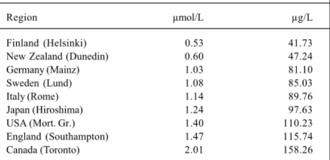
dietary allowances (RDA) in the United States of America are 55 µg/day for women and 70 µg/day for men. The se-lenium level in agricultural products is proportional to that nutrient’s content in the soil where the agricultural products are cultivated. The geochemical composition of certain regions, such as Finland, New Zealand, the eastern coast of the USA, and China, reveals selenium deficiency in the soil 10.
countries about the normal selenium serum level reported values ranging from 0.53 µmol/L in Finland to 2.01 µmol/L in Canada 11 (tab. I). As far as we know, no similar study has
been conducted in the Brazilian population, and, in our country, the geographical regions are as different as the composition of their soils.
The results of a number of studies about cardiac
di-sease induced by seleniumdeficiency, such as Keshan
disease that has been controlled with oral replacement of sodium selenite, have been published 12-14. Most of the
stu-dies reported patients who had undergone long-term paren-teral nutrition without selenium replacement, or patients with acquired immunodeficiency syndrome and severe un-dernourishment 15-17. However, the association between low
selenium levels and cardiovascular diseases remains con-troversial.
A low copper-content diet also results in cardiomyo-pathy in rats 18. The National Research Council in the USA
established the safe content of daily copper intake between 1.5 and 3.0 mg/day. The exact mechanisms of cellular injury in copper deficiency have not yet been clarified. However, because the superoxide dismutase enzyme activity is redu-ced with copper deficiency, we can assume that the lesions found in the myocardial basement membrane result from the loss of protection against the free oxygen radicals produced 18.
The treatment with antioxidants of rats with cardiac disease induced by copper deficiency resulted in a reduction in cardiac hypertrophy and milder mitochondrial injury 19. In
15-day-old pigs receiving a copper-deficient diet for 8 weeks, an increase in the heart weight/body weight ratio and electrocardiographic alterations were observed, confir-ming the findings in rats 20. In patients undergoing
myocar-dial revascularization, a lower concentration of microele-ments was observed, with the occurrence of heart failure in the early postoperative period, more frequent in patients with lower copper serum levels 21.
The present study aimed at assessing thiamin, sele-nium, and copper serum levels in patients with idiopathic dilated cardiomyopathy using diuretics, and their compari-son with those of an equal number of healthy individuals. The nutritional status of these patients with cardiac disease was assessed, as were the possible associations between undernourishment and the deficiency of the micronutrients
studied and between their serum levels and cardiac func-tion, which was evaluated on echocardiography.
Methods
From 1999 to 2000, we carried out an observational study comprising 30 patients with idiopathic dilated cardio-myopathy and an equal number of healthy controls. All par-ticipants were informed about the nature of the study and its objectives and provided written informed consent. The research project was submitted to and approved by the committee on ethics and research at the Hospital Universi-tário Pedro Ernesto - UERJ. The groups were paired by age, sex, and skin color (white and nonwhite). The patients had been diagnosed with idiopathic dilated cardiomyopathy 22
and were taking diuretics on a daily basis for a period ≥ 15 days. Patients with renal and hepatic dysfunction and alco-holic individuals were excluded from the study 23-25.
The patients underwent anamnesis, physical examina-tion, laboratory tests, electrocardiography, single- and 2-di-mensional echocardiography with Doppler, and nutritional assessment. The blood samples were withdrawn between 8 and 9 AM after a 12-hour fasting period. Routine biochemical
measurements, including all electrolytes, hepatic function test, and lipid profile were performed.
Thiamin assessment was performed by measuring the erythrocytic transketolase activity and the effect of thiamin pyrophosphate, as reported by Myron Brin 4. The following
values were considered as normal limits: erythrocytic trans-ketolase activity ≥ 800 µghexose.mL-1.h -1; effect of thiamin
pyrophosphate < 16%. Thiamin deficiency was defined as the simultaneous presence of low erythrocytic transketo-lase activity and the high effect of thiamin pyrophosphate 26.
The sera in which selenium and copper were measured were stored in a refrigerator. The serum levels of the micronu-trients were obtained by hydride generation and flame ato-mic absorption spectrophotometry, respectively according to the methodologies of Navarro et al 27 and Terrés-Martos
et al 28. The accuracy and precision of the method were tested
with 2 sera with internationally recognized standardized selenium and copper concentrations as follows: 1) Contox trace metal serum control - Kaulson Laboratory, Inc., NJ, USA; 2) Seronorm trace elements serum, produced by SERO AS, Billingstad, Norway.
Nutritional evaluation was performed through nutri-tional history, anthropometric assessment, measurement of serum albumin, lymphocyte count, and global subjective assessment 29,30.
The means were compared with the Student t test for independent samples. We adopted p≤0.05 as the significance level. The chi-square test was used to compare frequencies. To assess the association between numerical data, the cor-relation (r) and determination (r2) coefficients were calculated.
Results
The composition of the groups studied is shown in table II.
Table I - Mean selenium serum or plasma values in µmol/L and µg/L
in adults of different countries 6
Region µmol/L µg/L
Finland (Helsinki) 0.53 41.73
New Zealand (Dunedin) 0.60 47.24
Germany (Mainz) 1.03 81.10
Sweden (Lund) 1.08 85.03
Italy (Rome) 1.14 89.76
Japan (Hiroshima) 1.24 97.63
USA (Mort. Gr.) 1.40 110.23
England (Southampton) 1.47 115.74
Canada (Toronto) 2.01 158.26
Furosemide dosages ranged from 20 to 160mg/day. Only 1 patient was not taking furosemide but was taking thiazide and spironolactone, and it appeared in the prescription of 10 (33%) patients at the dosages of 25 (6 patients - 20%) or 100 mg/day (4 patients - 13%). Six (20%) patients took thiazides at the dosages of 12.5 (3%), 25 (7%), and 50mg/day (10%). In 5 (16%) of these patients, furosemide was added, and, in 2 (6%) of these patients, spironolactone was added. One (3%) patient took a combination of the 3 diuretics. Forty-six percent of the patients with cardiac disease took furosemide doses ≥ 80mg/day.
The following medications were also used in the patients with cardiac disease: digoxin (97%), angiotensin-converting en-zyme inhibitors (86%), amiodarone (13%), coumarin anticoagu-lants (13%), carvedilol (23%), and acetylsalicylic acid (6%). Eighty percent of the patients used the combination of digitalis, diuretics, and angiotensin-converting enzyme inhibitors.
The functional classes of the patients with cardiac di-sease (NYHA) were as follows:I, 17%; II, 53%; III, 23%; and IV, 7% (tab. III).
The groups had similar nutritional status according to both the objective and global subjective assessments, with cases of overweight and obesity.
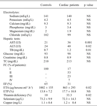
No significant alterations were observed in the elec-trolytes or in the hepatic and renal functional tests in either group. Dyslipidemia was highly prevalent both in patients with cardiac disease and in the controls (83.3% and 76.6%, respectively), table III.
Left ventricular ejection fraction (LVEF) in patients with cardiac disease ranged from 12 to 57% (mean = 31%). The subjective assessment of left ventricular function showed severe systolic dysfunction in 83.3% of the patients, moderate systolic dysfunction in 10%, and mild systolic dysfunction in 6.7% of the patients. Left bundle-branch block was observed in 60% of the patients with cardiac disease. Coronary angiography was performed in 50% of the patients with cardiac disease and was normal in all of them.
The means of the erythrocytic transketolase activity in the controls and in patients with cardiac disease were 1002±155 and 863±293 µg glucose.mL-1.h-1 (ranges: 706 to
1347 and 145 to 1344 µg glucose.mL-1.h-1), respectively
(p=0.02). The mean effect of thiamin pyrophosphate was 13.8±7.2% (1.4 to 31.5%) in the controls, and 17.7±10.8% (1.3 to 46.2%) in the patients with cardiac disease (p=0.12). Thiamin deficiency was observed in 3 controls and in 10 patients with cardiac disease (p=0.02), table III. No signifi-cant difference in the frequency of thiamin deficiency was observed between patients with cardiac disease taking furo-semide doses < 80mg/day and those taking furofuro-semide do-ses greater than that value (p=0.07). The use of spironolac-tone did not decrease thiamin deficiency frequency in the patients studied with cardiac disease (p=0.28). Thiamin de-ficiency was not more frequent in the patients taking the fu-rosemide/thiazide combination (p=0.17). No association was observed between thiamin serum level and left ventri-cular ejection fraction in patients with cardiac disease in the assessment performed either through erythrocytic transke-tolase activity (r = - 0.05) or through the effect of thiamin pyrophosphate (r=0.07). No significant difference was ob-served in the mean left ventricular ejection fraction of patients with cardiac disease who had thiamin deficiency (LVEF = 32.6%) and those who did not have thiamin defi-ciency (LVEF = 30.7%), p=0.68. None of the 10 patients with thiamin deficiency was undernourished.
In regard toselenium serum level, the accuracy (exact-ness) and precision (reproducibility) of the method are shown in table IV. With the reference material of Seronorm, 102.4% accuracy and 5.3% precision were obtained.
The mean selenium serum levels in controls and patients with cardiac disease were, respectively, 73.2±9.9 µg/ L (56.5 to 94.5 µg/L) and 72.3±14.3 µg/L (35.5 to 94 µg/L) with p=0.77. The results are visually compared in figure 1. No association was observed between selenium serum levels and left ventricular ejection fraction (r= - 0.10; r2 = 0.01).
In regard to copper serum level, the method’s accuracy and precision observed with the use of the reference mate-Table III - Results (means and percentages) of the groups studied
Controls Cardiac patients p value
Electrolytes:
Sodium (mEq/L) 141 140 NS
Potassium (mEq/L) 4.2 4.5 NS
Calcium (mg/dL) 9.3 9.3 NS
Phosphorus (mg/dL) 3 3.4 NS
Magnesium (mg/dL) 2 1.9 NS
Chloride (mEq/L) 102 99 NS
Hepatic tests:
AST (UI) 21 36 0.001
ALT (UI) 24 40 0.02
TB (mg/dL) 0.7 1.2 0.01
Glucose(mg/dL) 93 98 NS
Creatinine (mg/dL) 0.8 0.9 NS
TC(mg/dL) 210 213 NS
FC(% of patients):
I 100 17
II _ 53
III _ 23
IV _ 7
EF(%) 65 31
ETA (µg hexose.ml-1.h-1) 1002 ± 155 863 ± 293 0.02 ETP(%) 13.8 ± 7.2 17.7 ± 10.8 NS Thiamin deficiency(%) 10 33.3 0.02 Selenium(µg/L) 73.2 ± 9.9 72.3 ± 14.3 NS Copper(mg/L) 01.1 ± 0.4 01.2 ± 00.4 NS
ns- nonsignificant; AST- aspartate aminotransferase; ALT- alanine amino-transferase; TB- total bilirubin; TC- total cholesterol; CF- functional class (New York Heart Association); EF- ejection fraction; ETA- erythrocytic transketolase activity; ETP- effect of thiamin pyrophosphate.
Table II - Characteristics of the groups studied
Group Mean age Sex Color
(years) M F white nonwhite
Cardiac patients 43.2 * 23 7 22 8
Controls 39.6 * 23 7 22 8
rial Contox are shown in table IV. Accuracy of 98.7% and precision of 3.8% were obtained.
The mean copper serum levels in controls and patients with cardiac disease were, respectively, 1.1±0.4 mg/L (0.6 to 1.8mg/L) and 1.2±0.4mg/L (0.6 to 2.2mg/L), p=0.27. Visual comparison of the data is shown in figure 2. No association was observed between copper serum levels and left ventricular ejection fraction in patients with cardiac disease (r = -0.25; r2 = 0.06).
Discussion
Due to the lack of Brazilian studies reporting the nor-mal range in our population for erythrocytic transketolase activity, for the effect of thiamin pyrophosphate, and for se-lenium and copper serum levels, the formation of a control group with individuals with no evidence of disease or use of any medication paired by age, sex, skin color, and nutritional status was required.
From the clinical point of view, most patients with car-diac disease were compensated in NYHA functional classes I or II, even though they had severe myocardial disease ac-cording to echocardiographic data (83.3% had severe systo-lic dysfunction).
Both groups had similar nutritional profiles as follows: mean BMI of controls and patients with cardiac disease, res-pectively, 26.2 and 26kg/m2; mean body fat percentage,
res-pectively, 24.3 and 25%; mean CMB, resres-pectively, 26 and 24.4cm; and no significant alteration in serum albumin. Over-weight and obesity were present in both groups.
Three patients with cardiac disease had an increase in hepatic aminotransferases (AST and ALT) and total bilirubin greater than twice the normal limit. On echocardiography, these three patients had severe systolic dysfunction, 2 of whom had left ventricular ejection fraction < 20%. On physical examination, they had tender hepatomegaly and ju-gular venous distension, suggesting hepatic congestion as the cause of the enzymatic alteration, which is the reason why we did not find it necessary to measure hepatitis viral markers.
Assessment of thiamin levels should include erythro-cytic transketolase activity and the effect of thiamin pyro-phosphate on erythrocytic transketolase activity, which reflects the percentage of increase in this activity after the
addition of thiamin pyrophosphate to the hemolysate. Ho-wever, until the present time, no consensus exists in regard to the normal levels of erythrocytic transketolase activity due to the use of different methods of measurement by seve-ral laboratories 30. Two aspects should be considered in the
analysis of the erythrocytic transketolase activity 31: a) the
method involves the activity of other erythrocytic enzymes, such as ribose isomerase and xylulose epimerase, which do not depend on thiamin, and, in case of their deficiency, this may influence the result of the test, because no increase in erythrocytic transketolase activity will be detected (asses-sed by the formation of hexoses in the hemolysate), and oc-casional cases of thiamin deficiency will not be identified; b) thiamin deficiency may also cause a reduction in apotrans-ketolase and a weak response of that to thiamin pyrophos-phate, with the same consequence. These aspects show the importance of considering the simultaneous existence of low erythrocytic transketolase activity and the high effect of thiamin pyrophosphate as a criterion for the diagnosis of thiamin deficiency.
Dietary standardization was not adopted in this study because the objective of the study was to assess the status of the micronutrients in the nutritional reality of its participants. By limiting thiamin intake to 1.5mg/day, Seligmann et al 3
assessed the role played by diuretics in thiamin depletion, but they imposed a regimen of that micronutrient ingestion Table IV - Accuracy and precision of selenium and copper measurements
Material Concentration Accuracy Precision Certified Measure (%) RSD (%)
Selenium (mg/L) Seronorm 80 ± 13 81.9 ± 4.4 102.4 5.3 NIST-RM 3149
Copper (mg/L) Contox 00.8 ± 12 00.8 ± 0.03 98.7 3.8 RM-0148
RSD- relative standard deviation; NIST- National Institute of Standards; RM- reference material.
Fig. 1 - Selenium serum level. Two patients with cardiac disease had levels below 50 mg/L.
Selenium serum level (µg/L)
Controls Patients with cardiac disease
2 . 4
2 . 0
1 . 6
1 . 2
0 . 8
0 . 4
Fig. 2 - Copper serum level. The groups had similar copper serum levels.
Copper serum level (mg/L)
on study participants, which may not have matched the dietary reality of the patients.The usual ingestion of a higher thiamin content, both due to the presence of food richer in that nutrient or due to its conservation and prepa-ration avoiding the action of thiaminases, could overcome the losses imposed by the diuretics, reducing the clinical importance of the increased urinary excretion of thiamin in that population.
Daily ingestion of the nutrients was not estimated in the study because of the difficulty in precisely establishing the nutrient’s content in the food ingested, as well as their amount, in the absence of a standardized diet. The influen-ces of the proinfluen-cesses of food conservation and preparation in the bioavailability of thiamin also contributed making this assessment difficult 30, as did ignorance about the selenium
and copper contents in food cultivated in Brazil.
In a previous study 32, considering only the elevated
effect of thiamin pyrophosphate as a criterion for thiamin deficiency, a possible effect of spironolactone as a saver of thiamin spoliation induced by furosemide was observed. In the present study, a significant difference between the means of the effect of thiamin pyrophosphate in takers and nontakers of spironolactone was observed; however, adop-ting the new criterion here presented (low erythrocytic transketolase activity and elevated effect of thiamin pyrop-hosphate), no significant difference in the prevalence of thiamin deficiency between the groups was observed (p=0.28). Even though greater thiamin spoliation should be expected in patients taking the combination furosemide/ thiazide due to the increased urinary flow, it was not obser-ved in the present study, maybe because of the small number (5) of patients taking that combination.
The mean selenium serum level in controls (73.2± 9.9 µg/L) was similar to that reported by Navarro et al 27 in
Spain (74.90 µg/L); when compared with those shown in table I, it is among the lowest levels, above only those repor-ted in Finland and New Zealand, regions with selenium-poor soils. Despite the report by Inoko et al 33 of a case of
cardiomyopathy related to the selenium serum level of 62 µg/L, the most frequently reported levels in the literature are below 50 µg/L 11,13,15. In the Chinese cardiomyopathy cases,
the selenium serum levels reported were below 30 µg/L 11.
However, we do not know the role played by intermediate serum levels in worsening the already present cardiomyo-pathy or cardiomyocardiomyo-pathy with other etiologies. No associa-tion was observed between the selenium and copper levels in patients with cardiac disease and their cardiac function assessed by left ventricular ejection fraction. We believe
that this was due to the existence of another etiological me-chanism for the myocardial disease of the patients studied, in addition to the record of only 2 cases with low selenium serum levels, which did not allow a better evaluation of the possible influence of this deficiency in worsening the un-derlying disease.
The mean copper serum level in the control group (1.09±0.39mg/L) is similar to that reported by Terrés-Martos et al 28 (1.10mg/L), and to others observed in other countries,
such as the USA (0.9±0.03mg/L) 34 and Japan (0.98±
0.11mg/L) 35. No significant difference was observed between
the mean copper serum level in controls and that in patients with cardiac disease (p=0.27), suggesting a good content of that element in the food ingested by the patients studied. The preferentially hepatic excretion of copper minimizes the influence of the diuretics in copper spoliation.
In this study, thiamin deficiency was significantly more frequent in patients with cardiac disease, occurring in 1/3 of the patients, raising the possibility that the syste-matic replacement of that micronutrient might contribute to better myocardial performance in patients taking diu-retics for prolonged periods. However, it is worth empha-sizing the nonexistence in the Brazilian market of a com-mercial product with a thiamin unit dose adequate for that purpose. The only product available for oral administration has 300mg/tablet, which seems excessive, even though the minimum dose necessary to compensate for the losses induced by diuretics is yet to be defined. As thiamin de-ficiency has not been associated with protein-caloric un-dernourishment, we should consider the possibility of that vitamin deficiency even in the absence of the classical signs of undernourishment.
No significant difference in the mean selenium and copper serum levels was observed between controls and patients with cardiac disease. However, 2 patients with car-diac disease (6.6%) had selenium < 50 µg/L, which we consi-der significant, because such levels have already been re-ported as sufficient to induce myocardial dysfunction 6,8,10.
This finding raises the question of the possible benefit of selenium replacement in patients with idiopathic dilated car-diomyopathy refractory to the conventional therapeutic schemes. Even though serum measurement should precede replacement of the microelement, we should consider the difficulty of this procedure in Brazil, especially in public health units. However, the results here presented seem not to justify the systematic replacement ofselenium and cop-per in idiopathic dilated cardiomyopathy.
References
1. Yui Y, Fujiwara H, Mitsui H. Furosemide-induced thiamine deficiency. Jpn Circ J 1978; 4: 744.
2. Yui Y, Itokaua Y, Kawai C. Furosemide-induced thiamine deficiency. Cardiovasc Res 1980; 14: 537-40.
3. Seligmann H, Halkin H, Rauchfleisch S, et al. Thiamine deficiency in patients with
congestive heart failure receiving long-term furosemide therapy: a pilot study. Am J Med 1991; 91: 151-5.
5. Department of Medicine, Sheba Medical Center. Urinary thiamine excretion in the rat: effects of furosemide, other diuretics, and volume load. J Lab Clin Med 1999; 134: 232-7.
6. Rieck J, Halkin H, Almog S, et al. Urinary loss of thiamine is increased by low do-ses of furosemide in healthy volunteers. J Lab Clin Med 1999; 134: 238-43. 7. Shimon I, Shlomo A, Vered Z, et al. Improved left ventricular function after
thiamine supplementation in patients with congestive heart failure receiving long-term furosemide therapy. Am J Med 1995; 98: 485-90.
8. Dmerc, United Arab Emirates University, Al-Ain. Thiamin status in furosemide-treated rats. Pharmacol Res 2000; 42: 21-4.
9. Keshan Disease Research Group. Observations on effect of sodium selenite in prevention of Keshan disease. Chin Med J Engl 1979; 92: 471-6.
10. Burk RF, Levander OA. Selenium. In: Shils ME, Olson JA, Shike M, Ross AC -Modern Nutrition in Health and Disease, 9th ed.. Baltimore: Willians & Willians, 1999: 265-76.
11. Lockitch G. Selenium: clinical significance and analytical concepts. Crit Rev Clin Lab Sci 1989; 27: 483-541.
12. Reeves WC, Marcuard SP, Willis SE, Movahed A. Reversible cardiomyopathy due to selenium deficiency. J Parent Ent Nut 1989; 13: 663-5.
13. Oster O, Prellwitz W, Kasper W, Meinertz T. Congestive cardiomyopathy and se-lenium content of serum. Clin Chim Acta 1983; 128: 125-32.
14. Collipp PJ, Chen SY. Cardiomyopathy and selenium deficiency in a two-year-old girl. N Engl J Med 1981; 304: 1304-5.
15. Fleming CR, Lie JT, Maccall JT, et al. Selenium deficiency and fatal cardiomyopathy in a patient on home parenteral nutrition. Gastroenterology 1982; 83: 689-93. 16. Quercia RA, Korn S, O’neill D, et al. Selenium deficiency and fatal cardiomyopathy in
a patient receiving long-term home parenteral nutrition. Clin Pharm 1984; 3: 531-5. 17. Duorkin BM, Antonecchia PP, Smith F, et al. Reduced cardiac selenium content in the acquired imunodeficiency syndrome. J Parent Ent Nut 1989; 13: 644-7. 18. Medeiros DM, Davidson J, Jenkins JE. A unified perspective on copper
deficien-cy and cardiomyopathy. Proc Soc Exp Biol Med 1993; 203: 262-73. 19. Viestenz KE, Klevay LM. A randomized trial of copper therapy in rats with
eletrocar-diographic abnormalities due to copper deficiency. Am J Clin Nutr 1982; 35: 258-66. 20. Wildman RLH, Medeiros DM, Hamlin R, et al. Aspects of cardiomyopathy in copper-deficient pigs - electrocardiography, echocardiography, and ultrastruc-tural findings. Biol Trace Elem Res 1996; 55: 55-70.
21. Dementéva II, Adrianova MIL, Dzemeshkevich SL, Iavorovskii AG, Lokshin LS. Changes in the content of microelements - copper, zinc, iron - in the blood of patients following cardiopulmonary bypass. Anesteziol Reanimatol 1993; 4: 50-3.
22. Report of 1995 World Health Organization/International Society and Federa-tion of Cardiology Task Force on the definiFedera-tion and classificaFedera-tion of cardiomyo-pathies. Circulation 1996; 93: 841-3.
23. Burch GE, Gilles TD. Alcoholic cardiomyopathy: concept of disease and its treatment. Am J Med 1971; 50: 141-5.
24. Richardson PJ, Wodak AA. Alcohol-induced heart muscle disease. In: Symons C, Evans T, Mitchell AG. Specific Heart Muscle Disease. Bristol: Wright P.S.G., 1983: 99-122.
25. Gillet C, Julliere Y, Pirollet P, et al. Alcohol consumption and biological markers for alcoholism in idiopathic dilated cardiomyopathy:case-controlled study. Al-cohol AlAl-coholism 1992; 27: 353-8.
26. Tanphaichitr V, Wood B. Thiamin. In: Shils ME, Olson JA, Sihe M, Ross AC. Modern Nutrition in Health and Disease. 8th ed. Philadelphia: Lea & Febiger, 1994: 359-65.
27. Navarro M, Lopez H, Ruiz ML, et al. Determination of selenium in serum by hy-dride generation atomic absortion spectrometry for calculation of daily dietary intake. Sci Total Environ 1995; 175: 245-52.
28. Terrés-Martos C, Navarro-Alarcón M, Martín-Lagos F, GA H, López-Martinez MC. Determination of copper levels in serum of helthy subjects by atomic absorption spectrometry. Sci Total Environ 1997; 198: 97-103. 29. Baxter YC. Avaliação nutricional do cardiopata. Rev Soc Cardiol Est São Paulo
1997; 7: 445-57.
30. Dehoog S. The assessment of nutritional status. In: Mahan LK, Escott-Stump S. Food, Nutrition and Diet Therapy. 9th ed. Philadelphia: WB Saunders Co., 1996: 361-78.
31. Sauberlich HE. Biochimical alterations in thiamine deficiency - Their interpreta-tion. Am J Clin Nutr 1967; 20: 528-42.
32. Cunha S, Albanesi Fo FM. Espoliação por tiamina com o uso de diuréticos na car-diomiopatia dilatada. Arq Bras Cardiol 2000; 74(supl. 1): 80.
33. Inoko M, Konishi T, Matsusue S, Kobashi Y. Midmural fibrosis of left ventricle due to selenium deficiency. Circulation 1998; 98: 2638-9.