THE VALUE OF BRAINSTEM EVOKED POTENTIAL
IN CLINICAL DECISION OF A PATIENT WITH
HYPOXIC-ISCHEMIC ENCEPHALOPATHY
Anna Lecticia R. Pinto
1, Franciele C.S. Costa
2ABSTRACT - Establishing a prognosis for hypoxic-ischemic encephalopathy during the neonatal period is extremely difficult, as the neuroplasticity of the developing brain makes it almost impossible to measure the affected area. This case report describes a newborn with severe perinatal asphyxia and neonatal neu-rological syndrome including absent suck reflex. Normal brainstem auditory evoked potential led the di-agnosis towards a transitory dysfunction of deglutition, and the subject received daily stimulation in the hospital environment. Suck developed satisfactorily by day of life 30 and the patient was released without having to be tube fed. Neurophysiologic tests can be of value in the clinical decisions and analysis of func-tional prognosis of patients with hypoxic-ischemic encephalopathy.
KEY WORDS: hypoxic-ischemic encephalopathy, evoked potential, prognosis, newborn.
O valor do potencial evocado auditivo em decisão clínica em paciente com síndrome hipóxico-isquêmica
RESUMO - Estabelecer o prognóstico da encefalopatia hipóxico-isquêmica durante o período neonatal é extremamente difícil, devido à neuroplasticidade do cérebro em desenvolvimento que impede a medida exata das áreas afetadas. Este relato descreve um recém-nascido a termo com grave asfixia perinatal e sín-drome neurológica pós-natal, incluindo ausência do reflexo de sucção. O potencial evocado auditivo do tronco cerebral foi normal, sugerindo o diagnóstico de disfunção transitória da deglutição. Após estimu-lação diária no hospital a sucção foi obtida satisfatoriamente, e o paciente recebeu alta sem necessidade de alimentação enteral. Os testes neurofisiológicos podem ser de grande valor em decisões clínicas e aná-lise funcional prognóstica de pacientes com encefalopatia hipóxico-isquêmica.
PALAVRAS-CHAVE: encefalopatia hipóxico-isquêmica, potencial evocado, prognóstico, recém-nascido.
1MD,PhD, Pediatric Neurologist, Clínica Neurológica e Neurocirúrgica de Joinville, Professora do Departamento de Medicina da
Universidade da Região de Joinville SC, Brazil (UNIVILLE); 2Medical student, UNIVILLE.
Received 24 November 2006. Accepted 30 March 2007.
Dra. Anna Lecticia R. Pinto - Rua Plácido Olímpio de Oliveira 1244 - 89202-451 Joinville SC - Brasil. E-mail: anna.pinto@uol.com.br
Arq Neuropsiquiatr 2007;65(3-A):689-692
Perinatal asphyxia is considered one of the main risk factors for global developmental delay. The neo-natal neurological syndrome is frequently associated with severe asphyxia and is the major clinical indica-tion of brain tissue lesion due to insufficient oxygen supply, i.e. hypoxic-ischemic encephalopathy (HIE). Common symptoms are: variable change in level of alertness, including stupor or coma; ventilatory dis-turbances; disorders in brainstem vital reflexes; hypo-tonia; and seizures1
. Although neurologists frequent-ly perform prognostic evaluations of newborns with HIE, accurate prediction of neurologic outcome in sur-vivors remains difficult. One major obstacle is the lack of data for evaluating the intensity of insults, as the vast majority occur during the intrauterine period1-3.
Various studies have attempted to correlate outcome with indicators such as blood biochemical markers,
for example lactate and pH; Apgar scores; neonatal neurological syndrome characteristics; findings from neurodiagnostic methods such as ultrasound, CT scan, and MRI and results of neurophysiologic tests such as
electroencephalogram and evoked potentials3
. The latter neurophysiologic methods are particularly use-ful in evaluating brain maturity in both preterm and full-term newborns4-5
.
We present a full-term newborn with asphyxia at birth and neonatal neurological syndrome, in whom brainstem evoked potential proved useful in the prognosis of a transitory disturbance in deglutition.
CASE
690 Arq Neuropsiquiatr 2007;65(3-A)
of any other personal or familial medical condition. Elec-tronic fetal monitoring performed a short time prior to delivery revealed sustained fetal bradycardia and, a few minutes later, absence of heartbeat. Emergency caesarian section was performed secondary to possible abruptio pla-centae.
Apgar score was zero at 1 minute. Resuscitation consist-ed of positive pressure ventilation of O2 to 100%, cardiac
massage and two doses of adrenalin. Following resuscita-tion efforts, heartbeat rose to over 100 beats per minute. Apgar score was 2 at 5 minutes.
The subject was admitted to the neonatal intensive care unit (NICU) on mechanical ventilation, presenting with gen-eralized hypotonia and irregular respiratory movements. During the first hour of life, pH from systemic arterial blood was 6.75; CSF analysis was normal. Before 12 hours of life, the patient suffered an episode of clonic seizure activity which was successfully controlled with phenobarbital.
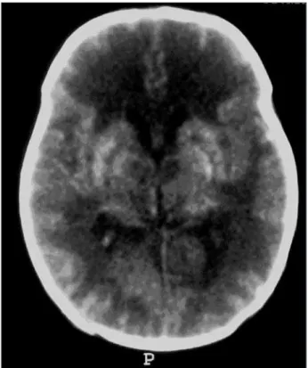
The subject was released from the NICU on day of life 14. After clearance of sedatives, neurologic evaluation re-vealed absent search and suck reflexes. On day of life 14, the subject underwent an evoked otoacoustic emissions test which showed normal cochlear activity. CT scan (Fig 1) performed on day of life 15 showed large areas of white matter hypodensity. EEG (Fig 2) performed on day of life 15 revealed low voltage and bursting of 1 to 2 seconds con-sisting of high-voltage sharp waves/spikes in background activity (suppression burst pattern), suggesting severe dif-fuse encephalopathy.
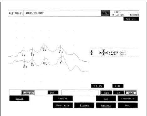
For prognostic purposes, brainstem auditory evoked po-tential exam (Fig 3) was conducted on day of life 24 and showed preserved brainstem activity. Stimulation of the
subject’s reflexes was subsequently intensified in order to develop the suck process, which was obtained by day of life 30. She was subsequently released from the hospital on day of life 39. A gastric feeding tube was not required.
Fig 1. Unenhanced CT showing large areas of the white matter hypodensity and bilateral basal ganglia hemorrhage.
Arq Neuropsiquiatr 2007;65(3-A) 691
DISCUSSION
Hypoxic-ischemic encephalopathy during the neo-natal period remains an important problem because of both its prevalence and its impact on neurode-velopment. Prognostic evaluation is challenging, as it is often not possible to measure the extent of the lesion nor to quantify the capacity of the brain to recover from the injury1-2
.
The first publications reporting evoked potential methods date back to the 1970s5. Some studies aimed
to verify the neurological prognostic value of evoked potentials in patients with HI insults. The relationship between degree of asphyxia and altered response in visual and somatosensory evoked potentials was well established6-8
. A study using a multimodality of evoked potentials demonstrated the accuracy of vi-sual and somatosensory evoked potentials in predict-ing the likelihood of neurologic sequelae9
. A study analyzing brainstem auditory evoked potential re-sponses in asphyxiated newborns found abnormali-ties in this exam in children with signs of brainstem dysfunction10. The signs described in this case showed
unequivocal evidence of HIE with apparent clinical damage to the brainstem. The suck reflex is a major clinical sign of vitality in the newborn. Sucking re-quires function of cranial nerves V, VII and XII; swal-lowing requires IX and X and the tongue requires XII. Feeding demands synchrony of sucking, swallowing, and the act of breathing. All of these functions are controlled by the brainstem and their elements are
intimately related1. In the present case, integrity of
the central auditory pathway indicated brainstem function in spite of physiologically evident clinical signs to the contrary. The disturbance was therefore interpreted as a temporary dysfunction secondary to ischemic events. As time is of the essence in initiating the oral feeding process, the results of the brainstem auditory evoked potential exam supported the plan of immediate treatment through increased stimula-tion. Risks associated with tube feeding, for example the increased risk of sudden infant death and the negative impact of tube feeding on the mother-child bond11-12, were thus avoided.
Anand et al.13
correlated brainstem auditory evok-ed potential to prognosis in children and newborns in coma after anoxic and/or traumatic encephalopathy. It was also related to poor prognosis when presented as abnormal; however, a normal test did not exclude the possibility of severe neurological sequelae13
. In the present case, follow-up of the subject after six months confirmed an abnormal neurologic outcome with global developmental delay and symptomatic epilepsy, although the wave latency values registered in the auditory evoked responses were within normal limits.
This report reinforces the importance of neuro-physiologic methods in the clinical and prognostic evaluation of hypoxic-ischemic encephalopathy. Re-sults of these exams can guide interventions to opti-mize developmental potential.
692 Arq Neuropsiquiatr 2007;65(3-A)
REFERENCES
1. Volpe JJ. Neurology of the Newborn. 4.Ed. Philadelphia: WB Saunders, 2001.
2. Shevell MI, Majnemer A, Miller SP. Neonatal neurologic prognostica-tion: the asphyxiated term newborn. Pediatr Neurol 1999;21:776-784. 3. Gonzalez de Dios J, Moya Benavent M, Izura Azanza V, Pastore
Olm-edo C. Electrophysiological studies in the follow-up of children with perinatal asphyxia history. An Esp Pediatr 1997;46:597-602.
4. Mercuri E, von Siebenthal �, Daniels �, et al. Multimodality evo�ed4. Mercuri E, von Siebenthal �, Daniels �, et al. Multimodality evo�edMultimodality evo�ed responses in the neurological assessment of the newborn. Eur J Pedi-atr 1994;153:622-631.
5. �rbe� A, �arlberg P, �jellmer I, et al. Clinical application of evo�ed electroencephalographic responses in newborn infants. I. Perinatal as-phyxia. Dev Med Child Neurol 1977;19:34-44.
6. Gibson NA, Graham M, Levene MI. Somatosensory evo�ed potentials and outcome in perinatal asphyxia. Arch Dis Child 1992;67:393-398. 7. Taylor MJ, Murphy WJ, Whyte �E. Prognostic reliability of
somato-sensory and visual evo�ed potentials of asphyxiated term infants. Dev Med Child Neurol 1992;34:507-515.
8. �arbord MG, Weston PF. Somatosensory evo�ed potentials predict neu-rologic outcome in full-term neonates with asphyxia. J Paediatr Child �ealth 1995;31:148-151.
9. Scalais E, Francois-Adant A, Nuttin C, et al. Multimodality evo�ed po-tentials as a prognostic tool in term asphyxiated newborns. Electroen-cephalogr Clin Neurophysiol 1998;108:199-207.
10. Fawer CL, Dubowitz LM, Levene MI, DubowitzV. Auditory brainstem responses in neurologically abnormal infants. Neuropediatrics 1983; 14:88-92.
11. �eusch�el RB, Fletcher �, �ill A, Buonomo C, Bousvaros A, Nur�o S. Isolated neonatal swallowing dysfunction: a case series and review of the literature. Dig Dis Sci 2003;48:30-35.
12. Guerriere DN, Mc�eever P, Llewellyn-Thomas �, Berall G. Mothers’ decisions about gastrostomy tube insertion in children: factors contrib-uting to uncertainty. Dev Med Child Neurol 2003;45:470-476. 13. Anand N�, Gupta A�, Raj �. Auditory brainstem response in neonates